. 2016 Aug 2;7(31):50131-50149.
doi: 10.18632/oncotarget.10321.

Inflammatory related gene IKKα, IKKβ, IKKγ cooperates to determine liver cancer stem cells progression by altering telomere via heterochromatin protein 1-HOTAIR axis

Affiliations

Inflammatory related gene IKKα, IKKβ, IKKγ cooperates to determine liver cancer stem cells progression by altering telomere via heterochromatin protein 1-HOTAIR axis

Jiahui An et al. Oncotarget. .

Abstract

Cancer stem cells are associated with tumor recurrence. IKK is a protein kinase that is composed of IKKα, IKKβ, IKKγ. Herein, we demonstrate that IKKα plus IKKβ promoted and IKKγ inhibited liver cancer stem cell growth in vitro and in vivo. Mechanistically, IKKα plus IKKβ enhanced and IKKγ inhibited the interplay among HP1α, HP1β and HP1γ that competes for the interaction among HP1α, SUZ12, HEZ2. Therefore, IKKα plus IKKβ inhibited and IKKγ enhanced the activity of H3K27 methyltransferase SUZ12 and EZH2, which methylates H3K27 immediately sites on HOTAIR promoter region. Therefore, IKKα plus IKKβ increased and IKKγ decreased the HOTAIR expression. Strikingly, IKKα plus IKKβ decreases and IKKγ increases the HP1α interplays with DNA methyltransferase DNMT3b, which increases or decreases TERRA promoter DNA methylation. Thus IKKα plus IKKβ reduces and IKKγ increases to recruit TRF1 and RNA polymerase II deposition and elongation on the TERRA promoter locus, which increases or decreases TERRA expression. Furthermore, IKKα plus IKKβ decreases/increases and IKKγ increases/decreases the interplay between TERT and TRRRA/between TERT and TREC. Ultimately, IKKα plus IKKβ increases and IKKγ decreases the telomerase activity. On the other hand, at the telomere locus, IKKα plus IKKβ increases/drcreases and IKKγ decreases/increases TRF2, POT1, pPOT1, Exo1, pExo1, SNM1B, pSNM1B/CST-AAF binding, which keep active telomere regulatory genes and poised for telomere length. Strikingly, HOTAIR is required for IKKα plus IKKβ and IKKγ to control telomerase activity and telomere length. These observations suggest that HOTAIR operates the action of IKKα, IKKβ, IKKγ in liver cancer stem cells. This study provides a novel basis to elucidate the oncogenic action of IKKα, IKKβ, IKKγ and prompts that IKKα, IKKβ, IKKγ cooperate to HOTAR to be used as a novel therapeutic targets for liver cancer.

Keywords: HOTAIR; IKKα; IKKβ; IKKγ; telomere.


Conflict of interest statement

The authors disclose no conflicts.

Figures

Figure 1
Figure 1. IKKα, IKKβ, IKKγ influence on human liver cancer stem cells (hLCSC) growth in vitro
A. The Western blotting analysis of CD133, CD24, Epcam in stable hLCSC cell lines transfected with pcDNA3.1-IKKα, pcDNA3.1-IKKβ, pcDNA3.1-IKKγ respectively.β-actin as internal control. B. The Western blotting analysis of IKKα, IKKβ, IKKγ in stable hLCSC cell lines transfected with pcDNA3.1-IKKα, pcDNA3.1-IKKβ, pcDNA3.1-IKKγ respectively.β-actin as internal control. C. Cells growth assay using CCK8. Each value was presented as mean±standard error of the mean (SEM). Data are means of value from three independent experiments, bar±SEM. **, P<0.01; *, P<0.05. D. S phase cells assay using BrdU. Each value was presented as mean±standard error of the mean (SEM). Data are means of value from three independent experiments, bar±SEM. **, P<0.01; *, P<0.05. E. Cells soft agar colony formation assay. Each value was presented as mean±standard error of the mean (SEM). Data are means of value from three independent experiments, bar±SEM. **, P<0.01; *, P<0.05.
Figure 2
Figure 2. IKKα, IKKβ, IKKγ influence on liver cancer stem cells growth in vivo
A. The mice were stratified and the tumors were recovered. The photography of xenograft tumor in the eight groups (indicated in left). B. The wet weight of each tumor was determined for each mouse. Each value was presented as mean±standard error of the mean (SEM). bar±SEM. **, P<0.01; *, P<0.05. C. The Xenograft appearance time (days). Each value was presented as mean±standard error of the mean (SEM). bar±SEM. **, P<0.01; *, P<0.05. D. A portion of each tumor was fixed in 4% paraformaldehyde and embedded in paraffin for histological hematoxylin-eosin (HE) staining (upper) and anti-PCNA immunostainning (lower). (original magnification×100). E. PCNA positive cells analysis. Each value was presented as mean±standard error of the mean (SEM). bar±SEM. **, P<0.01; *, P<0.05.
Figure 3
Figure 3. IKKα, IKKβ, IKKγ alter the methylation of HistoneH3 on lysine 27 dependent on HP1
A. Repeat Co-Immunoprecipitation (IP) with anti-HP1α, anti-HP1β followed by western blotting with, anti-HP1γ in liver cancer stem cells transfected with pcDNA3.1-IKKα, pcDNA3.1-IKKβ, pcDNA3.1-IKKγ, respectively. IgG IP as negative control. Western blotting with, anti-HP1γ as INPUT. B. Western blotting with anti-HP1α, anti-HP1β, anti-HP1γ in liver cancer stem cells transfected with pGFP-V-RS-HP1β or pGFP-V-RS-HP1γ, respectively. β-actin as internal control. C. Co-Immunoprecipitation (IP) with anti-HP1α or anti-HP1β followed by western blotting with, anti-HP1β or anti-HP1γ in liver cancer stem cells transfected with pGFP-V-RS-HP1β or pGFP-V-RS-HP1γ, respectively. IgG IP as negative control. Western blotting with anti-HP1β or anti-HP1γ as INPUT. D. Co-Immunoprecipitation (IP) with anti-SUZ12 followed by western blotting with anti-Histone H3, anti-EZH2, anti-HP1 α in liver cancer stem cells transfected with pGFP-V-RS-HP1β or pGFP-V-RS-HP1γ, respectively. IgG IP as negative control. Western blotting with anti-SUZ12 as INPUT. E. Co-Immunoprecipitation (IP) with anti-SUZ12 followed by western blotting with anti-Histone H3, anti-EZH2, anti-HP1 α in liver cancer stem cells transfected with pcDNA3.1-IKKα, pcDNA3.1-IKKβ, pcDNA3.1-IKKγ. respectively. IgG IP as negative control. Western blotting with anti-SUZ12 as INPUT. F. Western blotting with anti-H3K27me1, anti-H3K27me2, anti-H3K27me3, anti-H3K27Ac, anti-NFκB and anti-H3 in liver cancer stem cells transfected with pcDNA3.1-IKKα, pcDNA3.1-IKKβ, pcDNA3.1-IKKγ, respectively. β-actin as internal control.
Figure 4
Figure 4. IKKα, IKKβ, IKKγ regulate HOTAIR expression dependent on H3K27me3
A. Co-Immunoprecipitation (IP) with anti-HP1α followed by western blotting with, anti-H3K27me1, anti-H3K27me2, anti-H3K27me3 in liver cancer stem cells transfected with pcDNA3.1-IKKα, pcDNA3.1-IKKβ, pcDNA3.1-IKKγ respectively. IgG IP as negative control. Western blotting with anti-HP1 α as INPUT. B. Chromatin Immunoprecipitation (CHIP) with anti-H3K27me3 followed by PCR with HOTAIR promoter primers in liver cancer stem cells transfected with pcDNA3.1-IKKα, pcDNA3.1-IKKβ, pcDNA3.1-IKKγ respectively. IgG CHIP as negative control. PCR for HOTAIR promoter as INPUT. C. Chromatin Immunoprecipitation (CHIP) with anti-H3K27me3 followed by PCR with HOTAIR promoter primers in HP1 α depleted liver cancer stem cells transfected with pcDNA3.1-IKKα, pcDNA3.1-IKKβ, pcDNA3.1-IKKγ respectively. IgG CHIP as negative control. PCR for HOTAIR promoter as INPUT. D. HOTAIR promoter luciferase activity assay in liver cancer stem cells transfected with pcDNA3.1-IKKα, pcDNA3.1-IKKβ, pcDNA3.1-IKKγ, respectively. Data represent mean±SEM, n=3, **p<0.01, *p<0.05 represents difference significane. E. RT-PCR with HOTAIR primers in liver cancer stem cells transfected with pcDNA3.1-IKKα, pcDNA3.1-IKKβ, pcDNA3.1-IKKγ respectively. β-actin as internal control.
Figure 5
Figure 5. IKKα, IKKβ and IKKγ regulate TERRA expression
A. Co-Immunoprecipitation (IP) with anti-RNA polII or anti-DNMT3b followed by western blotting with, anti-TRF1 or anti-HP1α in liver cancer stem cells transfected with pcDNA3.1-IKKα, pcDNA3.1-IKKβ, pcDNA3.1-IKKγ respectively. IgG IP as negative control. Western blotting with anti-TRF1 or anti-HP1α as INPUT. B. Biotin-TERRA promoter DNA pulldown followed by Western blotting with anti-DNMT3b, anti-TRF1, anti-RNA polII in liver cancer stem cells transfected with pcDNA3.1-IKKα, pcDNA3.1-IKKβ, pcDNA3.1-IKKγ respectively. Biotin as INPUT and Histone as internal control. C. Chromatin Immunoprecipitation (CHIP) with anti-DNMT3b, anti-TRF1, anti-RNA polII followed by PCR with TERRA promoter primers in liver cancer stem cells transfected with pcDNA3.1-IKKα, pcDNA3.1-IKKβ, pcDNA3.1-IKKγ respectively. IgG CHIP as negative control. PCR for TERRA promoter as INPUT. D. TREEA promoter (Subtelomere region) methylation analysis by MspI plus BamHI digestion in liver cancer stem cells transfected with pcDNA3.1-IKKα, pcDNA3.1-IKKβ, pcDNA3.1-IKKγ respectively. E. TERRA promoter methylation analysis by Methylated DNA Immunoprecipitation (MeDIP)-Dot blot-western blotting with anti-5-Methylcytosine (5-mC) in expression in liver cancer stem cells transfected with pcDNA3.1-IKKα, pcDNA3.1-IKKβ, pcDNA3.1-IKKγ respectively. F. RT-PCR with TERRA primers in liver cancer stem cells transfected with pcDNA3.1-IKKα, pcDNA3.1-IKKβ, pcDNA3.1-IKKγ respectively. β-actin as internal control.
Figure 6
Figure 6. IKKα, IKKβ, IKKγ control telomerase activity
A. Super-EMSA assay with anti-TERT and biotin-TERRA probe in primers in liver cancer stem cells transfected with pcDNA3.1-IKKα, pcDNA3.1-IKKβ, pcDNA3.1-IKKγ respectively. Biotin as control. B. RNA Immunoprecipitation (RIP) with anti-TERT followed by RT-PCR with TERC and TERRA primers in liver cancer stem cells transfected with pcDNA3.1-IKKα, pcDNA3.1-IKKβ, pcDNA3.1-IKKγ respectively. IgG RIP as negative control. RT-PCR for TERC or TERRA as INPUT. C. Telomerase activity assay with TRAP method primers in liver cancer stem cells transfected with transfected with pcDNA3.1-IKKα, pcDNA3.1-IKKβ, pcDNA3.1-IKKγ respectively. Each value was presented as mean±standard error of the mean (SEM). **, P<0.01.
Figure 7
Figure 7. IKKα, IKKβ and IKKγ alter telomere length
A. Biotin-Telomere DNA pulldown followed by Western blotting with anti-POT1, anti-Exo1, anti-SNM1B, anti-TRF2, anti-CST/AAF in liver cancer stem cells transfected with pcDNA3.1-IKKα, pcDNA3.1-IKKβ, pcDNA3.1-IKKγ respectively. Biotin as INPUT and Histone as internal control. B. Chromatin Immunoprecipitation (CHIP) with anti-POT1, anti-Exo1, anti-SNM1B, anti-TRF2 followed by PCR with Telomere DNA primers in liver cancer stem cells transfected with pcDNA3.1-IKKα, pcDNA3.1-IKKβ, pcDNA3.1-IKKγ respectively. IgG CHIP as negative control. PCR for Ttelomere DNA promoter as INPUT. C. Super-EMSA assay with anti-TRF2 and biotin-Ttelomere DNA probe in primers in liver cancer stem cells transfected with pcDNA3.1-IKKα, pcDNA3.1-IKKβ, pcDNA3.1-IKKγ respectively. Biotin as control. D. The PCR detection of telomere repeat sequence in liver cancer stem cells transfected with pcDNA3.1-IKKα, pcDNA3.1-IKKβ, pcDNA3.1-IKKγ respectively. E. The real-time PCR detection of telomere length primers in liver cancer stem cells transfected with pcDNA3.1-IKKα, pcDNA3.1-IKKβ, pcDNA3.1-IKKγ, respectively. Each value was presented as mean±standard error of the mean (SEM). **, P<0.01.
Figure 8
Figure 8. HOTAIR depletion blocks IKKα, IKKβ, IKKγ function on telomere
A. (upper) Western blotting with anti-TERT in liver cancer stem cells transfected with pcDNA3.1, pcDNA3.1-IKKγ, pcDNA3.1-IKKγ plus pCMV6-A-HOTAIR, pcDNA3.1-IKKα plus pcDNA3.1-IKKβ, pcDNA3.1-IKKα plus pcDNA3.1-IKKβ plus pGFP–V-RS-HOTAIR, respectively. β-actin as internal control. (lower) RT-PCR with TERRA primers in liver cancer stem cells transfected with pcDNA3.1, pcDNA3.1-IKKγ, pcDNA3.1-IKKγ plus pCMV6-A-HOTAIR, pcDNA3.1-IKKα plus pcDNA3.1-IKKβ, pcDNA3.1-IKKα plus pcDNA3.1-IKKβ plus pGFP–V-RS-HOTAIR, respectively. β-actin as internal control. B. (upper) RNA Immunoprecipitation (RIP) with anti-TERT followed by RT-PCR with TERC or TERRA promoter primers in liver cancer stem cells transfected with pcDNA3.1, pcDNA3.1-IKKγ, pcDNA3.1-IKKγ plus pCMV6-A-HOTAIR, pcDNA3.1-IKKα plus pcDNA3.1-IKKβ, pcDNA3.1-IKKα plus pcDNA3.1-IKKβ plus pGFP–V-RS-HOTAIR, respectively. IgG RIP as negative control. RT-PCR for TERC or TERRA as INPUT. (lower) Co-Immunoprecipitation (IP) with anti-RNA polII or anti-DNMT3b followed by western blotting with, anti-TRF1 or anti-HP1α in liver cancer stem cells transfected with pcDNA3.1, pcDNA3.1-IKKγ, pcDNA3.1-IKKγ plus pCMV6-A-HOTAIR, pcDNA3.1-IKKα plus pcDNA3.1-IKKβ, pcDNA3.1-IKKα plus pcDNA3.1-IKKβ plus pGFP–V-RS-HOTAIR. IgG IP as negative control. Western blotting with anti-TRF1 or anti-HP1α as INPUT. C. (upper) RNA Immunoprecipitation (RIP) with anti-TERT followed by RT-PCR with TERC and TERRA primers in HOTAIR depleted liver cancer stem cells transfected with pcDNA3.1-IKKα, pcDNA3.1-IKKβ, pcDNA3.1-IKKγ respectively. IgG RIP as negative control. RT-PCR for TERC or TERRA as INPUT. (lower) Chromatin Immunoprecipitation (CHIP) with anti-TRF2 or antiCST/AAF followed by PCR with Telomere DNA primers in HOTAIR depleted liver cancer stem cells transfected with pcDNA3.1-IKKα, pcDNA3.1-IKKβ, pcDNA3.1-IKKγ respectively. IgG CHIP as negative control. PCR for Ttelomere DNA promoter as INPUT. D. Telomerase activity assay with TRAP method primers in liver cancer stem cells transfected with pcDNA3.1, pcDNA3.1-IKKγ, pcDNA3.1-IKKγ plus pCMV6-A-HOTAIR, pcDNA3.1-IKKα plus pcDNA3.1-IKKβ, pcDNA3.1-IKKα plus pcDNA3.1-IKKβ plus pGFP–V-RS-HOTAIR, respectively. Each value was presented as mean±standard error of the mean (SEM). **, P<0.01. E. The real-time PCR detection of telomere length primers in liver cancer stem cells transfected with pcDNA3.1, pcDNA3.1-IKKγ, pcDNA3.1-IKKγ plus pCMV6-A-HOTAIR, pcDNA3.1-IKKα plus pcDNA3.1-IKKβ, pcDNA3.1-IKKα plus pcDNA3.1-IKKβ plus pGFP–V-RS-HOTAIR, respectively. Each value was presented as mean±standard error of the mean (SEM). **, P<0.01.
Figure 9
Figure 9. HOTAIR operates the oncogenic action of IKKα, IKKβ, IKKγ
A. The Western blotting analysis of IKKα, IKKβ, IKKγ, TERT and RT-PCR analysis for HOTAIR, TERC, TERRA in HOTAIR depleted liver cancer stem cell lines transfected with pcDNA3.1-IKKα, pcDNA3.1-IKKβ, pcDNA3.1-IKKγ respectively.β-actin as internal control. B. Cells growth assay using CCK8. Each value was presented as mean±standard error of the mean (SEM). Data are means of value from three independent experiments, bar±SEM. **, P<0.01; *, P<0.05. C. Cells soft agar colony formation assay. Each value was presented as mean±standard error of the mean (SEM). Data are means of value from three independent experiments, bar±SEM. **, P<0.01; *, P<0.05. D. (left) The mice were stratified and the tumors were recovered. The photography of xenograft tumor in the eight groups. (right) The wet weight of each tumor was determined for each mouse. Each value was presented as mean±standard error of the mean (SEM). bar±SEM. **, P<0.01; *, P<0.05.
Figure 10
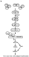
Figure 10. The schematic diagram illustrates a model that that IKKα plus IKKβ promoted and IKKγ inhibited liver cancer stem cell growth in vitro and in vivo
A. IKKα plus IKKβ enhanced and IKKγ inhibited the interplay among HP1α, HP1β and HP1γ that competes for the interaction among HP1α, SUZ12, HEZ2. Therefore, IKKα plus IKKβ inhibited and IKKγ enhanced the activity of H3K27 methyltransferase SUZ12 and EZH2, which methylates H3K27 immediately sites on HOTAIR promoter region. Therefore, IKKα plus IKKβ increased and IKKγ decreased the TOTAIR expression. Strikingly, IKKα plus IKKβ decreases and IKKγ increases the HP1α interplays with DNA methyltransferase DNMT3b, which increases or decreases TERRA promoter DNA methylation. So IKKα plus IKKβ reduces and IKKγ increases to recruit TRF1 and RNA polymerase II deposition and elongation on the TERRA promoter locus, which increases or decreases TREEA expression. Further on, IKKα plus IKKβ decreases/increases and IKKγ increases/decrease the interplay between TERT and TRRRA/between TERT and TREC. Ultimately, IKKα plus IKKβ increases and IKKγ decreases the telomerase activity. On the other hand, at the telomere locus, IKKα plus IKKβ increases/drcreases and IKKγ decreases/increases TRF2, POT1, pPOT1, Exo1, pExo1, SNM1B, pSNM1B/CST-AAF binding, which keep active telomere regulatory genes and poised for telomere length. B. HOTAIR is required for IKKα plus IKKβ and IKKγ to control telomerase activity and telomere length. HOTAIR depletion blocks the function of IKKα plusIKKβ, IKKγ on telomere.

Similar articles

Cited by

References

    1. Huang JJ, Chu HX, Jiang ZY, Zhang XJ, Sun HP, You QD. Recent advances in the structure-based and ligand-based design of IKKβ inhibitors as anti-inflammation and anti-cancer agents. Curr Med Chem. 2014;21:3893–91. - PubMed
    1. Shukla S, Kanwal R, Shankar E, Datt M, Chance MR, Fu P, MacLennan GT, Gupta S. Apigenin blocks IKKα activation and suppresses prostate cancer progression. Oncotarget. 2015;6:31216–32. doi: 10.18632/oncotarget.5157. - DOI - PMC - PubMed
    1. Li T, Wong VK, Jiang ZH, Jiang SP, Liu Y, Wang TY, Yao XJ, Su XH, Yan FG, Liu J, Leung EL, Yi XQ, Wong YF, et al. Mutation of cysteine 46 in IKK-beta increases inflammatory responses. Oncotarget. 2015;6:31805–19. doi: 10.18632/oncotarget.5567. - DOI - PMC - PubMed
    1. Singha B, Gatla HR, Phyo S, Patel A, Chen ZS, Vancurova I. IKK inhibition increases bortezomib effectiveness in ovarian cancer. Oncotarget. 2015;6:26347–58. doi: 10.18632/oncotarget.4613. - DOI - PMC - PubMed
    1. Li Z, Yang Z, Lapidus RG, Liu X, Cullen KJ, Dan HC. IKK phosphorylation of NF-κB at serine 536 contributes to acquired cisplatin resistance in head and neck squamous cell cancer. Am J Cancer Res. 2015;5:3098–110. - PMC - PubMed

MeSH terms