. 2016 Jul 1;11(7):e0158440.
doi: 10.1371/journal.pone.0158440. eCollection 2016.

MPT0B098, a Microtubule Inhibitor, Suppresses JAK2/STAT3 Signaling Pathway through Modulation of SOCS3 Stability in Oral Squamous Cell Carcinoma

Affiliations

MPT0B098, a Microtubule Inhibitor, Suppresses JAK2/STAT3 Signaling Pathway through Modulation of SOCS3 Stability in Oral Squamous Cell Carcinoma

Hsuan-Yu Peng et al. PLoS One. .

Abstract

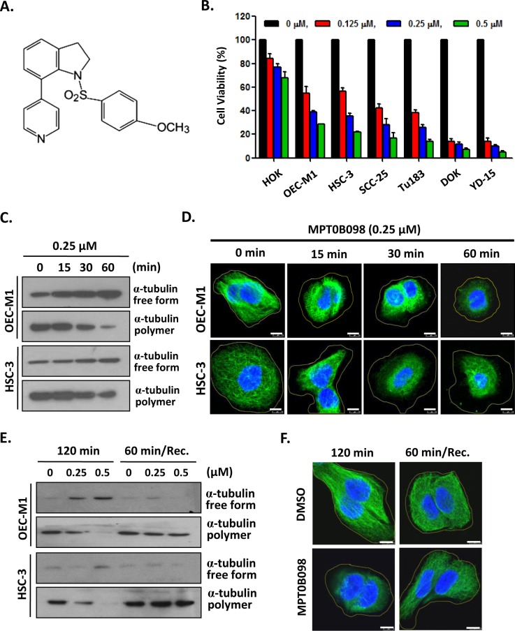
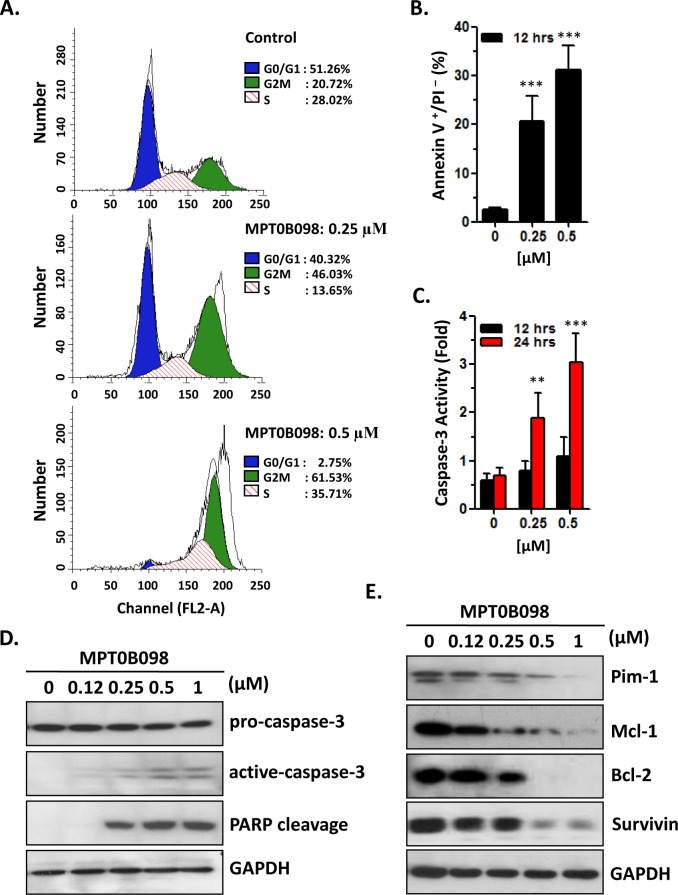
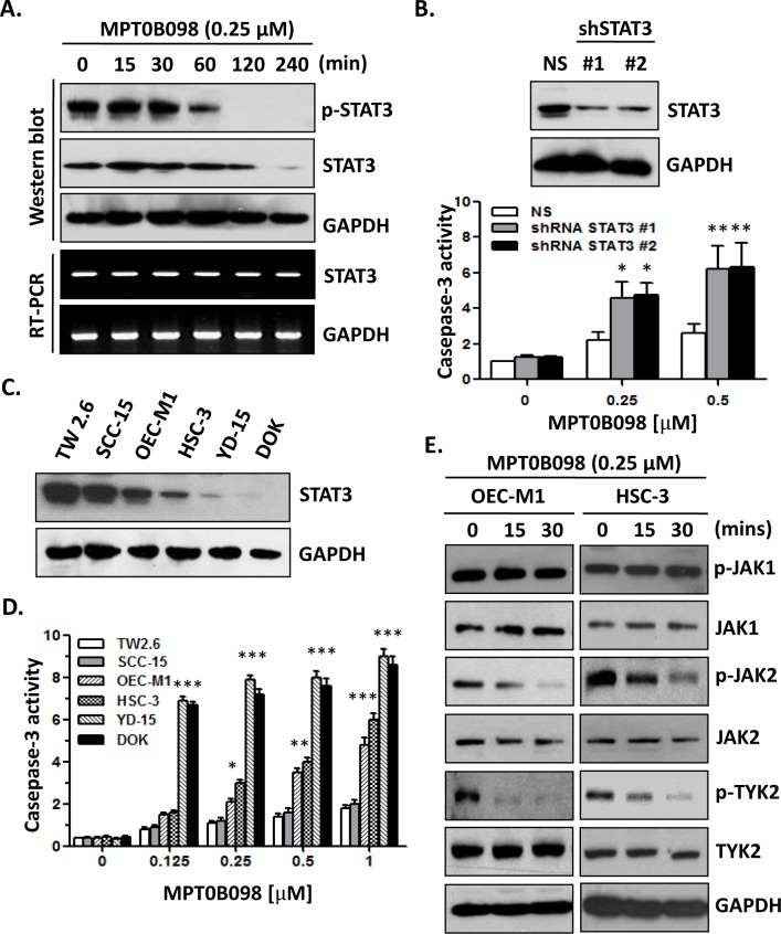
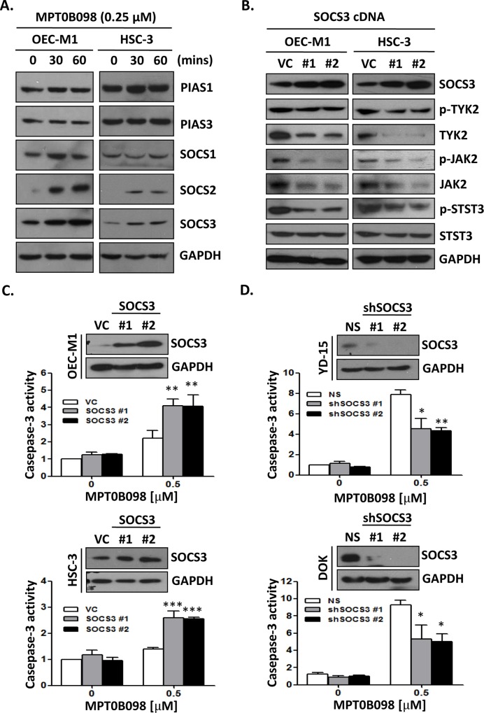
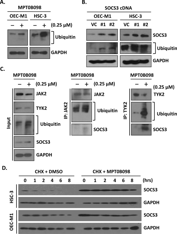
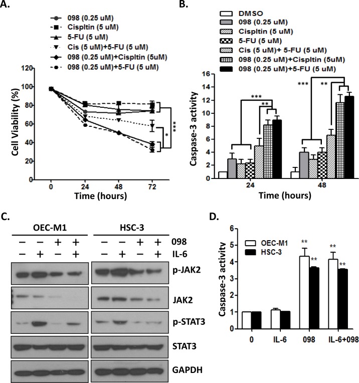
Microtubule inhibitors have been shown to inhibit Janus kinase 2/signal transducer and activator of transcription 3 (JAK2/STAT3) signal transduction pathway in various cancer cells. However, little is known of the mechanism by which the microtubule inhibitors inhibit STAT3 activity. In the present study, we examined the effect of a novel small-molecule microtubule inhibitor, MPT0B098, on STAT3 signaling in oral squamous cell carcinoma (OSCC). Treatment of various OSCC cells with MPT0B098 induced growth inhibition, cell cycle arrest and apoptosis, as well as increased the protein level of SOCS3. The accumulation of SOCS3 protein enhanced its binding to JAK2 and TYK2 which facilitated the ubiquitination and degradation of JAK2 and TYK2, resulting in a loss of STAT3 activity. The inhibition of STAT3 activity led to sensitization of OSCC cells to MPT0B098 cytotoxicity, indicating that STAT3 is a key mediator of drug resistance in oral carcinogenesis. Moreover, the combination of MPT0B098 with the clinical drug cisplatin or 5-FU significantly augmented growth inhibition and apoptosis in OSCC cells. Taken together, our results provide a novel mechanism for the action of MPT0B098 in which the JAK2/STAT3 signaling pathway is suppressed through the modulation of SOCS3 protein level. The findings also provide a promising combinational therapy of MPT0B098 for OSCC.


Conflict of interest statement

Competing Interests: The authors have declared that no competing interests exist.

Figures

Fig 1
Fig 1. MPT0B098 inhibits the proliferation and induces microtubules depolymerization in OSCC cells.
(A) Chemical structure of MPT0B098. (B) OSCC cells were treated with increasing concentrations of MPT0B098 for 72 hrs and the cell viability was assessed by MTT assay. Data are presents as mean ± SE relative to DMSO vehicle control (indicated as 0 μM) from three replicate experiments. *, p<0.05; **, p<0.01; ***, p<0.001. (C) OEC-M1 and HSC-3cells were incubated at 37°C from 0~60 min in the presence of 0.25 μM of MPT0B098. Free form and polymer form of microtubule were purified and assessed by Western blot analysis. (D) OEC-M1 and HSC-3 cells were treated with 0.25 μM of MPT0B098 from 0~60 min. Cells were fixed and then immunostained with anti-α-tubulin (green) antibody and then stained with DAPI (blue), followed by confocal microscopy. Scale bar = 7.5 μm. (E) OEC-M1 and HSC-3 cells were treated with MPT0B098 for 120 min at 37°C. For recovery (Rec.) assay, cells were treated with MPT0B098 for 60 min and then the drug was washed out to allow the microtubules to repolymerize for another 60 min. Cell lysates were analyzed by western blot using the anti-α-tubulin antibody. (F) The recovery assay was also done in OEC-M1 cells for immunostaining. After drug treatment, OEC-M1 cells were fixed and then immunostained with anti-α-tubulin (green) antibody and then stained with DAPI (blue), followed by confocal microscopy. Scale bar = 7.5 μm.
Fig 2
Fig 2. MPT0B098 induces the cell cycle arrest and apoptosis.
(A) OEC-M1 cells were treated with 0.25 or 0.5 μM of MPT0B098 for 12 hrs. Cells were then evaluated for effects on cell cycle using PI staining and analyzed by flow cytometry. Percentages of cells in different phases were shown. The data are representative of three independent experiments. (B) OEC-M1 cells were treated with different concentrations of MPT0B098 for 12 hrs. Apoptosis was assessed by annexin V/PI staining and analyzed by flow cytometry. The data are represented as mean ± SE; ***, p<0.001 versus vehicle control. (C) OEC-M1 cells were incubated with various concentrations of MPT0B098 for 12~24 hours and caspases-3 activity was assessed. The data are represented as mean ± SE; **, p<0.01; ***, p<0.001 versus vehicle control. (D) OEC-M1 cells were treated with different concentrations of MPT0B098 for 24 hrs. The proteolytic cleavage of caspase-3 and PARP were determined by Western blot analysis. GAPDH was used as protein loading control. (E) OEC-M1 cells were treated with different concentrations of MPT0B098 for 24 hrs. Effects on the expression of Pim-1, Mcl-1, Bcl-2 and survivin were determined by western blot analysis. GAPDH was used as protein loading control.
Fig 3
Fig 3. MPT0B098 modulates JAK2/STAT3 pathway in OSCC cells.
(A) OEC-M1 cells were treated with MPT0B098 (0.25 μM) for the indicated times. The phosphorylated STAT3 (pSTAT3) and total level of STAT3 were determined by Western blotting. The level of STAT3 mRNA was determined by RT-PCR. GAPDH was used as loading control. (B) Western blot analysis of STAT3 levels after 48 hrs transfection of OEC-M1 cells with control shRNA (NS) or two shRNA constructs (shSTAT3 #1, #2) against STAT3. GAPDH was used as loading control. These shRNA transfactants were treated MPT0B098 for 24 hrs and caspases-3 activity was assessed. The data are represented as mean ± SE; *, p<0.05; **, p<0.01 versus vehicle control. (C) Western blot analysis of endogenous STAT3 protein level in OSCC cells. GAPDH was used as loading control. (D) OSCC cells were incubated with various concentrations of MPT0B098 for 24 hrs and caspases-3 activity was assessed. Data are presents as mean ± SE relative to vehicle control from three replicate experiments. *, p<0.05; **, p<0.01; ***, p<0.001. (E) OEC-M1 and HSC-3 cells were treated with 0.25 μM of MPT0B098 for indicated times. The phosphorylated proteins (p-JAK1, p-JAK2 and p-TYK2) and total level of proteins (JAK1, JAK2 and TYK2) were determined by Western blotting. GAPDH was used as loading control.
Fig 4
Fig 4. MPT0B098 modulates JAK2/STAT3 pathway via SOCS3 accumulation.
(A) OEC-M1 and HSC-3 cells were treated with 0.25 μM of MPT0B098 for indicated times. The protein level of PIAS1, PIAS3, SOCS1, SOCS2, and SOCS3 were determined by Western blotting. GAPDH was used as loading control. (B) OEC-M1 and HSC-3 cells were tranfected with control vector (VC) and construct containing the SOCS3-coding region (SOCS3 cDNA #1, #2) for 48 hrs. The phosphorylated proteins (p-JAK2, p-TYK2 and p-STAT3) and total level of proteins (SOCS3, JAK2, TYK2 and STAT3) were determined by Western blotting. GAPDH was used as loading control. (C) Western blot analysis of SOCS3 levels after 48 hrs transfection of OEC-M1 and HSC-3 cells with control vector (VC) or two SOCS3 constructs (SOCS3 #1, #2). GAPDH was used as loading control. These SOCS3 overexpression transfactants were treated MPT0B098 for 24 hrs and caspases-3 activity was assessed. The data are represented as mean ± SE; **, p<0.01 versus vehicle control. (D) Western blot analysis of SOCS3 levels after 48 hrs transfection of YD-15 and DOK cells with control shRNA (NS) or two shSOCS3 constructs (shSOCS3 #1, #2). GAPDH was used as loading control. These shRNA transfactants were treated MPT0B098 for 24 hrs and caspases-3 activity was assessed. The data are represented as mean ± SE; ***, p<0.001 versus vehicle control.
Fig 5
Fig 5. MPT0B098-induced SOCS3 accumulation enhances the ubiquitination of JAK2 and TYK2.
(A) OEC-M1 and HSC-3 cells were treated with 0.25 μM MPT0B098 for 24 hrs and analyzed by Western blot for protein ubiquitination (ubiquitin). (B) Western blot analysis of SOCS3 and ubiquitin levels after 48 hrs transfection of OEC-M1 and HSC-3 cells with control vector (VC) or two SOCS3 constructs (SOCS3 #1, #2). (C) OEC-M1 cells were treated with 0.25 μM MPT0B098 for 24 hrs. The whole cell lysates (input) were immunoblotted with antibodies to JAK2, TYK2, ubiquitin and SOCS3 (left). Drug treated cell lysates were immunoprecipitated with anti-JAK2 (IP: JAK2) (middle) or anti-TYK2 (IP: TYK2) and then western blot for JAK2, TYK2, ubiquitin and SOCS3. (D) OEC-M1 and HSC-3 cells were treated with 10 μg/ml of cycloheximide in the presence of a vehicle (DMSO) or 0.25 μM MPT0B098 for the indicated times. The SOCS3 levels were determined by Western blotting. GAPDH was used as protein loading control.
Fig 6
Fig 6. Combination treatment significantly induces apoptosis in OSCC cells.
(A) OEC-M1 cells were treated with MPT0B098 (0.25 μM), cisplatin (5 μM), 5-fluorouracil (5-FU, 5 μM), cisplatin (5 μM)+5-FU (5 μM), 098 (0.25 μM)+ 5-FU (5 μM) and 098 (0.25 μM)+ cisplatin (5 μM) for the indicated times. The cell viability was assessed by MTT assay. (B) OEC-M1 cells were treated with MPT0B098 (0.25 μM), cisplatin (5 μM), 5-fluorouracil (5-FU, 5 μM), cisplatin (5 μM)+5-FU (5 μM), 098 (0.25 μM)+ 5-FU (5 μM) and 098 (0.25 μM)+ cisplatin (5 μM) for the indicated times and the caspase-3 activity was assessed. (C) OEC-M1 and HSC-3 cells were stimulated with IL-6 (10 ng/ml) in the presence or absence of MPT0B098 (0.25 μM) for the indicated times. Whole cell lysates were immunoblotted with antibodies to phosphorylated proteins (p-JAK2 and p-STAT3) and total level of proteins (JAK2 and STAT3). GAPDH was used as protein loading control. (D) OEC-M1 and HSC-3 cells were stimulated with IL-6 (10 ng/ml) in the presence or absence of MPT0B098 (0.25 μM) for 24 hrs and the caspase-3 activity was assessed. All data are presents as mean ± SE relative to DMSO vehicle control from three replicate experiments. *, p<0.05; **, p<0.01; ***, p<0.001.

Similar articles

Cited by

References

    1. Yu H, Jove R. The STATs of cancer—new molecular targets come of age. Nat Rev Cancer. 2004;4(2):97–105. 10.1038/nrc1275 . - DOI - PubMed
    1. Bromberg J. Stat proteins and oncogenesis. J Clin Invest. 2002;109(9):1139–42. 10.1172/JCI15617 . - DOI - PMC - PubMed
    1. Carpenter RL, Lo HW. STAT3 Target Genes Relevant to Human Cancers. Cancers. 2014;6(2):897–925. 10.3390/cancers6020897 . - DOI - PMC - PubMed
    1. Grandis JR, Drenning SD, Zeng Q, Watkins SC, Melhem MF, Endo S, et al. Constitutive activation of Stat3 signaling abrogates apoptosis in squamous cell carcinogenesis in vivo. Proc Natl Acad Sci U S A. 2000;97(8):4227–32. . - PMC - PubMed
    1. Macha MA, Matta A, Kaur J, Chauhan SS, Thakar A, Shukla NK, et al. Prognostic significance of nuclear pSTAT3 in oral cancer. Head & neck. 2011;33(4):482–9. 10.1002/hed.21468 . - DOI - PubMed

MeSH terms