Case Reports
. 2017 Jun;101(6):1461-1467.
doi: 10.1097/TP.0000000000001275.

A Difficult Decision: Atypical JC Polyomavirus Encephalopathy in a Kidney Transplant Recipient

Affiliations
Case Reports

A Difficult Decision: Atypical JC Polyomavirus Encephalopathy in a Kidney Transplant Recipient

Seweryn Bialasiewicz et al. Transplantation. 2017 Jun.

Abstract

Background: A number of cerebral manifestations are associated with JC polyomavirus (JCPyV) which are diagnosed by detection of JCPyV in cerebrospinal fluid (CSF), often with the support of cerebral imaging. Here we present an unusual case of a kidney transplant patient presenting with progressive neurological deterioration attributed to JCPyV encephalopathy.

Methods: Quantitative polymerase chain reaction JCPyV was used prospectively and retrospectively to track the viral load within the patient blood, urine, CSF, and kidney sections. A JCPyV VP1 enzyme-linked immunosorbent assay was used to measure patient and donor antibody titers. Immunohistochemical staining was used to identify active JCPyV infection within the kidney allograft.

Results: JC polyomavirus was detected in the CSF at the time of presentation. JC polyomavirus was not detected in pretransplant serum, however viral loads increased with time, peaking during the height of the neurological symptoms (1.5E copies/mL). No parenchymal brain lesions were evident on imaging, but transient cerebral venous sinus thrombosis was present. Progressive decline in neurological function necessitated immunotherapy cessation and allograft removal, which led to decreasing serum viral loads and resolution of neurological symptoms. JC polyomavirus was detected within the graft's collecting duct cells using quantitative polymerase chain reaction and immunohistochemical staining. The patient was JCPyV naive pretransplant, but showed high antibody titers during the neurological symptoms, with the IgM decrease paralleling the viral load after graft removal.

Conclusions: We report a case of atypical JCPyV encephalopathy associated with cerebral venous sinus thrombosis and disseminated primary JCPyV infection originating from the kidney allograft. Clinical improvement followed removal of the allograft and cessation of immunosuppression.


Figures
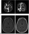
Figure 1
Figure 1
a) MR Venogram showing left transverse sinus thrombosis in axial oblique (left) and coronal oblique (right) views. b) MRI axial FLAIR (left) and T1 post gadolinium contrast injection (right), taken 2 weeks after presentation to hospital showing no evidence of meningeal enhancement or parenchymal lesions.
Figure 2
Figure 2
a) Timeline showing clinical progression and associated JCPyV loads. All viral loads are shown as JCPyV copies/mL of respective fluid. Asterisks denote which samples had their full JCPyV genomes sequenced. b) Progression of the patient's antibody responses, as determined by a VLP-based ELISA plotted against viral load in serum. Open circles in the viral load plots indicate viral loads extrapolated from whole blood, which equate to a 1 log decrease in serum, based on parallel sample evaluations (data not shown). Significant clinical history is denoted below the x-axis. ND = Not detected
Figure 2
Figure 2
a) Timeline showing clinical progression and associated JCPyV loads. All viral loads are shown as JCPyV copies/mL of respective fluid. Asterisks denote which samples had their full JCPyV genomes sequenced. b) Progression of the patient's antibody responses, as determined by a VLP-based ELISA plotted against viral load in serum. Open circles in the viral load plots indicate viral loads extrapolated from whole blood, which equate to a 1 log decrease in serum, based on parallel sample evaluations (data not shown). Significant clinical history is denoted below the x-axis. ND = Not detected
Figure 3
Figure 3
Immunuohistochemical SV40 LTAg staining within the explanted kidney medulla at 400× magnification showing staining within collecting duct cell nuclei.

Similar articles

Cited by

References

    1. Yuan CC, Miley W, Waters D. A quantification of human cells using an ERV-3 real time PCR assay. J Virol Methods. 2001;91(2):109–17. - PubMed
    1. Hoffman NG, Cook L, Atienza EE, Limaye AP, Jerome KR. Marked variability of BK virus load measurement using quantitative real-time PCR among commonly used assays. J Clin Microbiol. 2008;46(8):2671–80. doi: 10.1128/JCM.00258-08. - DOI - PMC - PubMed
    1. Pal A, Sirota L, Maudru T, Peden K, Lewis AM. Real-time, quantitative PCR assays for the detection of virus-specific DNA in samples with mixed populations of polyomaviruses. J Clin Microbiol. 2006;135(1):32–42. doi: 10.1016/j.jviromet.2006.01.018. - DOI - PubMed
    1. Randhawa PS, Gupta G, Vats A, Shapiro R, Viscidi RP. Immunoglobulin G, A, and M responses to BK virus in renal transplantation. Clin Vaccine Immunol. 2006;13(9):1057–63. doi: 10.1128/CVI.00114-06. - DOI - PMC - PubMed
    1. Gheuens S, Wüthrich C, Koralnik IJ. Progressive multifocal leukoencephalopathy: why gray and white matter. Annu Revg Pathol. 2013;8:189–215. - PubMed

Publication types

MeSH terms