. 2016 Jul 1;11(7):e0158097.
doi: 10.1371/journal.pone.0158097. eCollection 2016.

Low Doses of Oxygen Ion Irradiation Cause Acute Damage to Hematopoietic Cells in Mice

Affiliations

Low Doses of Oxygen Ion Irradiation Cause Acute Damage to Hematopoietic Cells in Mice

Jianhui Chang et al. PLoS One. .

Abstract

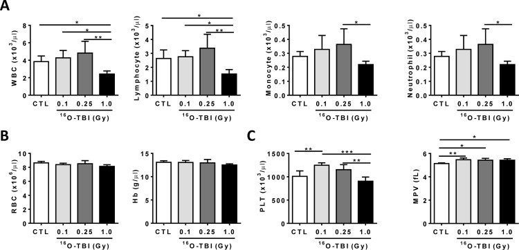
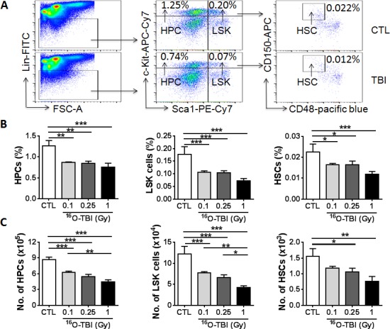
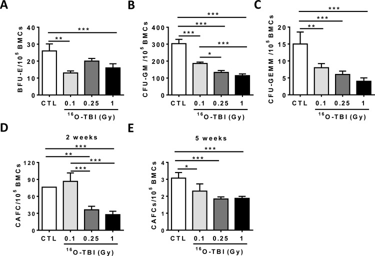
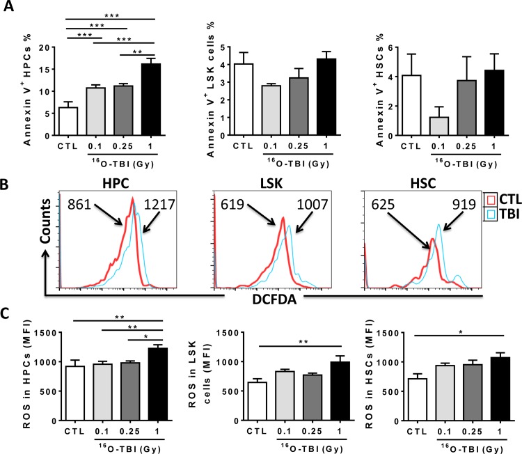
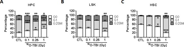
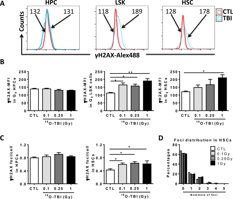
One of the major health risks to astronauts is radiation on long-duration space missions. Space radiation from sun and galactic cosmic rays consists primarily of 85% protons, 14% helium nuclei and 1% high-energy high-charge (HZE) particles, such as oxygen (16O), carbon, silicon, and iron ions. HZE particles exhibit dense linear tracks of ionization associated with clustered DNA damage and often high relative biological effectiveness (RBE). Therefore, new knowledge of risks from HZE particle exposures must be obtained. In the present study, we investigated the acute effects of low doses of 16O irradiation on the hematopoietic system. Specifically, we exposed C57BL/6J mice to 0.1, 0.25 and 1.0 Gy whole body 16O (600 MeV/n) irradiation and examined the effects on peripheral blood (PB) cells, and bone marrow (BM) hematopoietic stem cells (HSCs) and hematopoietic progenitor cells (HPCs) at two weeks after the exposure. The results showed that the numbers of white blood cells, lymphocytes, monocytes, neutrophils and platelets were significantly decreased in PB after exposure to 1.0 Gy, but not to 0.1 or 0.25 Gy. However, both the frequency and number of HPCs and HSCs were reduced in a radiation dose-dependent manner in comparison to un-irradiated controls. Furthermore, HPCs and HSCs from irradiated mice exhibited a significant reduction in clonogenic function determined by the colony-forming and cobblestone area-forming cell assays. These acute adverse effects of 16O irradiation on HSCs coincided with an increased production of reactive oxygen species (ROS), enhanced cell cycle entry of quiescent HSCs, and increased DNA damage. However, none of the 16O exposures induced apoptosis in HSCs. These data suggest that exposure to low doses of 16O irradiation induces acute BM injury in a dose-dependent manner primarily via increasing ROS production, cell cycling, and DNA damage in HSCs. This finding may aid in developing novel strategies in the protection of the hematopoietic system from space radiation.


Conflict of interest statement

Competing Interests: The authors have declared that no competing interests exist.

Figures

Fig 1
Fig 1. Blood cell counts were decreased under 1.0 Gy 16O exposure.
C57BL/6J mice were exposed to 0.1, 0.25 and 1.0 Gy doses of 16O irradiation or were sham irradiated as a control (CTL). The cell counts in peripheral blood were determined two weeks after radiation exposure. (A) The numbers of WBC, lymphocytes, monocytes, and neutrophils were significantly reduced after a dose of 1.0 Gy only. (B) The levels of RBC and Hb after 0.1, 0.25 and 1.0 Gy doses exposure were comparable to those in CTL mice. (C) The platelet counts (PLT) and mean platelet volume (MPV) were affected by all doses of 16O. The statistical significance for the difference between the control group and each of the irradiated groups is indicated by asterisks: *p<0.05, **p<0.01, ***p<0.001 as determined by one-way ANOVA.
Fig 2
Fig 2. 16O TBI causes reductions in numbers of HPCs and HSCs at two weeks after irradiation in a radiation dose dependent manner.
(A) Representative gating strategy of flow cytometric analysis for HPCs (Lin-Sca1-c-kit- cells), LSK cells (Lin-Sca1+c-kit+cells) and HSCs (Lin-Sca1+c-kit+CD150+CD48- cells) in BMCs is shown from 1.0 Gy 16O TBI and sham-irradiation (CTL). (B and C) Frequencies (panel B) and total numbers (panel C) of HPCs, LSK cells and HSCs from each mouse are presented as mean ± SD (n = 5). The statistical significance for the difference between the control group and each of the irradiated groups is indicated by asterisks. *p<0.05, **p<0.01, ***p<0.001 as determined by one-way ANOVA.
Fig 3
Fig 3. 16O TBI causes sustained reduction of HSC clonogenic function.
(A–C) At 2 weeks after TBI, BM-MNCs were isolated from irradiated and sham-irradiated (CTL) mice. A CFC assay was performed, and the results are presented as mean CFUs per 1x10⁵ BM-MNCs (n = 5). (D and E) Total BM cells (BMCs) were analyzed by CAFC assays, and the numbers of two and five week CAFCs were expressed as mean ± SD (n = 3 mice per group) per 1x10⁵ BMCs. The statistical significance for the difference between the control group and each of the irradiated groups is indicated by asterisks. *p<0.05, **p<0.01, ***p<0.001 as determined by one-way ANOVA.
Fig 4
Fig 4. 16O TBI causes an increase in ROS production in HPCs and HSCs two weeks after 1.0 Gy exposure and an increase in apoptosis in HPCs.
(A) Isolated Lin- cells were stained with Annexin V to determine cellular apoptosis, and percentages of Annexin V positive cells were presented as mean ± SD (n = 5). (B) Representative analysis of ROS production as measured by flow cytometry using DCFDA in BM HPCs and HSCs from control and 1.0 Gy16O TBI mice. The histograms indicate DCF MFI from a representative experiment. (C) The DCF MFI in BM HPCs and HSCs are presented as mean ± SD (n = 5). The statistical significance for the difference between the control group and each of the irradiated groups is indicated by asterisks. *p<0.05, **p<0.01, ***p<0.001 as determined by one-way ANOVA.
Fig 5
Fig 5. 16O TBI drives HSCs from quiescence into the cell cycle.
Lincells were isolated from control (CTL) and irradiated (TBI) mice 2 weeks after 0.1, 0.25 and 1.0 Gy TBI. Cell cycle was measured by flow cytometry using Ki-67 and 7-AAD double staining in BM HPCs and HSCs from control and irradiated mice. (A-C) The percentages of G0, G1 and G2SM phases in BM HPCs, LSK cells and HSCs after TBI are presented as mean ± SD (n = 5). The distribution of cell cycle phases in HPCs, LSK cells and HSCs was analyzed by Chi-Square test as indicated by X2 (HPC, X2 = 19.084, p<0.05; LSK cells, X2 = 6.486, p<0.05; HSCs, X2 = 33.853, p<0.05). The statistical significance for the difference in each cell cycle phase between the control groups and irradiated groups is indicated by asterisks. *p<0.05, **p<0.01, ***p<0.001 by one-way ANOVA analysis.
Fig 6
Fig 6. 16O TBI causes persistent increases in DNA damage in HSCs but not in HPCs two weeks after the exposure.
(A) Representative analysis of DNA damage measured in Lin- cells by flow cytometry using γH2AX staining in BM HPCs and HSCs from control and 1.0 Gy16O TBI mice. The histograms indicate γH2AX MFI from a representative experiment. (B) The γH2AX MFI in BM HPCs and HSCs after TBI are presented as mean ± SD. (C) Sorted HPCs and HSCs from irradiated and non-irradiated mice were stained with γH2AX antibody. The numbers of foci in each cell were counted and expressed as mean ± SD. (D) The distribution of foci was expressed as the percentages of different numbers of foci in control and irradiated HSCs. The statistical significance for the difference between the control groups and each of irradiated groups is indicated by asterisks. *p<0.05, **p<0.01 by one-way ANOVA analysis.

Similar articles

Cited by

References

    1. Cucinotta FA, Wu H, Shavers MR, George K (2003) Radiation dosimetry and biophysical models of space radiation effects. Gravit Space Biol Bull 16: 11–18. - PubMed
    1. Walker SA, Townsend LW, Norbury JW (2013) Heavy ion contributions to organ dose equivalent for the 1977 galactic cosmic ray spectrum. Advances in Space Research 51: 1792–1799.
    1. Waselenko JK, MacVittie TJ, Blakely WF, Pesik N, Wiley AL, Dickerson WE, et al. (2004) Medical management of the acute radiation syndrome: recommendations of the Strategic National Stockpile Radiation Working Group. Ann Intern Med 140: 1037–1051. - PubMed
    1. Moroni M, Coolbaugh TV, Lombardini E, Mitchell JM, Moccia KD, Shelton LJ, et al. (2011) Hematopoietic radiation syndrome in the Gottingen minipig. Radiat Res 176: 89–101. - PubMed
    1. Shao L, Sun Y, Zhang Z, Feng W, Gao Y, Cai Z, et al. (2010) Deletion of proapoptotic Puma selectively protects hematopoietic stem and progenitor cells against high-dose radiation. Blood 115: 4707–4714. 10.1182/blood-2009-10-248872 - DOI - PMC - PubMed