Review
. 2016 Jun 28;17(7):1018.
doi: 10.3390/ijms17071018.

Structure and Functional Diversity of GCN5-Related N-Acetyltransferases (GNAT)

Affiliations
Review

Structure and Functional Diversity of GCN5-Related N-Acetyltransferases (GNAT)

Abu Iftiaf Md Salah Ud-Din et al. Int J Mol Sci. .

Abstract

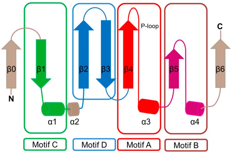
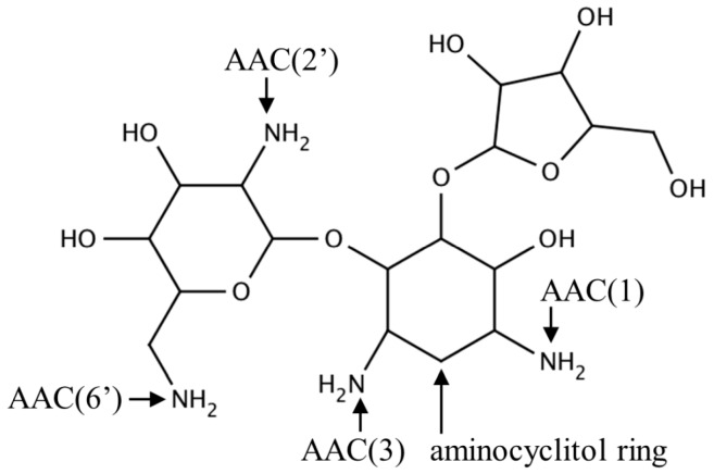
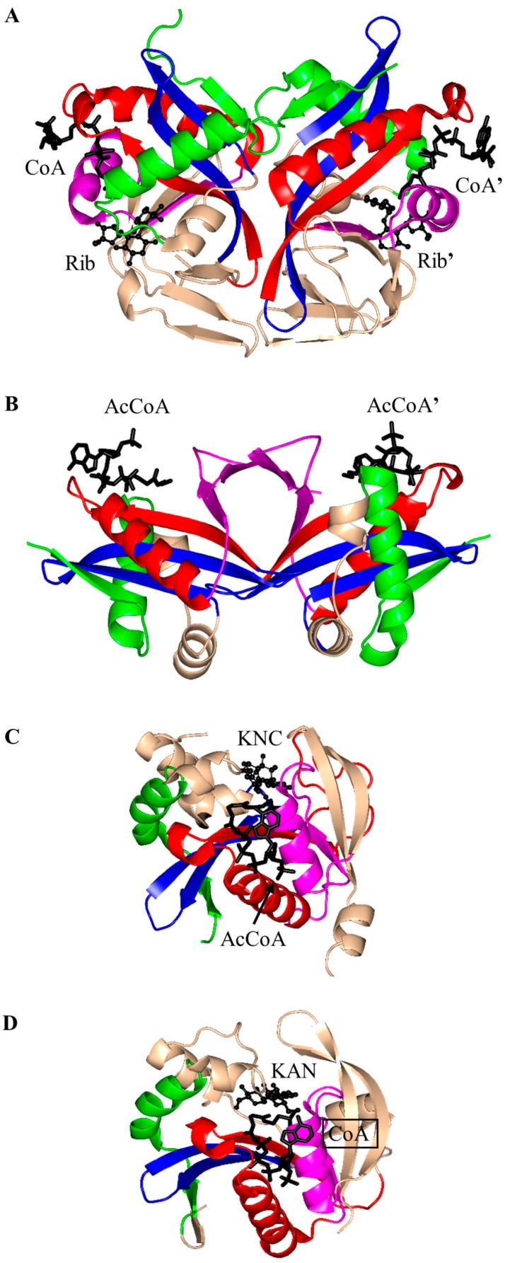
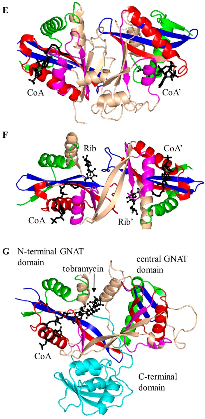
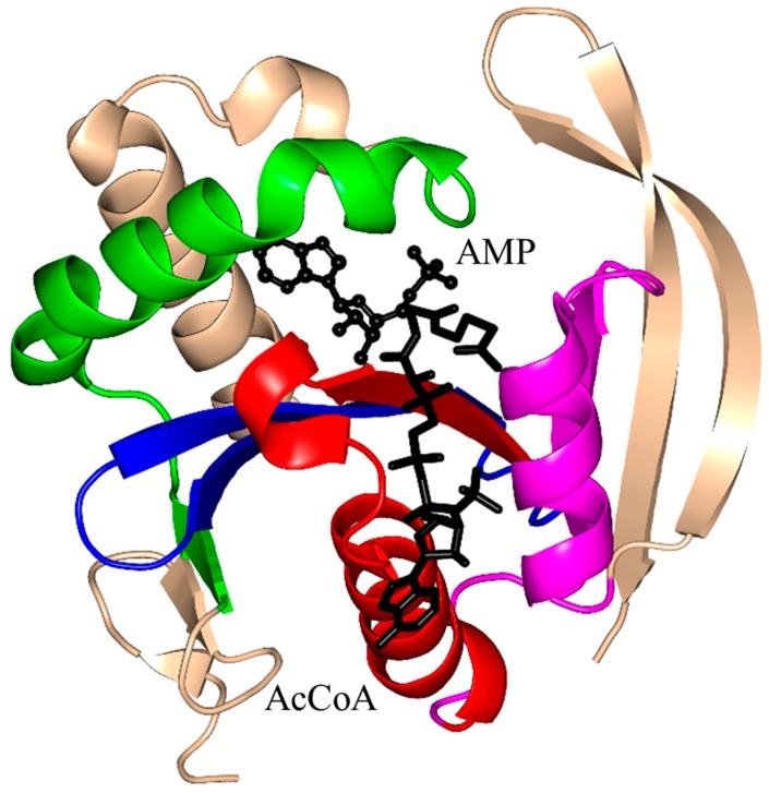
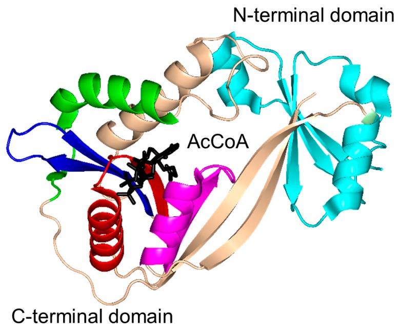
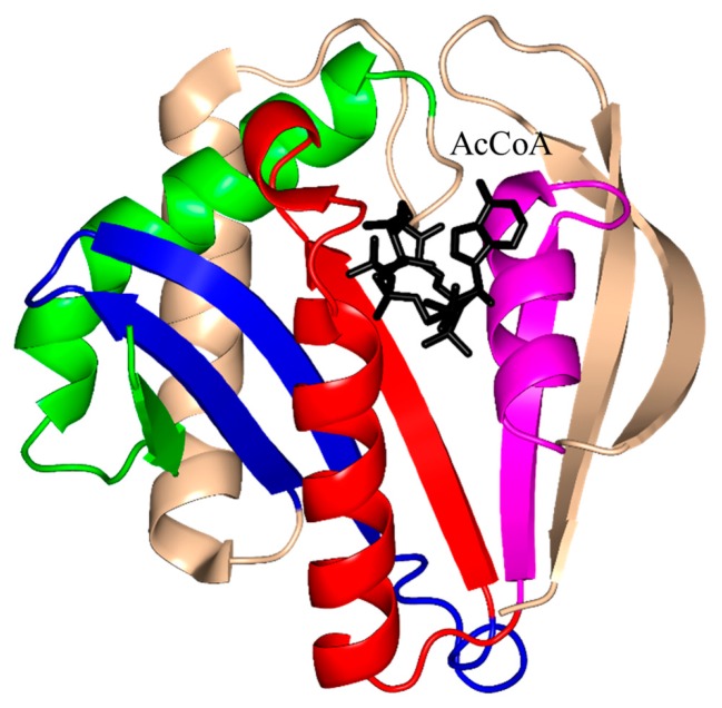
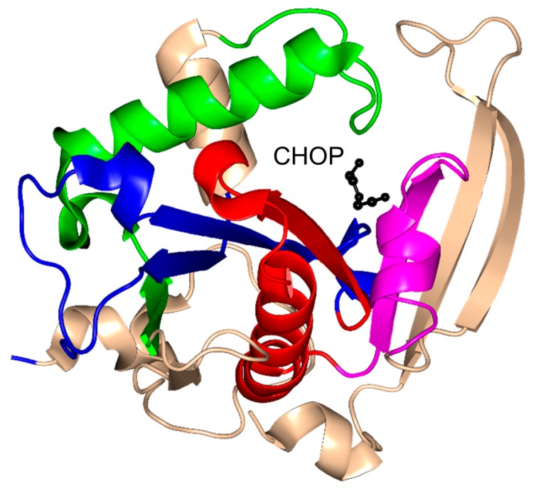
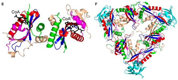
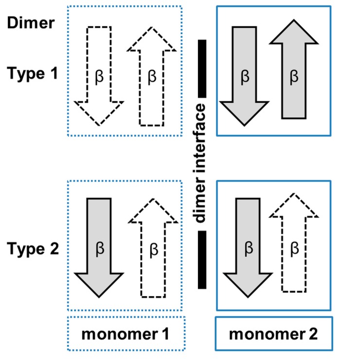
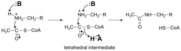
General control non-repressible 5 (GCN5)-related N-acetyltransferases (GNAT) catalyze the transfer of an acyl moiety from acyl coenzyme A (acyl-CoA) to a diverse group of substrates and are widely distributed in all domains of life. This review of the currently available data acquired on GNAT enzymes by a combination of structural, mutagenesis and kinetic methods summarizes the key similarities and differences between several distinctly different families within the GNAT superfamily, with an emphasis on the mechanistic insights obtained from the analysis of the complexes with substrates or inhibitors. It discusses the structural basis for the common acetyltransferase mechanism, outlines the factors important for the substrate recognition, and describes the mechanism of action of inhibitors of these enzymes. It is anticipated that understanding of the structural basis behind the reaction and substrate specificity of the enzymes from this superfamily can be exploited in the development of novel therapeutics to treat human diseases and combat emerging multidrug-resistant microbial infections.

Keywords: GNAT; acetyltransferase; catalytic residues; crystal structure; enzyme inhibitor; reaction mechanism.


Figures

Figure 1
Figure 1
Topology of the general control non-repressible 5 (GCN5)-related N-acetyltransferase (GNAT) enzymes. The secondary structure elements encompassing the conserved sequence motifs C (β1–α1), D (β2–β3), A (β4–α3) and B (β5–α4) are colored green, blue, red and magenta, respectively. The location of the conserved “P-loop” (β4–α3) is shown. The least conserved secondary structure elements (strands β0 and β6 and helix α2), that are absent in some GNAT proteins, are colored wheat.
Figure 2
Figure 2
Chemical structure of an aminoglycoside antibiotic (ribostamycin) showing the central aminocyclitol ring and acetyl group modification sites (1, 2′, 3 and 6′).
Figure 3
Figure 3
Cartoon representation of the structures of aminoglycoside N-acetyltransferases. (A) Aminoglycoside 2′-N-acetyltransferase-Ic from Mycobacterium tuberculosis in complex with CoA and ribostamycin (Rib) (PDB ID: 1M4G [23]); (B) aminoglycoside 3-N-acetyltransferase-Ia from Serratia marcescens in complex with AcCoA (PDB ID: 1BO4 [5]); (C) aminoglycoside 6′-N-acetyltransferase-Ib from Escherichia coli in complex with AcCoA and kanamycin C (KNC) (PDB ID: 1V0C [25]); (D) aminoglycoside 6′-N-acetyltransferase-Ie from Staphylococcus warneri complex with a sulfinic acid form of coenzyme A (CoA) and kanamycin A (KAN) (PDB ID: 4QC6 [24]); (E) aminoglycoside 6′-N-acetyltransferase-Ii from Enterococcus faecium in complex with CoA (PDB ID: 1N71 [34]); (F) Salmonella enterica aminoglycoside 6′-N-acetyltransferase-Iy in complex with CoA and ribostamycin (Rib) (PDB ID: 1S3Z [39]); and (G) M. tuberculosis enhanced intracellular survival (Eis) in complex with CoA and tobramycin (PDB ID: 4JD6 [19]). The conserved and non-conserved motifs are colored as in Figure 1 (motif C—green, motif D—blue, motif A—red, motif B—magenta, non-conserved N-terminal and C-terminal regions—wheat). The C-terminal animal sterol carrier domain of Eis is colored cyan. The AcCoA/CoA cofactor is drawn as black sticks, whereas the substrates (tobramycin and kanamycin) are shown in black using ball-and-stick representation.
Figure 3
Figure 3
Cartoon representation of the structures of aminoglycoside N-acetyltransferases. (A) Aminoglycoside 2′-N-acetyltransferase-Ic from Mycobacterium tuberculosis in complex with CoA and ribostamycin (Rib) (PDB ID: 1M4G [23]); (B) aminoglycoside 3-N-acetyltransferase-Ia from Serratia marcescens in complex with AcCoA (PDB ID: 1BO4 [5]); (C) aminoglycoside 6′-N-acetyltransferase-Ib from Escherichia coli in complex with AcCoA and kanamycin C (KNC) (PDB ID: 1V0C [25]); (D) aminoglycoside 6′-N-acetyltransferase-Ie from Staphylococcus warneri complex with a sulfinic acid form of coenzyme A (CoA) and kanamycin A (KAN) (PDB ID: 4QC6 [24]); (E) aminoglycoside 6′-N-acetyltransferase-Ii from Enterococcus faecium in complex with CoA (PDB ID: 1N71 [34]); (F) Salmonella enterica aminoglycoside 6′-N-acetyltransferase-Iy in complex with CoA and ribostamycin (Rib) (PDB ID: 1S3Z [39]); and (G) M. tuberculosis enhanced intracellular survival (Eis) in complex with CoA and tobramycin (PDB ID: 4JD6 [19]). The conserved and non-conserved motifs are colored as in Figure 1 (motif C—green, motif D—blue, motif A—red, motif B—magenta, non-conserved N-terminal and C-terminal regions—wheat). The C-terminal animal sterol carrier domain of Eis is colored cyan. The AcCoA/CoA cofactor is drawn as black sticks, whereas the substrates (tobramycin and kanamycin) are shown in black using ball-and-stick representation.
Figure 4
Figure 4
Cartoon representation of the structures of histone N-acetyltransferases. (A) Human HAT1 in complex with AcCoA and a histone H4 peptide (PDB ID: 2P0W [45]); (B) yeast Esa1 in complex with AcCoA (PDB ID: 1MJB [57]); (C) yeast GCN5 acetyltransferase in complex with CoA and a histone H3 peptide (PDB ID:1QSN [70]); (D) the histone acetyltransferase domain of hPCAF in complex with CoA (PDB ID: 1CM0 [73]); and (E) yeast Hpa2 histone acetyltransferase in complex with AcCoA (one dimer of the tetramer) (PDB ID: 1QSM [76]). In HAT1 and Esa1, the N-terminal domain is colored cyan, C-terminal domain is colored wheat, and conserved and non-conserved motifs of the central GNAT domain are colored as in Figure 1.
Figure 5
Figure 5
Cartoon representation of the structures of non-histone protein N-acetyltransferases. (A) Human α-tubulin acetyltransferase in complex with AcCoA (PDB ID: 4GS4 [79]); (B) M. tuberculosis MtPat in complex with cAMP and AcCoA (PDB ID: 4AB [81]); (C) N-acetyltransferase from Sulfolobus solfataricus in complex with CoA (PDB ID: 3F8K [78]); and (D) Human N-acetyltransferase Naa50p in complex with CoA and peptide MLGPEGGRWGRPVGRRRRP (PDB ID: 3TFY [80]). The conserved and non-conserved motifs of the GNAT domains, cofactors and substrates are colored as in Figure 3.
Figure 6
Figure 6
Cartoon representation of the structure of arylalkylamine N-acetyltransferase of Ovis aries in complex with a bisubstrate analog, CoA-S-acetyltryptamine (PDB ID: 1CJW [77]). The conserved and non-conserved motifs of the GNAT domain are colored as in Figure 3.
Figure 7
Figure 7
Cartoon representation of the structure of dimeric yeast glucosamine-6-phosphate N-acetyltransferase 1 in complex with CoA and N-acetyl-d-glucosamine-6-phosphate (GlcNAc-6P) (PDB ID: 1I1D [99]). The conserved and non-conserved motifs of the GNAT domain, cofactor and substrates are colored as in Figure 3.
Figure 8
Figure 8
Cartoon representation of the structure of the acetyltransferase domain of E. coli MccE in complex with AcCoA and adenosine monophosphate (AMP) (PDB ID: 3R96 [104]). The conserved and non-conserved motifs of the GNAT domain, cofactor and substrate are colored as in Figure 3.
Figure 9
Figure 9
Cartoon representation of the structure of Helicobacter pylori pseudaminic acid biosynthesis protein H in complex with AcCoA (PDB ID: 4RI1 [107]). The conserved and non-conserved motifs of the GNAT domain are colored as in Figure 3.
Figure 10
Figure 10
Cartoon representation of the structure of E. coli WecD in complex with AcCoA. The N-terminal partial GNAT domain is colored cyan (PDB ID: 2FT0 [113]). The conserved and non-conserved motifs of the GNAT domain are colored as in Figure 3.
Figure 11
Figure 11
Cartoon representation of the structure of the tabtoxin resistance protein from Pseudomonas syringae in complex with AcCoA (PDB ID: 1GHE [114]). The conserved and non-conserved motifs of the GNAT domain are colored as in Figure 3.
Figure 12
Figure 12
Cartoon representation of the structure of yeast Mpr1 in complex with cis-4-hydroxy-l-proline (CHOP) (PDB ID: 3W6X [119]). The conserved and non-conserved motifs of the GNAT domain are colored as in Figure 3.
Figure 13
Figure 13
Cartoon representation of the structure of Bacillus subtilis PaiA in complex with an oxidized CoA dimer (PDB ID: 1TIQ [126]). The conserved and non-conserved motifs of the GNAT domain are colored as in Figure 3.
Figure 14
Figure 14
Cartoon representation of the structure of Nε-lysine protein acetyltransferase. (A) Pseudomonas aeruginosa PA4794 in complex with AcCoA (PDB ID: 3PGP [137]); and (B) Rv1347c of M. tuberculosis (PDB IS: 1NYK3 [138]). The conserved and non-conserved motifs of the GNAT domains are colored as in Figure 3.
Figure 15
Figure 15
Cartoon representation of the structures of ribosomal protein Nα- acetyltransferases. (A). Salmonella typhimurium RimI in complex with a bisubstrate inhibitor, C-term-Arg-Arg-Phe-Tyr-Arg-Ala-N-α-AcCoA (PDB ID: 2CNM [147]); and (B) RimL from S. typhimurium in complex with CoA (PDB ID: 1S7N [108]). The conserved and non-conserved motifs of the GNAT domains are colored as in Figure 3.
Figure 16
Figure 16
Cartoon representation of the structure of putative M. tuberculosis succinyltransferase (Rv0802c) in complex with succinyl CoA (PDB ID: 2VZZ [4]). The conserved and non-conserved motifs of the GNAT domain are colored as in Figure 3.
Figure 17
Figure 17
Cartoon representation of the structures of aminoacyl transferases from the FemABX family. (A) Staphylococcus aureus FemA apoenzyme (PDB ID: 1LRZ [150]); (B) Weissella viridescens FemX in complex with UDP-MurNAc-pentapeptide (UM5P) substrate (PDB ID: 1P4N [152]); and (C) E. coli leucyl/phenylalanyl-tRNA protein transferase (EcLFT) in complex with puromycin (PDB ID: 2DPT [161]). The conserved and non-conserved motifs of the GNAT domains are colored as in Figure 3.
Figure 18
Figure 18
Cartoon representation of the structure of yeast N-myristoyltransferase in complex with myristoyl CoA and the non-peptide inhibitor (Z)-3-benzyl-5-(2-hydroxy-3-nitrobenzylidene)-2-thioxothiazolidin-4-one (PDB ID: 2P6F [169]). The conserved and non-conserved motifs of the GNAT domain, cofactor and inhibitor are colored as in Figure 3.
Figure 19
Figure 19
Cartoon representation of the structure of M. tuberculosis MshD in complex with desacetylmycothiol, AcCoA and CoA (PDB ID: 2C27 [187]). The conserved and non-conserved motifs of the GNAT domain, cofactor and substrate are colored as in Figure 3.
Figure 20
Figure 20
Representative oligomeric structures observed in the GNAT superfamily. (A) Monomeric ScMpr1; (B) dimeric StRimL where the β-strands from the two monomers at the dimer interface are arranged to form a continuous β-sheet; (C) dimeric interface where the C-terminal β-strands are interchanged between the monomers (ScGNA1); (D) a 12-strand β-barrel at the dimer interface of SmAAC(3)-Ia; (E) dimer interface involving α-helices from both monomers (hPCAF); and (F) one-half of the MtEis hexamer (two three-fold symmetrical trimers shown in this Figure assemble into an asymmetric “sandwich”). The conserved and non-conserved motifs of the GNAT domain, cofactors and substrates are colored as in Figure 3.
Figure 20
Figure 20
Representative oligomeric structures observed in the GNAT superfamily. (A) Monomeric ScMpr1; (B) dimeric StRimL where the β-strands from the two monomers at the dimer interface are arranged to form a continuous β-sheet; (C) dimeric interface where the C-terminal β-strands are interchanged between the monomers (ScGNA1); (D) a 12-strand β-barrel at the dimer interface of SmAAC(3)-Ia; (E) dimer interface involving α-helices from both monomers (hPCAF); and (F) one-half of the MtEis hexamer (two three-fold symmetrical trimers shown in this Figure assemble into an asymmetric “sandwich”). The conserved and non-conserved motifs of the GNAT domain, cofactors and substrates are colored as in Figure 3.
Figure 21
Figure 21
Schematic figure illustrating the topology of the β-strands found in the two most common types of dimer interface in GNAT proteins. Strands from one monomer are colored white, and those from the second monomer are colored gray.
Figure 22
Figure 22
A common general direct acyltransfer mechanism of GNAT superfamily members.

Similar articles

Cited by

References

    1. Neuwald A.F., Landsnan D. GCN5-related histone N-acetyltransferases belong to a diverse superfamily that includes the yeast SPT10 protein. Proc. Natl. Acad. Sci. USA. 1997;51:154–155. doi: 10.1016/S0968-0004(97)01034-7. - DOI - PubMed
    1. Dyda F., Klein D.C., Hickman A.B. GCN5-related N-acetyltransferases: A structural overview. Annu. Rev. Biophys. Biomol. Struct. 2000;29:81–103. doi: 10.1146/annurev.biophys.29.1.81. - DOI - PMC - PubMed
    1. Farazi T.A., Waksman G., Gordon J.I. Structures of Saccharomyces cerevisiae N-myristoyltransferase with bound myristoylCoA and peptide provide insights about substrate recognition and catalysis. Biochemistry. 2001;40:6335–6343. doi: 10.1021/bi0101401. - DOI - PubMed
    1. Vetting M.W., Errey J.C., Blanchard J.S. Rv0802c from Mycobacterium tuberculosis: The first structure of a succinyltransferase with the GNAT fold. Acta Crystallogr. F Struct. Biol. Commun. 2008;64:978–985. doi: 10.1107/S1744309108031679. - DOI - PMC - PubMed
    1. Wolf E., Vassilev A., Makino Y., Sali A., Nakatani Y., Burley S.K. Crystal structure of a GCN5-related N-acetyltransferase: Serratia marcescens aminoglycoside 3-N-acetyltransferase. Cell. 1998;94:439–449. doi: 10.1016/S0092-8674(00)81585-8. - DOI - PubMed

MeSH terms

LinkOut - more resources