Review
. 2016 Jun 29;17(7):1029.
doi: 10.3390/ijms17071029.

Aquaporins and Brain Tumors

Affiliations
Review

Aquaporins and Brain Tumors

Rosario Maugeri et al. Int J Mol Sci. .

Abstract

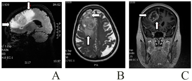
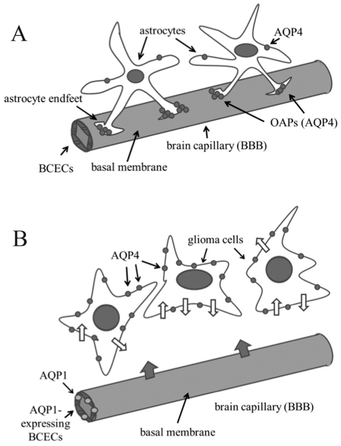
Brain primary tumors are among the most diverse and complex human cancers, and they are normally classified on the basis of the cell-type and/or the grade of malignancy (the most malignant being glioblastoma multiforme (GBM), grade IV). Glioma cells are able to migrate throughout the brain and to stimulate angiogenesis, by inducing brain capillary endothelial cell proliferation. This in turn causes loss of tight junctions and fragility of the blood-brain barrier, which becomes leaky. As a consequence, the most serious clinical complication of glioblastoma is the vasogenic brain edema. Both glioma cell migration and edema have been correlated with modification of the expression/localization of different isoforms of aquaporins (AQPs), a family of water channels, some of which are also involved in the transport of other small molecules, such as glycerol and urea. In this review, we discuss relationships among expression/localization of AQPs and brain tumors/edema, also focusing on the possible role of these molecules as both diagnostic biomarkers of cancer progression, and therapeutic targets. Finally, we will discuss the possibility that AQPs, together with other cancer promoting factors, can be exchanged among brain cells via extracellular vesicles (EVs).

Keywords: aquaporins (AQPs); blood–brain barrier (BBB); brain tumors; extracellular vesicles (EVs); glioblastoma multiforme.


Figures

Figure 1
Figure 1
Patient with a right frontal glioblastoma and a marked area of peritumoral edema. Images from a Brain Magnetic Resonance. The arrows point to the peritumoral edema close to the glioma: (A) Sagittal T1-weighted; (B) Axial T2-weighted; and (C) Coronal T1-weighted contrast-enhanced with gadolinium.
Figure 2
Figure 2
Alteration of expression and/or localization of aquaporin 1 (AQP1) and AQP4 in brain cancer cells respect to normal glial cells. (A) In normal brain, brain capillary endothelial cells (BCECs) do not express AQP1, while AQP4 is mainly present in the astrocyte endfeet, where it forms orthogonal arrays of particles (OAPs); (B) In gliomas, BCECs express AQP1, thus probably allowing an increase of blood–brain-barrier (BBB) permeability (large grey arrows indicate water flux from blood to brain), while AQP4 is delocalized: endfeet (and OAPs) are no more visible and AQP4 is found throughout the plasma membrane of glioma cells, thus probably increasing water trafficking across the cell membranes (narrow white arrows on glioma cell membrane).

Similar articles

Cited by

References

    1. Crocetti E., Trama A., Stiller C., Caldarella A., Soffietti R., Jaal J., Weber D.C., Ricardi U., Slowinski J., Brandes A., et al. Epidemiology of glial and non-glial brain tumours in Europe. Eur. J. Cancer. 2012;48:1532–1542. doi: 10.1016/j.ejca.2011.12.013. - DOI - PubMed
    1. Fuller G.N., Scheithauer B.W. The 2007 revised World Health Organization (WHO) classification of tumours of the central nervous system: Newly codified entities. Brain Pathol. 2007;17:304–307. doi: 10.1111/j.1750-3639.2007.00084.x. - DOI - PMC - PubMed
    1. Louis D.N., Ohgaki H., Wiestler O.D., Cavenee W.K., Burger P.C., Jouvet A., Scheithauer B.W., Kleihues P. The 2007 WHO classification of tumours of the central nervous system. Acta Neuropathol. 2007;114:97–109. doi: 10.1007/s00401-007-0243-4. - DOI - PMC - PubMed
    1. Jovcevska I. Glioma and glioblastoma—How much do we (not) know? Mol. Clin. Oncol. 2013;1:935–941. doi: 10.3892/mco.2013.172. - DOI - PMC - PubMed
    1. Burton E.C., Lamborn K.R., Feuerstein B.G., Prados M., Scott J., Forsyth P., Passe S., Jenkins R.B., Aldape K.D. Genetic aberrations defined by comparative genomic hybridization distinguish long-term from typical survivors of glioblastoma. Cancer Res. 2002;62:6205–6210. - PubMed

Substances