. 2016 Jun 28;8(7):181.
doi: 10.3390/v8070181.

Development of Neutralization Assay Using an eGFP Chikungunya Virus

Affiliations

Development of Neutralization Assay Using an eGFP Chikungunya Virus

Cheng-Lin Deng et al. Viruses. .

Abstract

Chikungunya virus (CHIKV), a member of the Alphavirus genus, is an important human emerging/re-emerging pathogen. Currently, there are no effective antiviral drugs or vaccines against CHIKV infection. Herein, we construct an infectious clone of CHIKV and an eGFP reporter CHIKV (eGFP-CHIKV) with an isolated strain (assigned to Asian lineage) from CHIKV-infected patients. The eGFP-CHIKV reporter virus allows for direct visualization of viral replication through the levels of eGFP expression. Using a known CHIKV inhibitor, ribavirin, we confirmed that the eGFP-CHIKV reporter virus could be used to identify inhibitors against CHIKV. Importantly, we developed a novel and reliable eGFP-CHIKV reporter virus-based neutralization assay that could be used for rapid screening neutralizing antibodies against CHIKV.

Keywords: Chikungunya virus; antiviral; neutralization antibody; reporter virus.


Figures

Figure 1
Figure 1
Isolation and characterization of CHIKV from clinical human cases. (a) the flow chart of virus isolation on C6/36 cells from human serum; (b) the CHIKV strain that was passaged for seven rounds on C6/36 cells showed apparent CPE on BHK-21 cells; (c) plaque morphology of the CHIKV strain on BHK-21 cells on the four days post-inoculation; and (d) phylogenetic analyses of CHIKV genome sequences using the neighbor-joining method. The newly isolated CHIKV strain is highlighted in red. ECSA lineage = the East, Central and South African lineage.
Figure 2
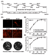
Figure 2
Construction and characterization of the full-length cDNA clone of CHIKV. (a) six cDNA fragments represented by thick lines were synthesized from genomic RNA through RT–PCR to cover the complete CHIKV genome and unique restriction sites as well as their nucleotide numbers are shown. Genome organization is depicted below. Individual fragments were assembled to form the full-length cDNA clone of CHIKV (pACYC-CHIKV). The complete CHIKV cDNA is under the control of T7 promoter elements for in vitro transcription. The numbers are the nucleotide positions based on the CHIKV genome sequences we identified in this study (KC488650); (b) IFA of viral protein expression in BHK-21 cells transfected with the full-length CHIKV RNA transcript. The transfected cells were analyzed by IFA at the indicated time points post-transfection; (c) plaque morphology of parental and recombinant CHIKV on BHK-21 cells; (d,e) comparison of the growth kinetics of recombinant and parental CHIKV in BHK-21 and C6/36 cells, respectively. The virus growth curves were compared at MOI = 0.1 on both cells. Three independent experiments were performed in duplicate, and the representative data were presented.
Figure 3
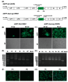
Figure 3
Construction and stability of CHIKV reporter viruses. (a) schematic of the construction of two versions of CHIKV reporter virus (eGFP-2A-CHIKV and eGFP-dual-sg-CHIKV). An infectious cDNA clone of pACYC-CHIKV was used as a backbone for the construction of CHIKV reporter viruses; (b,c) eGFP expressions in cells transfected with different versions of CHIKV reporter RNA transcripts. The data for eGFP-2A-CHIKV and eGFP-dual-sg-CHIKV were present on left and right panels, respectively, and thereafter. The expression of eGFP in transfected BHK-21 cells was analyzed by fluorescent microscopy at the indicated time points post-transfection. The lower panels were the images visualized under differential interference contrast microscopy; (d,e) detection of the eGFP gene during virus passage. Viral RNAs were extracted from culture supernatants of the indicated passages, respectively. RT–PCR was performed with primer pairs to cover the complete eGFP gene region. The resulting RT–PCR products were resolved by 1% agarose gel electrophoresis; and (f,g) detection of eGFP expression in BHK-21 cells infected with different passages of reporter viruses. eGFP expressions were observed under fluorescent microscope at 48 hpi. Both RT-PCR and eGFP expression results indicated that the reporter gene still maintained in eGFP-dual-sg-CHIKV. The eGFP-dual-sg-CHIKV was designated as eGFP-CHIKV.
Figure 4
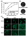
Figure 4
Characterization of eGFP-CHIKV reporter viruses. (a) comparison of the growth kinetics of recombinant WT CHIKV and eGFP-CHIKV in BHK-21 cells. The representative data were presented from three independent experiments; (b) plaque morphology of recombinant WT CHIKV and eGFP-CHIKV on BHK-21 cells; and (c) correlation between eGFP expression and different MOIs infection. BHK-21 cells were infected with eGFP-CHIKV at the indicated MOI and eGFP expression were observed at 24, 36 and 48 hpi.
Figure 5
Figure 5
Antiviral activity of ribavirin on eGFP-CHIKV reporter viruses. (a) detection of eGFP expression by fluorescent microscope at different concentrations of ribavirin; (b) viral titer reduction assay of CHIKV with different concentrations of ribavirin. BHK-21 cells were infected with eGFP-CHIKV at MOI of 0.05 and treated with various concentrations of ribavirin. The viral titers were quantified by plaque assay at 48 hpi; and (c) viral titer reduction assay of WT CHIKV at different concentrations of ribavirin. The experiments were performed in triplicate, and the representative data were presented.
Figure 6
Figure 6
Confirmation of the availability of eGFP-CHIKV in neutralization assay with the serum samples from CHIKV-infected patients. (a) detection of eGFP expression by fluorescent microscope with different dilutions of human sera. The neutralization assay was performed in a 12-well plate. Serially 4-fold diluted human sera were incubated with eGFP-CHIKV at an MOI of 0.05 for 1 h at 37 °C before adding the mixture to the monolayer of BHK-21 cells in the 24-well plates. The readout of eGFP expression was recorded at 48 hpi. The serum from health person was used as a negative control; (b) quantification of neutralization assay with increasing serum dilution. The percentage inhibition of infectivity was normalized to eGFP-CHIKV infection without serum incubation by quantification of eGFP expression levels; (c) neutralizing activity of the human sera against WT CHIKV based on PRNT assay. WT CHIKV was incubated with 4-fold dilutions of individual sera before infecting BHK-21 cells. The percentage inhibition of infectivity for each dilution was normalized to WT CHIKV infection alone by counting the number of plaque forming units (PFU); (d) detection of CHIKV genome copy number in BHK-21 cells treated with virus or virus-antiserum mixture by real-time RT-PCR. The results are positive if Ct value ≤34, otherwise negative if Ct value >34. The genome copy numbers of #34 and #81 were significantly decreased than those of control samples (t-test, p < 0.05). CHIKV only = BHK-21 cells were infected by WT CHIKV without serum incubation; mock-infected = BHK-21 cells were neither infected by WT CHIKV nor incubated with serum; NA = data not available. All experiments were performed in triplicate, and the representative data were presented.
Figure 7
Figure 7
Neutralization assay with sera from mice using eGFP-CHIKV. (a) detection of eGFP expression by fluorescent microscope with different dilutions of sera from mice immunized with different antigens. #1 and #3 sera were from two mice immunized with formalin-inactivated CHIKV, and anti-E2 serum was obtained from one mouse immunized with SDS-PAGE purified CHIKV E2 protein. NT assay was performed as described for CHIKV-infected patient sera; (b) the reactivity of the tested sera against CHIKV through an IFA. All three sera (#1, #3 and E2 sera) were diluted at 1:200. Mock and CHIKV infected BHK-21 cells were used to performed IFA.

Similar articles

Cited by

References

    1. Burt F.J., Rolph M.S., Rulli N.E., Mahalingam S., Heise M.T. Chikungunya: A re-emerging virus. Lancet. 2012;379:662–671. doi: 10.1016/S0140-6736(11)60281-X. - DOI - PubMed
    1. Morrison T.E. Reemergence of Chikungunya Virus. J. Virol. 2014;88:11644–11647. doi: 10.1128/JVI.01432-14. - DOI - PMC - PubMed
    1. Cruz D.J.M., Bonotto R.M., Gomes R.G.B., da Silva C.T., Taniguchi J.B., No J.H., Lombardot B., Schwartz O., Hansen M.A.E., Freitas-Junior L.H. Identification of novel compounds inhibiting Chikungunya virus-induced cell death by high throughput screening of a kinase inhibitor library. PLoS Neglect. Trop. Dis. 2013;7:181. doi: 10.1371/journal.pntd.0002471. - DOI - PMC - PubMed
    1. Ziegler S.A., Nuckols J., McGee C.E., Huang Y.-J.S., Vanlandingham D.L., Tesh R.B., Higgs S. In vivo imaging of Chikungunya virus in mice and Aedes mosquitoes using a Renilla luciferase clone. Vector Borne Zoonotic Dis. 2011;11:1471–1477. doi: 10.1089/vbz.2011.0648. - DOI - PMC - PubMed
    1. Weaver S.C., Reisen W.K. Present and future arboviral threats. Antivir. Res. 2010;85:328–345. doi: 10.1016/j.antiviral.2009.10.008. - DOI - PMC - PubMed

Publication types

MeSH terms

LinkOut - more resources