Review
. 2016 Jun 29;5(3):24.
doi: 10.3390/antibiotics5030024.

Ribosomal Antibiotics: Contemporary Challenges

Affiliations
Review

Ribosomal Antibiotics: Contemporary Challenges

Tamar Auerbach-Nevo et al. Antibiotics (Basel). .

Abstract

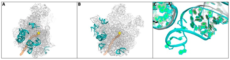
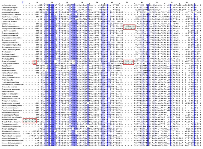
Most ribosomal antibiotics obstruct distinct ribosomal functions. In selected cases, in addition to paralyzing vital ribosomal tasks, some ribosomal antibiotics are involved in cellular regulation. Owing to the global rapid increase in the appearance of multi-drug resistance in pathogenic bacterial strains, and to the extremely slow progress in developing new antibiotics worldwide, it seems that, in addition to the traditional attempts at improving current antibiotics and the intensive screening for additional natural compounds, this field should undergo substantial conceptual revision. Here, we highlight several contemporary issues, including challenging the common preference of broad-range antibiotics; the marginal attention to alterations in the microbiome population resulting from antibiotics usage, and the insufficient awareness of ecological and environmental aspects of antibiotics usage. We also highlight recent advances in the identification of species-specific structural motifs that may be exploited for the design and the creation of novel, environmental friendly, degradable, antibiotic types, with a better distinction between pathogens and useful bacterial species in the microbiome. Thus, these studies are leading towards the design of "pathogen-specific antibiotics," in contrast to the current preference of broad range antibiotics, partially because it requires significant efforts in speeding up the discovery of the unique species motifs as well as the clinical pathogen identification.

Keywords: microbiome; multi-drug resistance; novel degradable antibiotics; species-specific antibiotics susceptibility.


Conflict of interest statement

The authors declare no conflict of interest.

Figures

Figure 1
Figure 1
Overall structure of the bacterial ribosome showing the two subunits: the small (SSU) and the large (LSU), the mRNA, the PTC, and the nascent protein exit tunnel.
Figure 2
Figure 2
Main antibiotics binding sites shown on the skeletons of the large (left); and the small (right) ribosomal subunits.
Figure 3
Figure 3
The pleuromutilins binding pocket in SA50S. Color code: SA rRNA-light brown, BC3205-violet, SB571579-green, Retapamulin-cyan, Tiamulin-slate, SB280080-yellow. The pleuromutilins antibiotics are superposed based on the locations of the rRNA of their binding pocket. The critical (additional) H = bond is shown.
Figure 4
Figure 4
View into the PTC (rRNA backbone in grey), in which the approximate peptide bond formation position is marked by a blue circle, and the A- and P-sites tRNA 3′-ends are marked by A and P. Shown also are the locations of two components of Synercid™ (chemical formulas shown on the right side) within the PTC and in the tunnel’s entrance (dalfopristin in cyan and quinupristin in green). Note the swung location, out of the active site of nucleotide U2585 (in red).
Figure 5
Figure 5
Left: The backbone of the large ribosomal subunit of SA50S is shown in gray from two views (face on (A) and a rotation of 90° (B)). The polypeptide exit tunnel is shown in gold and the PTC location is marked by a yellow star. The rRNA regions with fold variability compared with all other known structures on the SA50S surface are shown in cyan. Right (C): The stem loop of helix H63 in SA (cyan) and of Deinococcus radiodurans D50S (grey). Panel A & B are adapted from [16].
Figure 6
Figure 6
Multiple sequence alignment of protein uL3 from different bacterial species clearly showing its unique additional insertion (framed in red). The conserved sequences are highlighted in purple.

Similar articles

Cited by

References

    1. Fischbach M.A., Walsh C.T. Antibiotics for emerging pathogens. Science. 2009;325:1089–1093. doi: 10.1126/science.1176667. - DOI - PMC - PubMed
    1. Gibbons A. Resistance to antibiotics found in isolated Amazonian tribe. Science AAAS. 2015 doi: 10.1126/science.aab2509. - DOI
    1. Schlunzen F., Zarivach R., Harms J., Bashan A., Tocilj A., Albrecht R., Yonath A., Franceschi F. Structural basis for the interaction of antibiotics with the peptidyl transferase centre in eubacteria. Nature. 2001;413:814–821. doi: 10.1038/35101544. - DOI - PubMed
    1. Auerbach T., Bashan A., Harms J., Schluenzen F., Zarivach R., Bartels H., Agmon I., Kessler M., Pioletti M., Franceschi F., et al. Antibiotics targeting ribosomes: Crystallographic studies. Curr. Drug Targets Infect. Disord. 2002;2:169–186. doi: 10.2174/1568005023342506. - DOI - PubMed
    1. Hansen J.L., Moore P.B., Steitz T.A. Structures of five antibiotics bound at the peptidyl transferase center of the large ribosomal subunit. J. Mol. Biol. 2003;330:1061–1075. doi: 10.1016/S0022-2836(03)00668-5. - DOI - PubMed

LinkOut - more resources