

Urea transport and clinical potential of urearetics
- PMID: 27367911
- PMCID: PMC4974121
- DOI: 10.1097/MNH.0000000000000252
Urea transport and clinical potential of urearetics
Abstract
Purpose of review: Urea is transported by urea transporter proteins in kidney, erythrocytes, and other tissues. Mice in which different urea transporters have been knocked out have urine-concentrating defects, which has led to the development and testing of urea transporters Slc14A2 (UT-A) and Slc14A1 (UT-B) inhibitors as urearetics. This review summarizes the knowledge gained during the past year on urea transporter regulation and investigations into the clinical potential of urearetics.
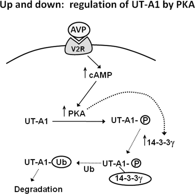
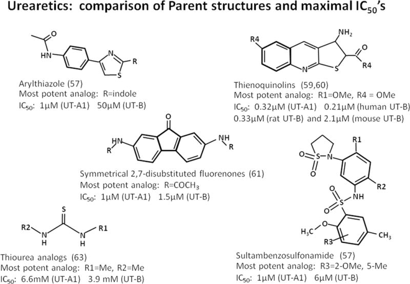
Recent findings: UT-A1 undergoes several posttranslational modifications that increase its function by increasing UT-A1 accumulation in the apical plasma membrane. UT-A1 is phosphorylated by protein kinase A, exchange protein activated by cyclic AMP, protein kinase Cα, and AMP-activated protein kinase, all at different serine residues. UT-A1 is also regulated by 14-3-3, which contributes to UT-A1 removal from the membrane. UT-A1 is glycosylated with various glycan moieties in animal models of diabetes mellitus. Transgenic expression of UT-A1 into UT-A1/UT-A3 knockout mice restores urine-concentrating ability. UT-B is present in descending vasa recta and urinary bladder, and is linked to bladder cancer. Inhibitors of UT-A and UT-B have been developed that result in diuresis with fewer abnormalities in serum electrolytes than conventional diuretics.
Summary: Urea transporters play critical roles in the urine-concentrating mechanism. Urea transport inhibitors are a promising new class of diuretic agent.
Conflict of interest statement
Conflicts of interest
None
Figures
Similar articles
-
Urea transport in the kidney.Compr Physiol. 2011 Apr;1(2):699-729. doi: 10.1002/cphy.c100030. Compr Physiol. 2011. PMID: 23737200 Review.
-
The urea transporter UT-A1 plays a predominant role in a urea-dependent urine-concentrating mechanism.J Biol Chem. 2020 Jul 17;295(29):9893-9900. doi: 10.1074/jbc.RA120.013628. Epub 2020 May 27. J Biol Chem. 2020. PMID: 32461256 Free PMC article.
-
The SLC14 gene family of urea transporters.Pflugers Arch. 2004 Feb;447(5):603-9. doi: 10.1007/s00424-003-1124-x. Epub 2003 Jul 11. Pflugers Arch. 2004. PMID: 12856182 Review.
-
Renal urea transporters.Curr Opin Nephrol Hypertens. 2004 Sep;13(5):525-32. doi: 10.1097/00041552-200409000-00008. Curr Opin Nephrol Hypertens. 2004. PMID: 15300159 Review.
-
Urea transporters and renal function: lessons from knockout mice.Curr Opin Nephrol Hypertens. 2008 Sep;17(5):513-8. doi: 10.1097/MNH.0b013e3283050969. Curr Opin Nephrol Hypertens. 2008. PMID: 18695393 Review.
Cited by
-
Current State of SLC and ABC Transporters in the Skin and Their Relation to Sweat Metabolites and Skin Diseases.Proteomes. 2021 May 16;9(2):23. doi: 10.3390/proteomes9020023. Proteomes. 2021. PMID: 34065737 Free PMC article. Review.
-
The Expression of AQP5 and UTs in the Sweat Glands of Uremic Patients.Biomed Res Int. 2017;2017:8629783. doi: 10.1155/2017/8629783. Epub 2017 Nov 27. Biomed Res Int. 2017. PMID: 29279852 Free PMC article.
-
UT-A1/A3 knockout mice show reduced fibrosis following unilateral ureteral obstruction.Am J Physiol Renal Physiol. 2020 May 1;318(5):F1160-F1166. doi: 10.1152/ajprenal.00008.2020. Epub 2020 Mar 16. Am J Physiol Renal Physiol. 2020. PMID: 32174141 Free PMC article.
-
Mammalian urine concentration: a review of renal medullary architecture and membrane transporters.J Comp Physiol B. 2018 Nov;188(6):899-918. doi: 10.1007/s00360-018-1164-3. Epub 2018 May 24. J Comp Physiol B. 2018. PMID: 29797052 Free PMC article. Review.
-
Discovery of novel diarylamides as orally active diuretics targeting urea transporters.Acta Pharm Sin B. 2021 Jan;11(1):181-202. doi: 10.1016/j.apsb.2020.06.001. Epub 2020 Jun 14. Acta Pharm Sin B. 2021. PMID: 33532188 Free PMC article.
References
-
- Galluci E, Micelli S, Lippe C. Non-electrolyte permeability across thin lipid membranes. Arch Int Physiol Biochim. 1971;79:881–7. - PubMed
-
- Sands JM, Nonoguchi H, Knepper MA. Vasopressin effects on urea and H2O transport in inner medullary collecting duct subsegments. Am J Physiol. 1987;253:F823–F32. - PubMed
-
- Klein JD, Blount MA, Sands JM. Urea transport in the kidney. Compr Physiol. 2011;1(2):699–729. - PubMed
-
- Yang B, Sands JM. In: Urea Transporters. Harris R, editor. New York: Springer; 2014. p. 265.
Publication types
MeSH terms
Substances
Grants and funding
LinkOut - more resources
Full Text Sources
Other Literature Sources
Research Materials
Miscellaneous
