

18F-fluorodeoxyglucose positron emission tomography and the risk of subsequent aortic complications in giant-cell arteritis: A multicenter cohort of 130 patients
- PMID: 27367985
- PMCID: PMC4937899
- DOI: 10.1097/MD.0000000000003851
18F-fluorodeoxyglucose positron emission tomography and the risk of subsequent aortic complications in giant-cell arteritis: A multicenter cohort of 130 patients
Abstract
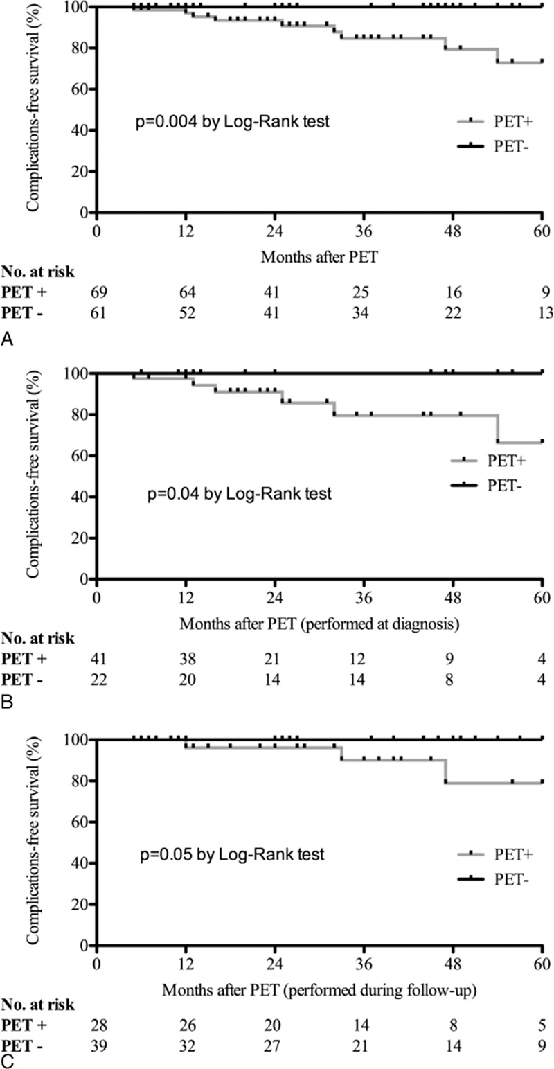
Previous studies reported a 2- to 17-fold higher risk of aortic complications (dilation or dissection) in patients with giant-cell arteritis (GCA). We aimed to determine whether or not GCA patients with large-vessel involvement demonstrated by positron emission tomography with F-fluorodeoxyglucose combined with computed tomography (FDG-PET/CT) have a higher risk of aortic complications. We conducted a retrospective multicenter study between 1995 and 2014. Patients were included if they fulfilled at least 3 American College of Rheumatology criteria for GCA, or 2 criteria associated with extratemporal biopsy-proven giant-cell vasculitis; they underwent at least 1 FDG-PET/CT scan at diagnosis or during follow-up; and the morphology of the aorta was assessed by medical imaging at diagnosis. Patients with an aortic complication at the time of diagnosis were excluded. Of the 130 patients included [85 women (65%), median age 70 (50-86)], GCA was biopsy proven in 77 (59%). FDG-PET/CT was performed at diagnosis in 63 (48%) patients and during the follow-up period in the 67 (52%) remaining patients. FDG-PET/CT was positive in 38/63 (60%) patients at diagnosis and in 31/67 (46%) patients when performed during follow-up (P = NS). One hundred four patients (80%) underwent at least 1 morphological assessment of the aorta during follow-up. Nine (9%) patients developed aortic complications (dilation in all and dissection in 1) at a median time of 33 (6-129) months after diagnosis. All of them displayed large-vessel inflammation on previous FDG-PET/CT. A positive FDG-PET/CT was significantly associated with a higher risk of aortic complications (P = 0.004).In our study, a positive FDG-PET/CT was associated with an increased risk of aortic complications at 5 years.
Conflict of interest statement
The authors have no conflicts of interest to disclose.
Figures
References
-
- Agard C, Barrier JH, Dupas B, et al. Aortic involvement in recent-onset giant cell (temporal) arteritis: a case-control prospective study using helical aortic computed tomodensitometric scan. Arthritis Rheum 2008; 59:670–676. - PubMed
-
- Bongartz T, Matteson EL. Large-vessel involvement in giant cell arteritis. Curr Opin Rhematol 2006; 18:10–17. - PubMed
-
- Klein RG, Hunder GG, Stanson AW, et al. Large artery involvement in giant cell (temporal) arteritis. Ann Intern Med 1975; 83:806–812. - PubMed
-
- Prieto-Gonzalez S, Arguis P, Garcia-Martinez A, et al. Large vessel involvement in biopsy-proven giant cell arteritis: prospective study in 40 newly diagnosed patients using CT angiography. Ann Rheum Dis 2012; 71:1170–1176. - PubMed
-
- Brack A, Martinez-Taboada V, Stanson A, et al. Disease pattern in cranial and large-vessel giant cell arteritis. Arthritis Rheum 1999; 42:311–317. - PubMed
Publication types
MeSH terms
Substances
LinkOut - more resources
Full Text Sources
Other Literature Sources
Medical
Miscellaneous
