Peptide-drug conjugates as effective prodrug strategies for targeted delivery
- PMID: 27370248
- PMCID: PMC5199637
- DOI: 10.1016/j.addr.2016.06.015
Peptide-drug conjugates as effective prodrug strategies for targeted delivery
Abstract
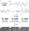
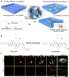
Peptide-drug conjugates (PDCs) represent an important class of therapeutic agents that combine one or more drug molecules with a short peptide through a biodegradable linker. This prodrug strategy uniquely and specifically exploits the biological activities and self-assembling potential of small-molecule peptides to improve the treatment efficacy of medicinal compounds. We review here the recent progress in the design and synthesis of peptide-drug conjugates in the context of targeted drug delivery and cancer chemotherapy. We analyze carefully the key design features in choosing the peptide sequence and linker chemistry for the drug of interest, as well as the strategies to optimize the conjugate design. We highlight the recent progress in the design and synthesis of self-assembling peptide-drug amphiphiles to construct supramolecular nanomedicine and nanofiber hydrogels for both systemic and topical delivery of active pharmaceutical ingredients.
Keywords: Cancer; Conjugated chemistry; Drug amphiphiles; Drug delivery; Peptide–drug conjugates.
Copyright © 2016 Elsevier B.V. All rights reserved.
Figures


Similar articles
-
Self-Assembling Peptide Nanofibrous Hydrogel as a Versatile Drug Delivery Platform.Curr Pharm Des. 2015;21(29):4342-54. doi: 10.2174/1381612821666150901104821. Curr Pharm Des. 2015. PMID: 26323419 Review.
-
Methotrexate-based amphiphilic prodrug nanoaggregates for co-administration of multiple therapeutics and synergistic cancer therapy.Acta Biomater. 2018 Sep 1;77:228-239. doi: 10.1016/j.actbio.2018.07.014. Epub 2018 Jul 10. Acta Biomater. 2018. PMID: 30006314
-
Recent advances in self-assembled peptides: Implications for targeted drug delivery and vaccine engineering.Adv Drug Deliv Rev. 2017 Feb;110-111:169-187. doi: 10.1016/j.addr.2016.06.013. Epub 2016 Jun 26. Adv Drug Deliv Rev. 2017. PMID: 27356149 Review.
-
Current progress and remaining challenges of peptide-drug conjugates (PDCs): next generation of antibody-drug conjugates (ADCs)?J Nanobiotechnology. 2025 Apr 22;23(1):305. doi: 10.1186/s12951-025-03277-2. J Nanobiotechnology. 2025. PMID: 40259322 Free PMC article. Review.
-
Peptide-Based Prodrug Approaches for Cancer Nanomedicine.ACS Appl Bio Mater. 2024 Dec 16;7(12):8163-8176. doi: 10.1021/acsabm.4c01364. Epub 2024 Nov 27. ACS Appl Bio Mater. 2024. PMID: 39601471 Review.
Cited by
-
Antimicrobial nanomedicine for ocular bacterial and fungal infection.Drug Deliv Transl Res. 2021 Aug;11(4):1352-1375. doi: 10.1007/s13346-021-00966-x. Epub 2021 Apr 11. Drug Deliv Transl Res. 2021. PMID: 33840082 Review.
-
Chemistries and applications of DNA-natural product conjugate.Front Chem. 2022 Sep 6;10:984916. doi: 10.3389/fchem.2022.984916. eCollection 2022. Front Chem. 2022. PMID: 36147254 Free PMC article. Review.
-
Carrier-free cryptotanshinone-peptide conjugates self-assembled nanoparticles: An efficient and low-risk strategy for acne vulgaris.Asian J Pharm Sci. 2024 Aug;19(4):100946. doi: 10.1016/j.ajps.2024.100946. Epub 2024 Jul 20. Asian J Pharm Sci. 2024. PMID: 39246508 Free PMC article.
-
Improved In Vivo Anti-Tumor and Anti-Metastatic Effect of GnRH-III-Daunorubicin Analogs on Colorectal and Breast Carcinoma Bearing Mice.Int J Mol Sci. 2019 Sep 25;20(19):4763. doi: 10.3390/ijms20194763. Int J Mol Sci. 2019. PMID: 31557968 Free PMC article.
-
Energy Landscapes of Supramolecular Peptide-Drug Conjugates Directed by Linker Selection and Drug Topology.ACS Nano. 2022 Jun 28;16(6):9546-9558. doi: 10.1021/acsnano.2c02804. Epub 2022 May 31. ACS Nano. 2022. PMID: 35639629 Free PMC article.
References
-
- Peppas NA, Hilt JZ, Khademhosseini A, Langer R. Hydrogels in biology and medicine: From molecular principles to bionanotechnology. Adv Mater. 2006;18:1345–1360.
-
- Edwards DA, Hanes J, Caponetti G, Hrkach J, BenJebria A, Eskew ML, Mintzes J, Deaver D, Lotan N, Langer R. Large porous particles for pulmonary drug delivery. Science. 1997;276:1868–1871. - PubMed
-
- Peer D, Karp JM, Hong S, FaroKHzad OC, Margalit R, Langer R. Nanocarriers as an emerging platform for cancer therapy. Nat Nanotechnol. 2007;2:751–760. - PubMed
-
- Langer R. Drug delivery and targeting. Nature. 1998;392:5–10. - PubMed
Publication types
MeSH terms
Substances
Grants and funding
LinkOut - more resources
Full Text Sources
Other Literature Sources

