

Tissue Inhibitor of Metalloproteinase-1 Is Confined to Tumor-Associated Myofibroblasts and Is Increased With Progression in Gastric Adenocarcinoma
- PMID: 27370797
- PMCID: PMC4971780
- DOI: 10.1369/0022155416656173
Tissue Inhibitor of Metalloproteinase-1 Is Confined to Tumor-Associated Myofibroblasts and Is Increased With Progression in Gastric Adenocarcinoma
Abstract
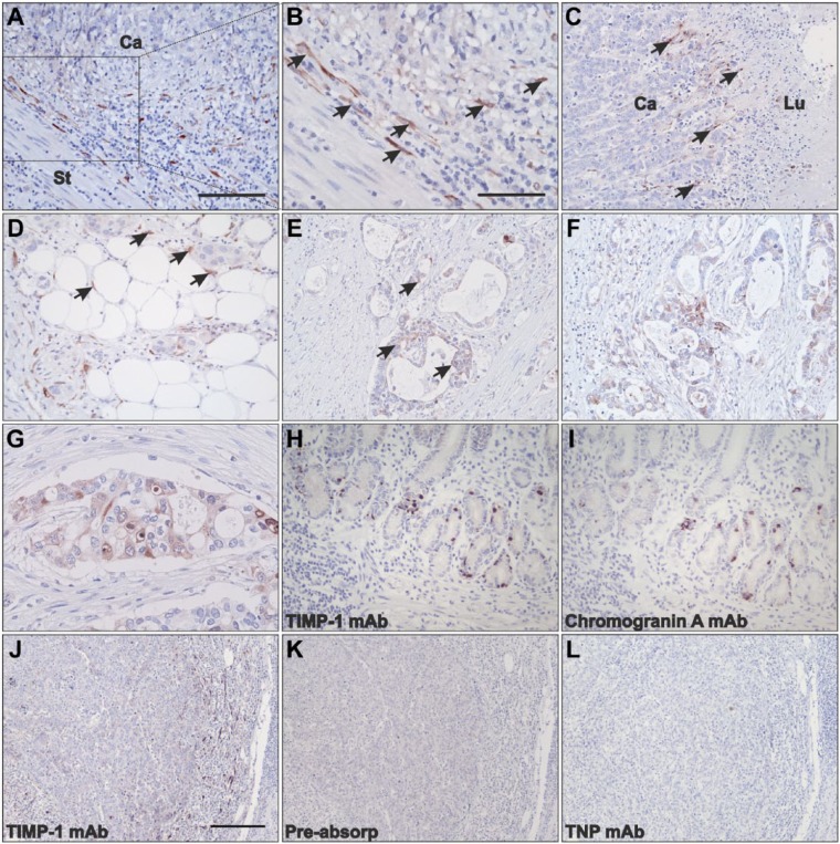
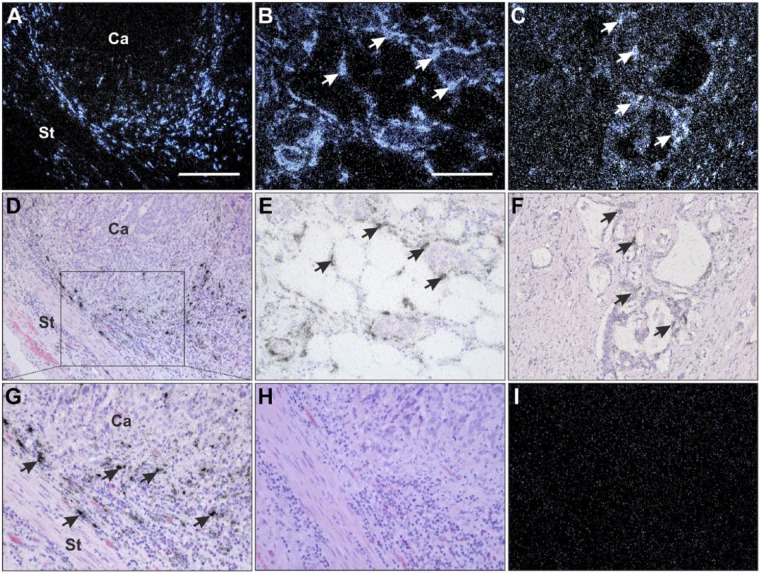
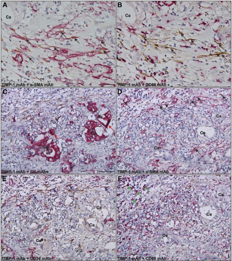
The tissue inhibitor of metalloproteinase-1 (TIMP-1) inhibits the extracellular matrix-degrading activity of several matrix metalloproteinases, thereby regulating cancer cell invasion and metastasis. Studies describing the expression pattern and cellular localization of TIMP-1 in gastric cancer are, however, highly discordant. We addressed these inconsistencies by performing immunohistochemistry and in situ hybridization analyses in a set of 49 gastric cancer lesions to reexamine the TIMP-1 localization. In addition, we correlated these findings to clinicopathological parameters. We show that strong expression of TIMP-1 protein and mRNA was observed in a subpopulation of stromal fibroblast-like cells at the periphery of the cancer lesions. In a few cases, a small fraction of cancer cells showed weak expression of TIMP-1 protein and mRNA. The stromal TIMP-1-expressing cells were mainly tumor-associated myofibroblasts. In the normal-appearing mucosa, scattered TIMP-1 protein was only found in chromogranin A positive cells. TIMP-1-positive myofibroblasts at the invasive front of the tumors were more frequently seen in intestinal than in diffuse histological subtype cases (p=0.009). A significant trend to a higher number of cases showing TIMP-1 staining in myofibroblasts with increasing tumor, node, metastasis (TNM) stage was also revealed (p=0.041). In conclusion, tumor-associated myofibroblasts are the main source of increased TIMP-1 expression in gastric cancer.
Keywords: TIMP-1; gastric cancer; immunohistochemistry; in situ hybridization; invasion; myofibroblasts; neuroendocrine cells.
© 2016 The Histochemical Society.
Conflict of interest statement
Figures
Similar articles
-
Imbalance between expression of matrix metalloproteinase-9 and tissue inhibitor of metalloproteinase-1 in invasiveness and metastasis of human gastric carcinoma.World J Gastroenterol. 2003 May;9(5):899-904. doi: 10.3748/wjg.v9.i5.899. World J Gastroenterol. 2003. PMID: 12717827 Free PMC article.
-
Localization of tissue inhibitor of metalloproteinases 1 (TIMP-1) in human colorectal adenoma and adenocarcinoma.Int J Cancer. 2005 Jan 10;113(2):198-206. doi: 10.1002/ijc.20566. Int J Cancer. 2005. PMID: 15386409
-
[Relationship of expression unbalance of matrix metalloproteinase and tissue inhibitor of metalloproteinase to invasiveness and metastasis in gastric carcinomas].Ai Zheng. 2002 Mar;21(3):305-10. Ai Zheng. 2002. PMID: 12452001 Chinese.
-
[The significance of metalloproteinases and their inhibitors in gastric cancer].Postepy Hig Med Dosw (Online). 2009 Jun 5;63:258-65. Postepy Hig Med Dosw (Online). 2009. PMID: 19502687 Review. Polish.
-
Matrix-degrading metalloproteinases in tumor progression.Princess Takamatsu Symp. 1994;24:152-61. Princess Takamatsu Symp. 1994. PMID: 8983072 Review.
Cited by
-
The Many Facets of Metzincins and Their Endogenous Inhibitors: Perspectives on Ovarian Cancer Progression.Int J Mol Sci. 2018 Feb 2;19(2):450. doi: 10.3390/ijms19020450. Int J Mol Sci. 2018. PMID: 29393911 Free PMC article. Review.
-
Total saponins from Lilium lancifolium: a promising alternative to inhibit the growth of gastric carcinoma cells.J Cancer. 2020 Apr 27;11(14):4261-4273. doi: 10.7150/jca.42285. eCollection 2020. J Cancer. 2020. PMID: 32368309 Free PMC article.
-
Mature and progenitor endothelial cells perform angiogenesis also under protease inhibition: the amoeboid angiogenesis.J Exp Clin Cancer Res. 2018 Apr 3;37(1):74. doi: 10.1186/s13046-018-0742-2. J Exp Clin Cancer Res. 2018. PMID: 29615071 Free PMC article.
-
Identification of Potential Key Genes Associated With the Pathogenesis and Prognosis of Gastric Cancer Based on Integrated Bioinformatics Analysis.Front Genet. 2018 Jul 17;9:265. doi: 10.3389/fgene.2018.00265. eCollection 2018. Front Genet. 2018. PMID: 30065754 Free PMC article.
-
A common molecular signature of intestinal-type gastric carcinoma indicates processes related to gastric carcinogenesis.Oncotarget. 2017 Dec 27;9(7):7359-7371. doi: 10.18632/oncotarget.23670. eCollection 2018 Jan 26. Oncotarget. 2017. PMID: 29484116 Free PMC article.
References
-
- Egeblad M, Werb Z. New functions for the matrix metalloproteinases in cancer progression. Nat Rev Cancer. 2002;2:161–74. - PubMed
-
- Baker AH, Edwards DR, Murphy G. Metalloproteinase inhibitors: biological actions and therapeutic opportunities. J Cell Sci. 2002;115:3719–27. - PubMed
-
- Holten-Andersen MN, Hansen U, Brünner N, Nielsen HJ, Illemann M, Nielsen BS. Localization of tissue inhibitor of metalloproteinases 1 (TIMP-1) in human colorectal adenoma and adenocarcinoma. Int J Cancer. 2005;113:198–206. - PubMed
Publication types
MeSH terms
Substances
LinkOut - more resources
Full Text Sources
Other Literature Sources
Medical
Research Materials
Miscellaneous
