. 2016 Jul:9:195-206.
doi: 10.1016/j.ebiom.2016.06.015. Epub 2016 Jun 11.

Regulation of Retinoic Acid Inducible Gene-I (RIG-I) Activation by the Histone Deacetylase 6

Affiliations

Regulation of Retinoic Acid Inducible Gene-I (RIG-I) Activation by the Histone Deacetylase 6

Helene Minyi Liu et al. EBioMedicine. 2016 Jul.

Abstract

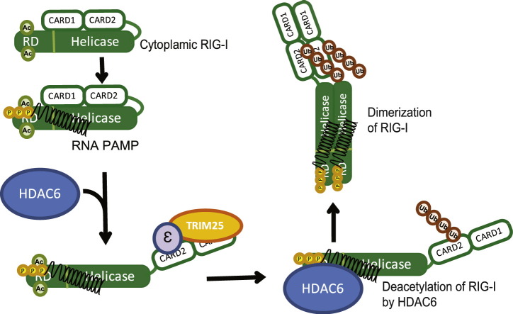
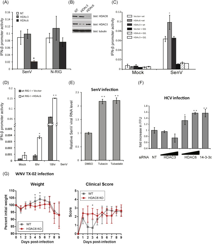
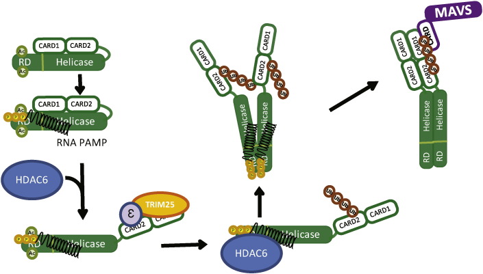
Retinoic acid inducible gene-I (RIG-I) is a cytosolic pathogen recognition receptor that initiates the immune response against many RNA viruses. Upon RNA ligand binding, RIG-I undergoes a conformational change facilitating its homo-oligomerization and activation that results in its translocation from the cytosol to intracellular membranes to bind its signaling adaptor protein, mitochondrial antiviral-signaling protein (MAVS). Here we show that RIG-I activation is regulated by reversible acetylation. Acetyl-mimetic mutants of RIG-I do not form virus-induced homo-oligomers, revealing that acetyl-lysine residues of the RIG-I repressor domain prevent assembly to active homo-oligomers. During acute infection, deacetylation of RIG-I promotes its oligomerization upon ligand binding. We identify histone deacetylase 6 (HDAC6) as the deacetylase that promotes RIG-I activation and innate antiviral immunity to recognize and restrict RNA virus infection.

Keywords: Deacetylation; HCV; HDAC6; Innate immunity; Interferon; RIG-I; West Nile virus.


Figures

Image 1
Graphical abstract
Supplementary Fig. 1
(a) RIG-I acetylation at K858 and K909 of the C-terminal RD. Ectopically expressed Flag-tagged RIG-I and indicated mutants were immunoprecipitated (IP) from mock or SenV-infected cell lystate. Acetyl-lysine levels were detected by immunoblot assay. C-RIG and N-RIG positions are indicated by filled or open arrows respectively. Non-specific bands are indicated by stars. (b) Virus infection-inducible interaction between MAVS and WT RIG-I but not the acetyl-mimetic RIG-I mutant. Flag-tagged WT RIG-I or RIG-I K858-909Q were ectopically expressed in Huh7 cells then infected with SenV for 6 h. Cell lysates were subjected to immunoprecipitation with rabbit anti-MAVS antibodies. Flag-tagged WT RIG-I was co-immunoprecipitated with MAVS during SenV infection, whereas RIG-I K858-909Q could not form significant amout of virus-inducible complex with MAVS.
Supplementary Fig. 2
(a) Graphical representation of specific RNA binding by WT RIG-I or RIG-I mutants over a dose range of RIG-I concentration. (b) K63-linked ubquitination of the RIG-I CARDs in RIG-I acetyl-mimetic mutants remains intact. WT or mutant Flag-RIG were co-expressed with HA-tagged K63ubiquitin in Huh7 cells. Cell extracts were subject to immunoprecipitation with anti-Flag antibody. Ubiquitination level of RIG-I products was evaluated by anti-HA immunoblot. Immnoblotting of input lysates is shown in the right panel, respective ubiquitin product for each lane of upper left panel is presented in the lower left panel. (c) Constiutive IFN-β promoter activity in resting/noninfected RIG-I knock-down Huh7 cells complemented with RIG-I WT and RIG-I mutants. *: p value < 0.005. (d) Flag-tagged WT or HA-tagged RIG-I mutants were co-trasnfected with WT HA-RIG-I in Huh7 cells. Cells mock-infected (−) or infeted with SenV (+) for 16 h, extracts prepared and subjected to co-immunoprecipitation with anti-Flag antibody. Input (lysate) and IP products were assessed by immunoblot assay with anti-HA or anti-Flag antibodfy.
Supplementary Fig. 3
Increaseing RIG-I acetylation levels in cells treated Tubastatin A in a dose-dependent manner. Huh7 cells were treated with increasing dose of Tubastatin A for 6 h. Cells were harvested and subjected to immunoblot assay with anti-RIG-I or anti-acetyl-lysine antibodies. A dominant band in the lysate detected in the acetyl-lyine blot at the molecular weight around 55 kDa, overlapping with endogenous tubulin as detected by anti-tubulin antibodiesy. Tubastatin A treatment increases the levels of acyl RIG-I and acetyl-tubulin (control).
Supplementary Fig. 4
(a) The deacetylase activity of HDAC6 was required to enhance RIG-I dependent IFN-β promoter activity. HDAC6-DC, the catalytic site defective mutant of HDAC6, could not boost RIG-I signaling through IFN-β promoter above level sof cells expressing vector alone. *: p value < 0.05. (b) Mapping of the RIG-I interaction domain of HDAC6. Cells were cotransfected with Myc-RIG-I and Flag vector control or the indicated Flag-HDAC6 WT or mutant construct. FL, full-length HDAC6; dBUZ, HDAC6 with BUZ domain deletion; N80, the first 80 amino acid of HDAC6 from the N-terminus. IP products were assesed by immunoblot assay.
Fig. 1
Fig. 1
RIG-I is acetylated in resting cells. (a) Endogenous RIG-I was immunoprecipitated (IP) from mock or TSA treated cell lystate. Acetyl-RIG-I level was detected by anti-acetyl-lysine antibody. (b) Illustration of RIG-I mutants used in (c) to (g). (c) Ectopically expressed Flag-tagged RIG-I and mutants were recovered by IP from mock or SenV-infected cell lystate. Acetyl-lysine levels were detected by immunoblotting. Relative intensities of acetyl-RIG-I to total Flag-RIG-I was measured and calculated by Image J software and expressed as a value relative to the corresponding uninfected control. (d) Ectopically expressed Flag-tagged RIG-I and mutants were recovered by IP and analyzed for acetylation levels by immunoblot (blot) assay. GFP-RD-RR referred to Flag-tagged GFP-fused RIG-I RD with K858-909R mutations. (e) Ectopically expressed Flag-tagged RIG-I and mutants were recovered by IP from mock or SenV-infected cell lystate. Acetyl-lysine levels were detected by immunoblot assay. (f) SenV-induced IFN-β promoter induction in RIG-I knock-down Huh7 cells complemented with WT RIG-I or RIG-I acetyl-mimetic mutants. (g) Membrane (M) localization of RIG-I to the MAM from a resting, cyosolic (C) state during SenV infection. RIG-I acetyl-mimetic mutants were not detected in the membrane fractions, whereas WT RIG-I was redistributed to the MAM as marked by mitofusin 2, a MAM protein during SenV infection. Tubulin marks the cytosolic fraction.
Fig. 2
Fig. 2
Oligomerization difficiency of RIG-I acetyl-mimetic mutants. (a) in vitro transcribed biotinylated-PAMP RNA from HCV 3′UTR was transfected into cells expressing the indicated Flag-tagged RIG-I mutants. After crosslinking, PAMP RNA/RIG-I complexes were recovered by anti-Flag IP and blotted onto nitracellulose membrane. RNA is visualized using Streptavidin-HRP. Relative RNA binding activities were quantified using Image J software, and expressed as a value relative to the corresponding WT control. (b) Representative gel-shift assays for WT and RIG-I mutants in the presence of 18-bp dsRNA. The oligomer formation efficiency of RIG-I-RNA (2:1) complex by band intensities was quantified and plotted. (c) Relative RNA-stimulated ATP hydrolysis of WT RIG-I and three RIG-I mutants. Data were normalized to WT RIG-I and shown as the relative level ATPase rate. Error bars represent the standard deviation of triplicate measurements. (d) Impaired oligomerization of RIG-I acetyl-mimetic mutants during SenV infection. Flag-tagged wt RIG-I and HA-tagged RIG-I acetyl-mimetic mutants were co-transfected into HEK293 cells. After SenV infection, cell extracts were subjected to IP by anti-Flag antibody. RIG-I oligomerization activities were shown by the levels of co-IP HA-tagged RIG-I. Arrows indicate Flag-RIG-I oligomers. (e) RIG-I translocon formation. HEK293 cells expressing Flag-tagged WT or mutant RIG-I were co-expressed with Myc-14-3-3ε and cells were infected with 100 HAU SenV for 16 h followed by anti-Flag IP of cell extracts. The products were analyzed by immunoblot with anti-TRIM25 antibody or anti-Myc antibody.
Fig. 3
Fig. 3
RIG-I deacetylation by the cytoplasmic deacetylase HDAC6 during viral infection. (a) and (b) RIG-I signaling to the IFN-β promoter was measured by promoter-luciferase assay in mock-infected or SenV-infected Huh7 cells that were treated with DMSO control or HDAC inhibitor. (a) cells were treated with increasing dose of Tricostatin A (TSA), Valproate (VPA), or Bufexamac (Bufex). (b) Huh7 cells were transfected with vector lone or the constitutively active N-RIG mutant and cultured in the presence of treatment with DMSO (control), TSA, or Bufexamac. 12 h later the cells were harvested and assessed for IFN-β-luciferase activity. *: p value < 0.05. (c) Screening of RIG-I interacting HDACs. Flag-tagged HDACs and Myc-tagged RIG-I were co-transfected into HEK293 cells. Cells were mock or SenV infected and extracts prepared 16 h later. Proteins were recovered by anti-Myc co-IP and identified by anti-Flag or anti-Myc immunoblot analysis. (d) Endogenous RIG-I and HDAC6 form virus-inducible complexes. Mock or SenV-infected Huh7 cell lysates were used for IP by anti-RIG-I antibody. Recovered proteins were identified by immunoblot assay for RIG-I, HDAC6 and HDAC 3. (e) Huh7 cells were mock infected or infected with SenV for 16 h in the presence of DMSO (control) or Tubastatin A treatment. 16 h later the cells were co-stained with DAPI to detect the nucleus and anti-IRF3 antibody followed by anti-rabbit flourescein secondary antibody for immunostain analysis of IRF3 subcellular distribution. (f) WT and IFNAR KO MEFs were treated with DMSO control or with increasing levels of HDAC6 specific inhibitor, Tubastatin A or Tubacin, during mock infection or SenV infection. 16 h later the cells were harvested and the level of IFIT2 mRNA was quantified by RT-qPCR assay. *: p value < 0.05.
Fig. 4
Fig. 4
HDAC6 deacetylase activity is required for RIG-I signaling induction. (a) Huh7 cells were transfected with nontargeting siRNA control (NT) or siRNA specific for knockdown of HDAC3 or HDAC6. Cells were then infected with SenV or transfected with vector or N-RIG to induce signaling in the presence of the IFN-β-luciferase promoter construct. IFN-β promoter activity was measured 16 h later. (b) HDAC6 and HDAC3 protein levels from siRNA treated cells in A. (c) WT or mutant RIG-I was ectopically expressed with vector control, HDAC3 or HDAC6 in combination in the presence of the IFN-β-luciferase promoter in Huh7 cells. Cells were mock infected or infected with SenV and harvested for assessment of IFN-β promoter activity 16 h later. (d) WT RIG-I was ectopically expressed with vector control or HDAC6 in combination in the presence of the IFN-β-luciferase promoter in Huh7 cells. Cells were mock infected or infected with SenV and harvested at the indicated time for assessment of IFN-β promoter activity. (e) Huh7 cells were treated with DMSo control, Tubastatin A or Tubacin and mock infected or infected with SenV. Cells were harvested and viral RNA measured by RT-qPCR assay 24 h later. (f) Huh7 cells were treated with NT siRNA or increasing levels of siRNA for knockdown of HDAC3 or HDAC6. Cells were infected with HCV. Infectious virus in supernatant was determined at 48 h post-infection by viral focus forming unit assay. *: p value < 0.05; **: p value < 0.01. (G) WT and HDAC KO mice were challenged with WNV and monitored for clinical scores and weight loss. Statistical significance was determined using the Holm-Sidak method of multiple t-tests, with alpha = 5.000%. *: p value < 0.05.
Fig. 5
Fig. 5
Model of HDAC6-depende t RIG-I activation. Details are described in the text.

Similar articles

Cited by

References

    1. Bantscheff M., Hopf C., Savitski M.M., Dittmann A., Grandi P., Michon A.M., Schlegl J., Abraham Y., Becher I., Bergamini G., Boesche M., Delling M., Dumpelfeld B., Eberhard D., Huthmacher C., Mathieson T., Poeckel D., Reader V., Strunk K., Sweetman G., Kruse U., Neubauer G., Ramsden N.G., Drewes G. Chemoproteomics profiling of HDAC inhibitors reveals selective targeting of HDAC complexes. Nat. Biotechnol. 2011;29:255–265. - PubMed
    1. Bhatia R., Shaffer T.H., Hossain J., Fisher A.O., Horner L.M., Rodriguez M.E., Penfil S., Theroux M.C. Surfactant administration prior to one lung ventilation: physiological and inflammatory correlates in a piglet model. Pediatr. Pulmonol. 2011;46:1069–1078. - PMC - PubMed
    1. Blander J.M. A long-awaited merger of the pathways mediating host defence and programmed cell death. Nat. Rev. Immunol. 2014;14:601–618. - PubMed
    1. Boyault C., Zhang Y., Fritah S., Caron C., Gilquin B., Kwon S.H., Garrido C., Yao T.P., Vourc'h C., Matthias P., Khochbin S. HDAC6 controls major cell response pathways to cytotoxic accumulation of protein aggregates. Genes Dev. 2007;21:2172–2181. - PMC - PubMed
    1. Chan Y.K., Gack M.U. RIG-I-like receptor regulation in virus infection and immunity. Curr. Opin. Virol. 2015;12:7–14. - PMC - PubMed

MeSH terms