. 2016 Jul 12;16(2):288-294.
doi: 10.1016/j.celrep.2016.06.014. Epub 2016 Jun 30.

2.8-Å Cryo-EM Structure of the Large Ribosomal Subunit from the Eukaryotic Parasite Leishmania

Affiliations

2.8-Å Cryo-EM Structure of the Large Ribosomal Subunit from the Eukaryotic Parasite Leishmania

Moran Shalev-Benami et al. Cell Rep. .

Abstract

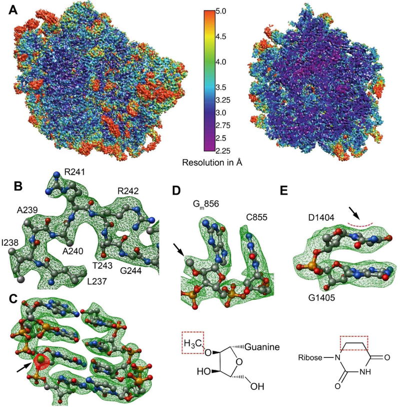
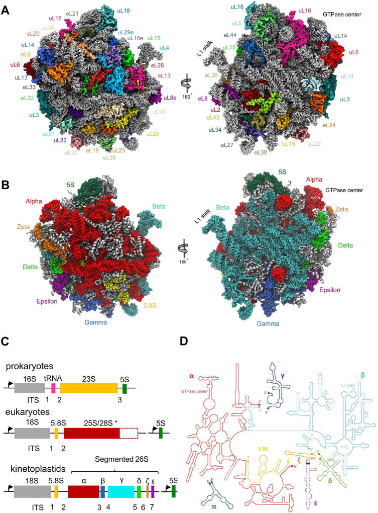
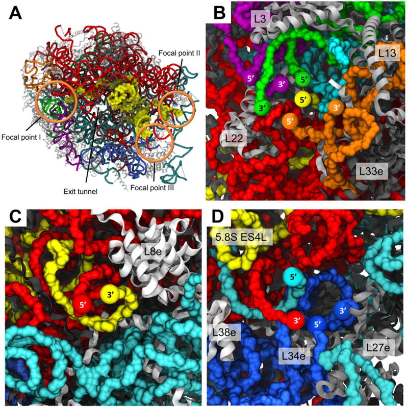
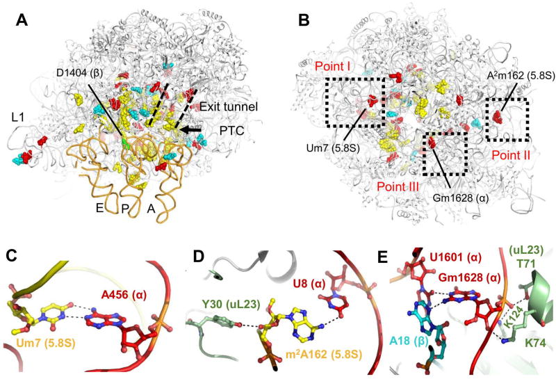
Leishmania is a single-cell eukaryotic parasite of the Trypanosomatidae family, whose members cause an array of tropical diseases. The often fatal outcome of infections, lack of effective vaccines, limited selection of therapeutic drugs, and emerging resistant strains, underline the need to develop strategies to combat these pathogens. The Trypanosomatid ribosome has recently been highlighted as a promising therapeutic target due to structural features that are distinct from other eukaryotes. Here, we present the 2.8-Å resolution structure of the Leishmania donovani large ribosomal subunit (LSU) derived from a cryo-EM map, further enabling the structural observation of eukaryotic rRNA modifications that play a significant role in ribosome assembly and function. The structure illustrates the unique fragmented nature of leishmanial LSU rRNA and highlights the irregular distribution of rRNA modifications in Leishmania, a characteristic with implications for anti-parasitic drug development.


Figures

Figure 1
Figure 1. Cryo-EM map and modeling of L. donovani LSU
(A) Surface (left) and cross-section (right) representations of the cryo-EM density map colored according to local resolution distribution. (B–C) Snapshots of map vs. model from segments of protein uL2 (B) and 26S rRNA chain alpha (C). The arrow in (C) points to the density (red) corresponding to a Mg+2 ion (green) interacting with the rRNA phosphate backbone. (D–E) Two examples of rRNA modifications observed. (D) The 2’-O methyl group of G856 of alpha 26S rRNA. A total of 46 2’-O methylated residues were modeled in the map. A detailed list of rRNA modifications is supplemented to the manuscript (Table S4). (E) Dihydrouridine distorted ring in 26S rRNA residue U1404 of beta 26S. The arrows in (D) and (E) point to the densities for methyl group and ring distortion, respectively.
Figure 2
Figure 2. Structure of L. donovani LSU
(A) Protein distribution from two LSU views: solvent facing side (left) and subunit interface (right). Ribosomal proteins are shown as variably colored cartoon representations and rRNA as light grey ribbon. (B) Tertiary structure of rRNA distribution in the leishmanial ribosome LSU as viewed from either the solvent exposed side (left) or the inter-subunit interface (right). 5S rRNA is shown in dark green, 5.8S in yellow, and 26S rRNA segments α, β, γ, δ, ε and ζ are shown in red, cyan, blue, green, purple and orange, respectively. (C) Typical rRNA encoding operons in prokaryotes, eukaryotes and kinetoplastids. *The largest LSU component (25–28S) varies greatly in length within different species. In all panels, promoters are marked as arrows pointing in the direction of transcription. Internal Transcribed Segments (ITSs) are numbered according to their order in the transcription unit. (D) Schematic diagram of rRNA secondary structure in L. donovani LSU. Functional sites are highlighted.
Figure 3
Figure 3. Focal points of LSU rRNA segmentation
26S rRNA segment terminals in the leishmanial ribosome converge to three focal points, all located on the solvent exposed side of the LSU. (A) LSU surface with localization of the three focal points in regards to the ribosome exit tunnel. The 5.8S rRNA chain is presented as yellow surface and all other chains are represented as cartoon tubes. (B) Focal point I includes the 5’ and 3’ ends of delta, epsilon and zeta, as well as the 5’ end of the 5.8S rRNA. (C) Focal point II includes alpha 5’ and 5.8S 3’ ends. (D) Focal point III includes both terminals of the gamma unit as well as alpha 3’ and beta 5’ ends. 5S rRNA is shown in dark green, 5.8S in yellow, and 26S segments α, β, γ, δ, ε and ζ are shown in red, cyan, blue, green, purple and orange, respectively. Ribosomal proteins are illustrated as grey cartoons. Proteins in close proximity to the focal point cavity are labeled accordingly.
Figure 4
Figure 4. LSU rRNA modifications
Leishmania LSU rRNA is heavily modified by multiple 2’-O methyl groups and pseudouridins. Modified residues are mainly localized at functional ribosomal sites such as the PTC, the L1 stalk, the protein exit tunnel and all three tRNA binding sites (A) and are mostly distributed within internal parts of the ribosome (B). The predicted modified nucleotides as observed from two ribosomal views, one from the tRNA binding site in the subunit interface (A) and the other from the solvent exposed surface (B). 2’-O methylation sites that are unique to trypanosomatids are highlighted in red, pseudouridinylations in cyan and dihydrouridine in green. Eukaryote conserved residues are highlighted in yellow. A, P and E site-tRNAs are presented as orange tubes and were docked to their binding sites from previously reported yeast and rabbit cryo-EM structures of ribosomes at high resolution (Schmidt, et al., 2016; Brown, et al., 2015) Some rRNA modifications are also located in close proximity to the three focal points and interact with residues belonging to other chains. (C–E) Close up of modifications and their interaction networks in focal points I–III, respectively (also outlined in panel B). rRNA chains are colored in red, cyan, and yellow for alpha, beta, and 5.8S, respectively, and protein uL23 is shown in light green. Interacting residues are presented in a ball-and-stick representation and atoms are colored according to their identity. Polar contacts are presented as black dashed lines.

References

    1. Afonine PV, Headd JJ, Terwilliger TC, Adams PD. New tool: phenix.real_space_refine. Computational Crystallography Newsletter. 2013;4:43–44.
    1. Ben-Shem A, Garreau de Loubresse N, Melnikov S, Jenner L, Yusupova G, Yusupov M. The structure of the eukaryotic ribosome at 3.0 Å resolution. Science. 2011;334:1524–1529. - PubMed
    1. Brown A, Shao S, Murray J, Hegde RS, Ramakrishnan V. Structural basis for stop codon recognition in eukaryotes. Nature. 2015;524:493–496. - PMC - PubMed
    1. Demirci H, Murphy F, Belardinelli R, Kelley AC, Ramakrishnan V, Gregory ST, Dahlberg AE, Jogl G. Modification of 16S ribosomal RNA by the KsgA methyltransferase restructures the 30S subunit to optimize ribosome function. RNA. 2010;16:2319–24. - PMC - PubMed
    1. Eliaz D, Doniger T, Tkacz ID, Biswas VK, Gupta SK, Kolev NG, Unger R, Ullu E, Tschudi C, Michaeli S, et al. Genome-wide analysis of small nucleolar RNAs of Leishmania major reveals a rich repertoire of RNAs involved in modification and processing of rRNA. RNA biology. 2015;12:1222–1255. - PMC - PubMed

MeSH terms