. 2016 Jul 21;63(2):277-292.
doi: 10.1016/j.molcel.2016.05.038. Epub 2016 Jun 30.

BRCA1/FANCD2/BRG1-Driven DNA Repair Stabilizes the Differentiation State of Human Mammary Epithelial Cells

Affiliations

BRCA1/FANCD2/BRG1-Driven DNA Repair Stabilizes the Differentiation State of Human Mammary Epithelial Cells

Hua Wang et al. Mol Cell. .

Abstract

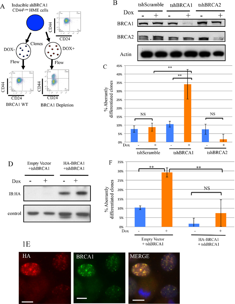
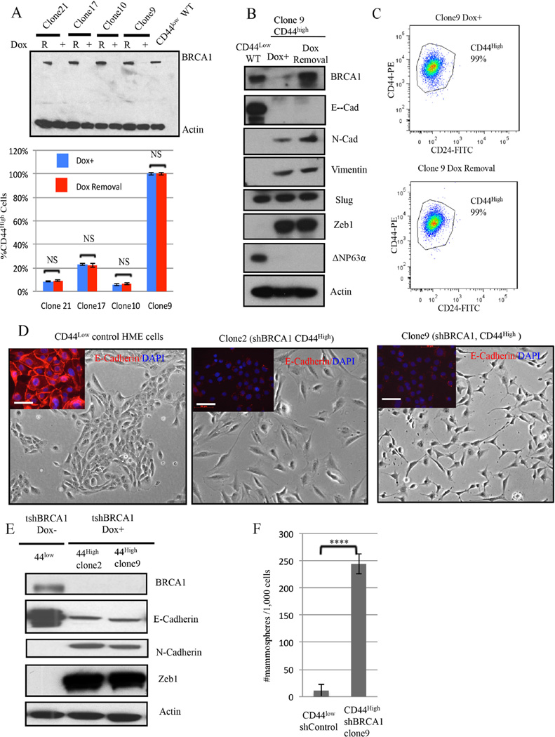
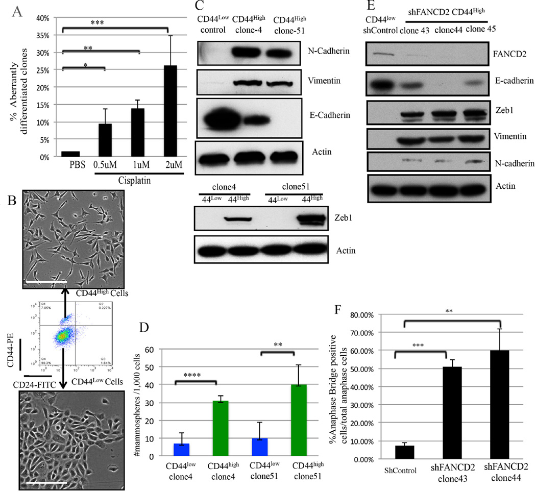
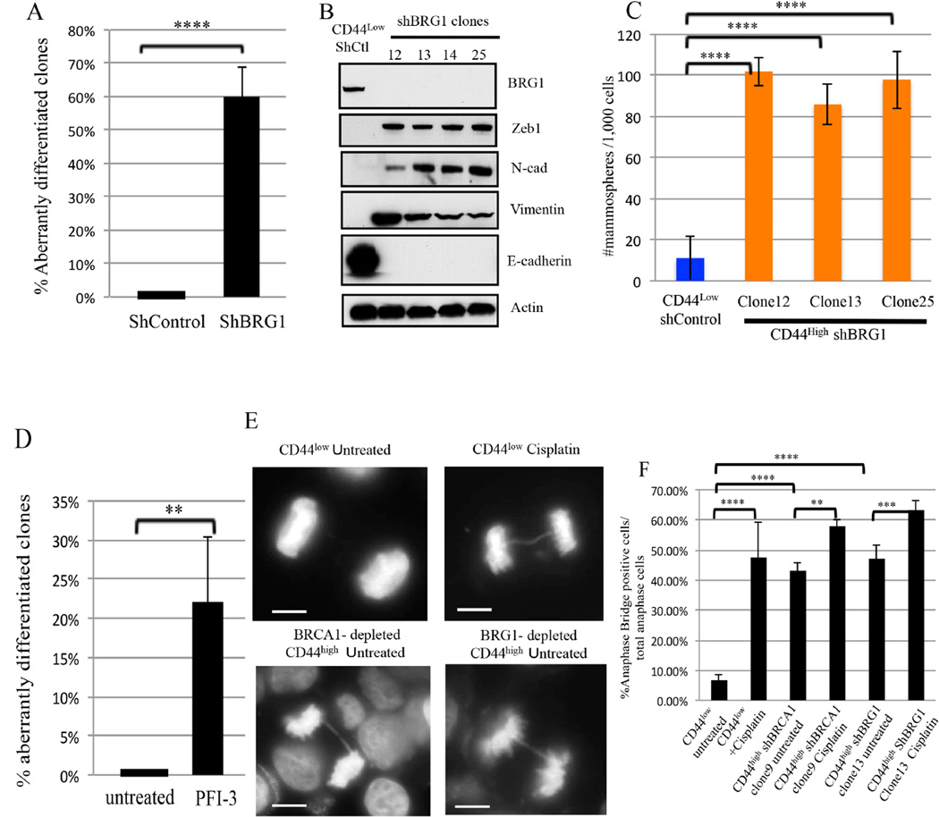
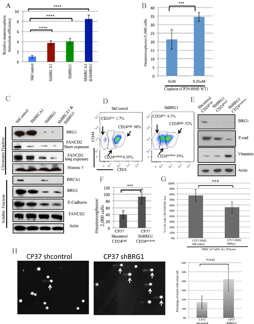
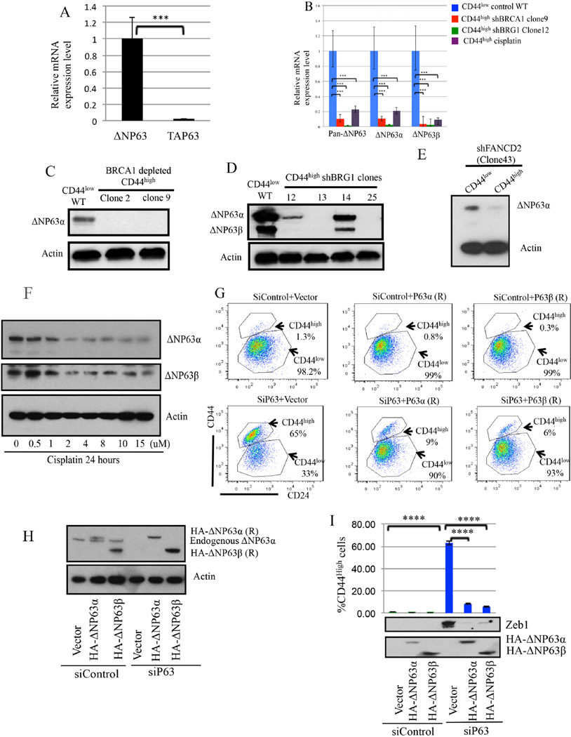
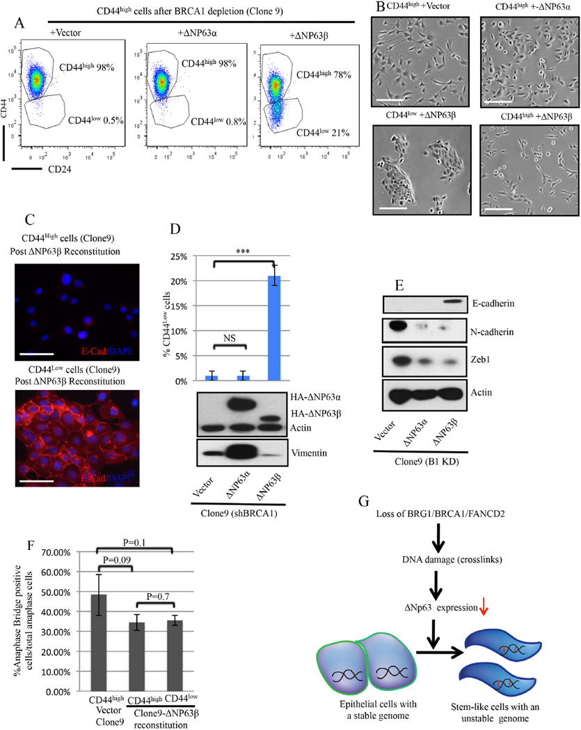
An abnormal differentiation state is common in BRCA1-deficient mammary epithelial cells, but the underlying mechanism is unclear. Here, we report a convergence between DNA repair and normal, cultured human mammary epithelial (HME) cell differentiation. Surprisingly, depleting BRCA1 or FANCD2 (Fanconi anemia [FA] proteins) or BRG1, a mSWI/SNF subunit, caused HME cells to undergo spontaneous epithelial-to-mesenchymal transition (EMT) and aberrant differentiation. This also occurred when wild-type HMEs were exposed to chemicals that generate DNA interstrand crosslinks (repaired by FA proteins), but not in response to double-strand breaks. Suppressed expression of ΔNP63 also occurred in each of these settings, an effect that links DNA damage to the aberrant differentiation outcome. Taken together with somatic breast cancer genome data, these results point to a breakdown in a BRCA/FA-mSWI/SNF-ΔNP63-mediated DNA repair and differentiation maintenance process in mammary epithelial cells that may contribute to sporadic breast cancer development.

Keywords: BRCA1; BRG1; EMT; FANCD2; breast cancer; cisplatin; crosslink repair.


Figures

Figure 1
Figure 1. BRCA1 contributes to the maintenance of differentiation in CD44low human mammary epithelial (HME) cells (Also see Figure S1)
1A) Experimental scheme to search for a differentiation marker expression change in CD44low HME cell clones after BRCA1 depletion. 1B) Immunoblotting for BRCA1 or BRCA2 in CD44low HME cells infected with the indicated tet-on shRNA vectors before and after doxycycline exposure. 1C) Summary of the analysis of aberrantly differentiated CD44high clones (that contain ≥5% CD44high cells) emerging after BRCA1 or BRCA2 depletion in CD44low HME cells. The data represented are mean values ±S.D. **P<0.01 and NS: Not significant. 1D) Immunoblotting for shRNA-resistant HA-BRCA1 in CD44low HME cells before and after endogenous BRCA1 depletion. A nonspecific band serves as the loading control. 1E) Formation of nuclear foci by shRNA-resistant HA-BRCA1 in CD44low cells after depleting endogenous BRCA1. Cells were treated with cisplatin (1µM, 24hours). Scale bar: 10µm. 1F) Prevention of the emergence of aberrantly differentiated clones by shRNA-resistant BRCA1 in CD44low HME cells following shBRCA1 expression. The data represented are mean values ±S.D. **P<0.01.
Figure 2
Figure 2. Aberrant differentiation of HME cells following BRCA1 depletion
2A) (Top panel) Immunoblotting for BRCA1 after Doxycycline-induced BRCA1 depletion (DOX +) and after BRCA1 re-accumulation (R) for 4 weeks after Dox removal. (Lower panel)The abundance of CD44high cells was measured before and 4 weeks after doxycycline removal in four, independent, BRCA1- depleted clones. The data represented are mean values ±S.D. NS: Not significant. 2B) Immunoblotting for BRCA1 and EMT markers in CD44high HME cells (clone9) after BRCA1 depletion or reexpression. 2C) Representative CD24 and CD44 FACS profiles of clone 9 derived after BRCA1 depletion (clone 9 Dox+) and 4 weeks after subsequent Doxcycline removal. 2D) Respective phase-contrast images of CD44low parental and of CD44high cells (clones 2 and 9) isolated after Dox-induced BRCA1 depletion in CD44low cells. The inserts depict the results of anti-E-cadherin (red) and DAPI (blue) staining. Scale bar: 50 µm 2E) Immunoblotting for EMT marker proteins in two, independent, aberrantly differentiated CD44high clones (clones 2 and 9) that appeared after BRCA1 depletion in CD44low cells. 2F) Mammosphere assays were performed on a clone (clone9) containing uniformly CD44high HME cells derived after BRCA1 depletion of CD44low cells or on shluc CD44low control cells. The data are represented as mean values ±S.D. ****P<0.0001.
Figure 3
Figure 3. DNA Crosslink damage affects the differentiation of CD44low HME cells (Also see Figures S2)
3A) Summary of the effect of Cisplatin exposure upon the state of HME cell differentiation as reflected by the abundance of clones containing CD44high cells (≥5%). The data are represented as mean values ±S.D. *P<0.05, **P<0.01 and ***P<0.001. 3B) Phase-contrast images of FACS-purified CD44high and CD44low cells derived from an HME clone (Clone 4) following cisplatin-treatment. Scale Bar: 100µm 3C) Immunoblotting for EMT markers in FACS-purified CD44low and CD44high cells (clones 4 and 51) following cisplatin treatment of CD44low (control) cells. 3D) Mammosphere assays performed on cisplatin-treated, FACS-purified CD44high cells compared with their CD44low counterparts in two independent clones. The data represented are mean values ±S.D. **P<0.01 and ****P<0.0001. 3E) Immunoblotting for FANCD2 and EMT markers in CD44high cells from three, independent, FANCD2-depleted clones. 3F) Statistical analysis of anaphase bridge-positive cells in two, independent FANCD2-depleted clones (clone43 and clone44). The data represented are mean values ±S.D. **P<0.01 and ***P<0.001
Figure 4
Figure 4. BRG1 suppresses DNA damage-associated aberrant differentiation (Also see Figures S3 and S4)
4A) Summary of the effect of BRG1 depletion on the differentiation state of CD44low HME cells. ****P<0.0001. 4B) Immunoblotting for EMT markers from four, independent CD44high clones following BRG1 -depletion. 4C) Mammosphere assays were performed on CD44high HME cells that appeared after BRG1 depletion and on Shluc CD44low control cells. The data represented are mean±S.D. ****P<0.0001. 4D) Summary of the BRG1 inhibitor, PFI-3 (50 µM), exposure on the differentiation state in CD44low HME cells. . The data represented are mean values ±S.D. **P<0.01. **P<0.01. 4E) Representative images of normal anaphase chromosomes and anaphase bridges in untreated and cisplatin-treated (1µM, 24hr) CD44low cells, respectively. Other panels depict drug-free CD44high cells following long term, hairpin-mediated BRCA1 (clone 9) or BRG1 (Clone 13) depletion. Scale Bar: 10 µM. 4F) Analysis of anaphase bridge-positive cells in drug-free CD44low, shBRCA1 clone 9, and shBRG1 clone 13 CD44high cells and after exposure of these cells to cisplatin (1 µM, 24 hours). The data represented are mean±S.D.
Figure 5
Figure 5. BRCA1 and BRG1 cooperate in crosslink repair and differentiation maintenance. (Also see Figures S5 and S6)
5A) Effect of BRCA1, BRG1, and BRCA1+BRG1 co-depletion on mammosphere-forming efficiency in CP29-HME cells. The mammosphere number was corrected for clonogenic survival efficiency on plastic to measure the relative mammosphere formation efficiency. The data represented are mean±S.D. **** P<0.0001. 5B) A mammosphere assay was performed on wt CP29-HME cells before and after cisplatin treatment. The data represented are mean±S.D. *** P<0.001 5C) Immunoblotting for chromatin-associated and soluble FANCD2 and E-cadherin following hairpin depletion of BRCA1, BRG1, or both in primary, telomerase-immortalized BRCA1+/+: p53 +/+ CP29-HME cells. 5D) CD24 and CD44 expression profiles following BRG1 depletion in CP37-HME cells. 5E) Immunoblotting for BRG1 and for EMT markers following BRG1 depletion in CP37- HME cells. FACS- purified CD24high and CD24medium cells were analyzed here. 5F) A mammospshere assay of CP37-HME cells following BRG1 depletion. The data represented are mean±S.D. *** P<0.001 5G) Quantification of FANCD2-containing DNA damage foci in CP37-HME cells after mitomycin C exposure. The data are represented as mean values ±S.D. *** P<0.001. 5H) Percent of cells with comet tails in BRG1-depleted CP37-HME cells compared to shcontrol cells. The data represented are mean values±S.D.
Figure 6
Figure 6. ΔNp63 depletion is associated with aberrant HME cell differentiation (Also see Figure S7)
6A) ΔNp63 is the primary p63 isoform in CD44low HME cells. The data represented are mean values±S.D. *** P<0.001 6B) ΔNP63α and ΔNP63β mRNA expression were significantly reduced in aberrantly differentiated CD44high cells after BRCA1 or BRG1 depletion or after Cisplatin treatment. The data represented are mean values±S.D. ***P<0.001. 6C-6E) Immunoblotting for ΔNp63 in CD44high cells after BRCA1/BRG1/FANCD2 depletion. 6F) Cisplatin suppresses endogenous ΔNP63 expression in CD44low HME cells. 6G-6I) ΔNP63 is necessary and sufficient to suppress the aberrant differentiation of CD44low cells. After siP63 transfection, the percentage of CD44high cells was measured, and expression of the EMT marker, Zeb1, and of the two HA-tagged ΔNP63 vectors was analyzed. ****P<0.0001.
Figure 7
Figure 7. ΔNP63β expression promotes differentiation of BRCA1-deficient CD44high HME cells
7A) Representative CD24 and CD44 profiles after ΔNP63 α vs β reconstitution of a BRCA1-depleted clone (clone9) composed, almost uniformly, of CD44high cells when the experiment was initiated. 7B) Phase-contrast images of CD44high cells after empty vector or HA-ΔNP63α transduction of CD44high cells (clone9) and phase contrast images of CD44high and CD44low cells isolated by FACS after HA-ΔNP63β transduction of CD44high cells (clone9). Scale bar: 100µm. 7C) CD44low cells arising after HA-ΔNP63β reconstitution were positive for E-Cadherin while CD44high cells remained E-cadherin negative. Scale bar: 50µm. 7D) Percentage of CD44low cells after ΔNP63β, ΔNP63α,β or empty vector reconstitution of CD44high cells (clone9). 7E) Immuoblotting for EMT markers after ΔNP63β or ΔNP63αβ or empty vector reconstitution in CD44high cells (clone9). 7F) Percentage of anaphase bridge-positive cells in ΔNp63β- reconstituted CD44high and CD44low BRCA-depleted clone 9 cells. The data represented are mean values±S.D.. 7G) A model of the mechanism underlying BRCA1/BRG1/FANCD2 dependent differentiation maintenance in HME cells in response to DNA damage. The short red bars represent interstrand DNA cross links.

References

    1. Adorno M, Cordenonsi M, Montagner M, Dupont S, Wong C, Hann B, Solari A, Bobisse S, Rondina MB, Guzzardo V, et al. A Mutant-p53/Smad complex opposes p63 to empower TGFbeta-induced metastasis. Cell. 2009;137:87–98. - PubMed
    1. Aguilera A, Garcia-Muse T. R loops: from transcription byproducts to threats to genome stability. Molecular cell. 2012;46:115–124. - PubMed
    1. Al-Hajj M, Wicha MS, Benito-Hernandez A, Morrison SJ, Clarke MF. Prospective identification of tumorigenic breast cancer cells. Proceedings of the National Academy of Sciences of the United States of America. 2003;100:3983–3988. - PMC - PubMed
    1. Bochar DA, Wang L, Beniya H, Kinev A, Xue Y, Lane WS, Wang W, Kashanchi F, Shiekhattar R. BRCA1 is associated with a human SWI/SNF-related complex: linking chromatin remodeling to breast cancer. Cell. 2000;102:257–265. - PubMed
    1. Buckley NE, Conlon SJ, Jirstrom K, Kay EW, Crawford NT, O’Grady A, Sheehan K, Mc Dade SS, Wang CW, McCance DJ, et al. The DeltaNp63 proteins are key allies of BRCA1 in the prevention of basal-like breast cancer. Cancer research. 2011;71:1933–1944. - PubMed

Publication types

MeSH terms