

Lens glutathione homeostasis: Discrepancies and gaps in knowledge standing in the way of novel therapeutic approaches
- PMID: 27373973
- PMCID: PMC5199622
- DOI: 10.1016/j.exer.2016.06.018
Lens glutathione homeostasis: Discrepancies and gaps in knowledge standing in the way of novel therapeutic approaches
Abstract
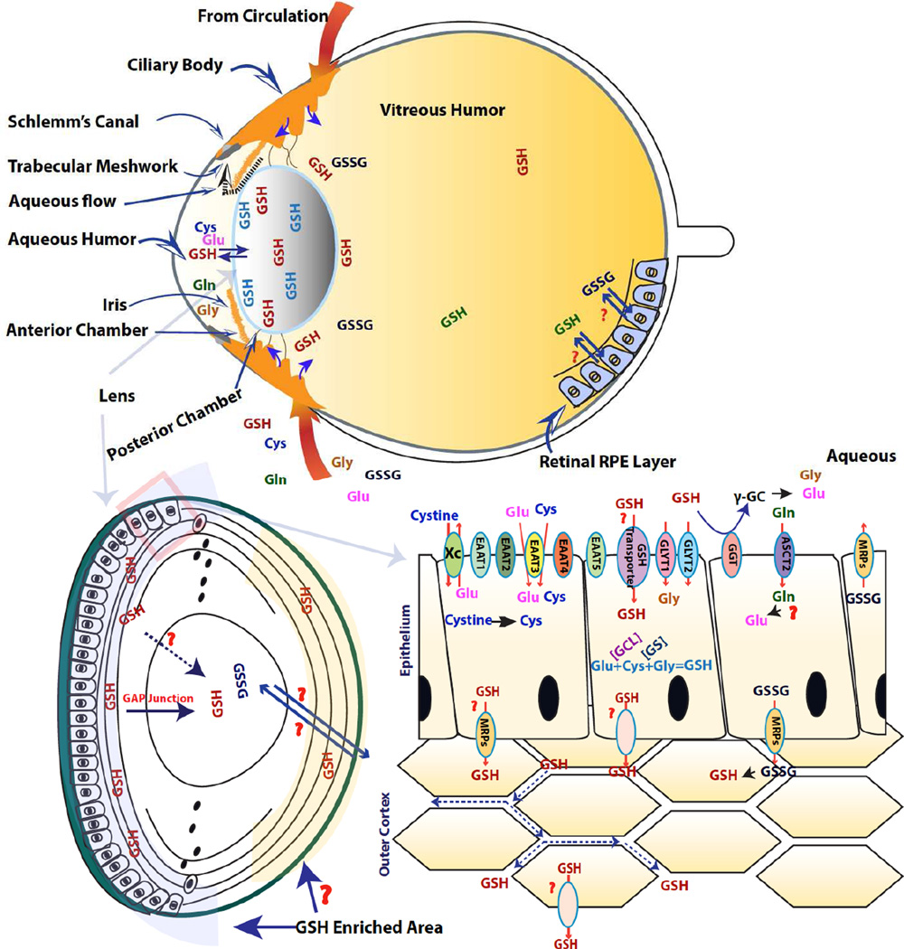
Cataract is the major cause of blindness worldwide. The WHO has estimated around 20 million people have bilateral blindness from cataract, and that number is expected to reach 50 million in 2050. The cataract surgery is currently the main treatment approach, though often associated with complications, such as Posterior Capsule Opacification (PCO)-also known as secondary cataract. The lens is an avascular ocular structure equipped with an unusually high level of glutathione (GSH), which plays a vital role in maintaining lens transparency by regulating lenticular redox state. The lens epithelium and outer cortex are thought to be responsible for providing the majority of lens GSH via GSH de novo synthesis, assisted by a continuous supply of constituent amino acids from the aqueous humor, as well as extracellular GSH recycling from the gamma-glutamyl cycle. However, when de novo synthesis is impaired, in the presence of low GSH levels, as in the aging human lens, compensatory mechanisms exist, suggesting that the lens is able to uptake GSH from the surrounding ocular tissues. However, these uptake mechanisms, and the GSH source and its origin, are largely unknown. The lens nucleus does not have the ability to synthesize its own GSH and fully relies on transport from the outer cortex by yet unknown mechanisms. Understanding how aging reduces GSH levels, particularly in the lens nucleus, how it is associated with age-related nuclear cataract (ARNC), and how the lens compensates for GSH loss via external uptake should be a major research priority. The intent of this review, which is dedicated to the memory of David C. Beebe, is to summarize our current understanding of lens GSH homeostasis and highlight discrepancies and gaps in knowledge that stand in the way of pharmacologically minimizing the impact of declining GSH content in the prevention of age-related cataract.
Keywords: Aging; Cataract; Glutathione; Homeostasis; Lens; Transporter.
Copyright © 2016 Elsevier Ltd. All rights reserved.
Figures
References
-
- Aoyama K, Nakaki T. Neuroprotective properties of the excitatory amino acid carrier 1 (EAAC1) Amino acids. 2013;45:133–142. - PubMed
-
- Aoyama K, Suh SW, Hamby AM, Liu J, Chan WY, Chen Y, Swanson RA. Neuronal glutathione deficiency and age-dependent neurodegeneration in the EAAC1 deficient mouse. Nat Neurosci. 2006;9:119–126. - PubMed
-
- Aoyama K, Watabe M, Nakaki T. Modulation of neuronal glutathione synthesis by EAAC1 and its interacting protein GTRAP3–18. Amino acids. 2012;42:163–169. - PubMed
-
- Bachhawat AK, Thakur A, Kaur J, Zulkifli M. Glutathione transporters. Biochimica et biophysica acta. 2013;1830:3154–3164. - PubMed
-
- Barber GW. Free amino acids in senile cataractous lenses: possible osmotic etiology. Investigative ophthalmology. 1968;7:564–583. - PubMed
Publication types
MeSH terms
Substances
Grants and funding
LinkOut - more resources
Full Text Sources
Other Literature Sources
Medical
