. 2016 Jul 11;30(1):120-135.
doi: 10.1016/j.ccell.2016.06.001. Epub 2016 Jun 30.

Origin and Role of a Subset of Tumor-Associated Neutrophils with Antigen-Presenting Cell Features in Early-Stage Human Lung Cancer

Affiliations

Origin and Role of a Subset of Tumor-Associated Neutrophils with Antigen-Presenting Cell Features in Early-Stage Human Lung Cancer

Sunil Singhal et al. Cancer Cell. .

Abstract

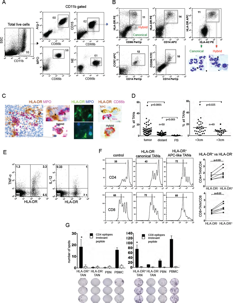
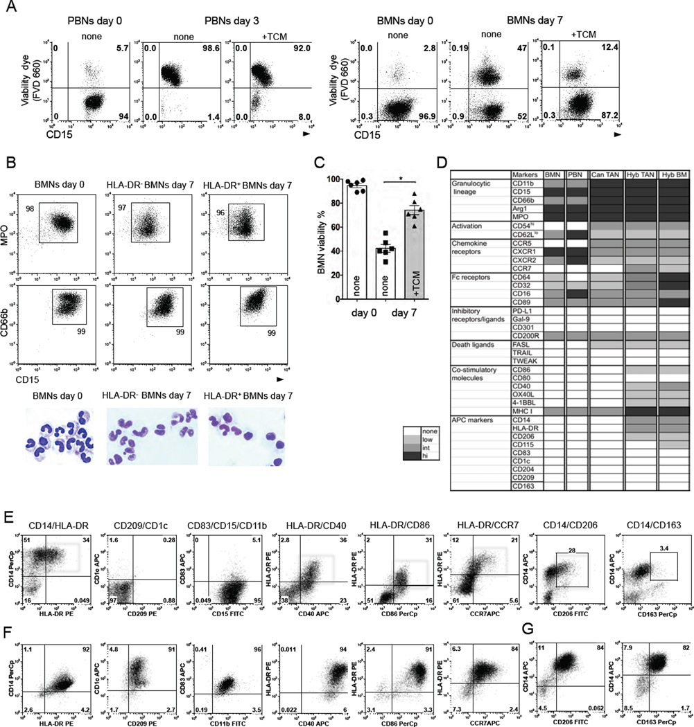
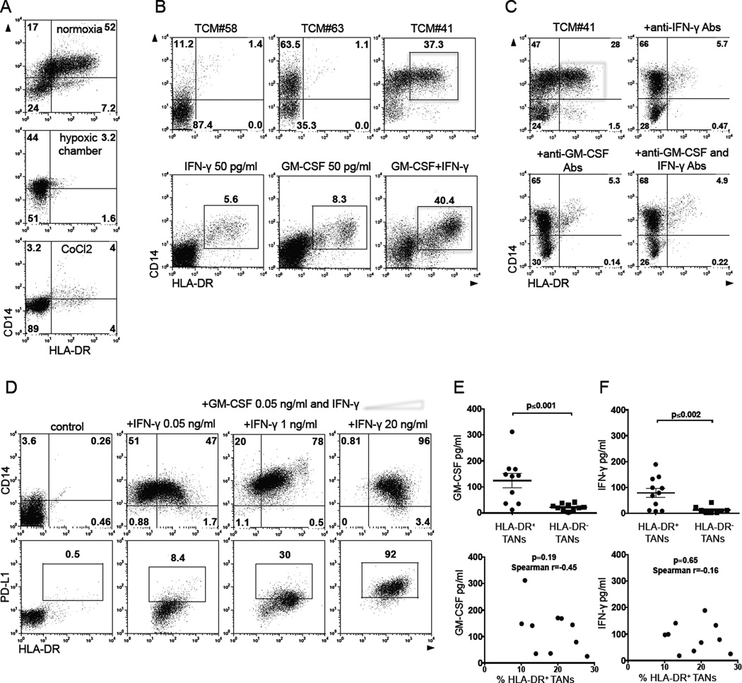
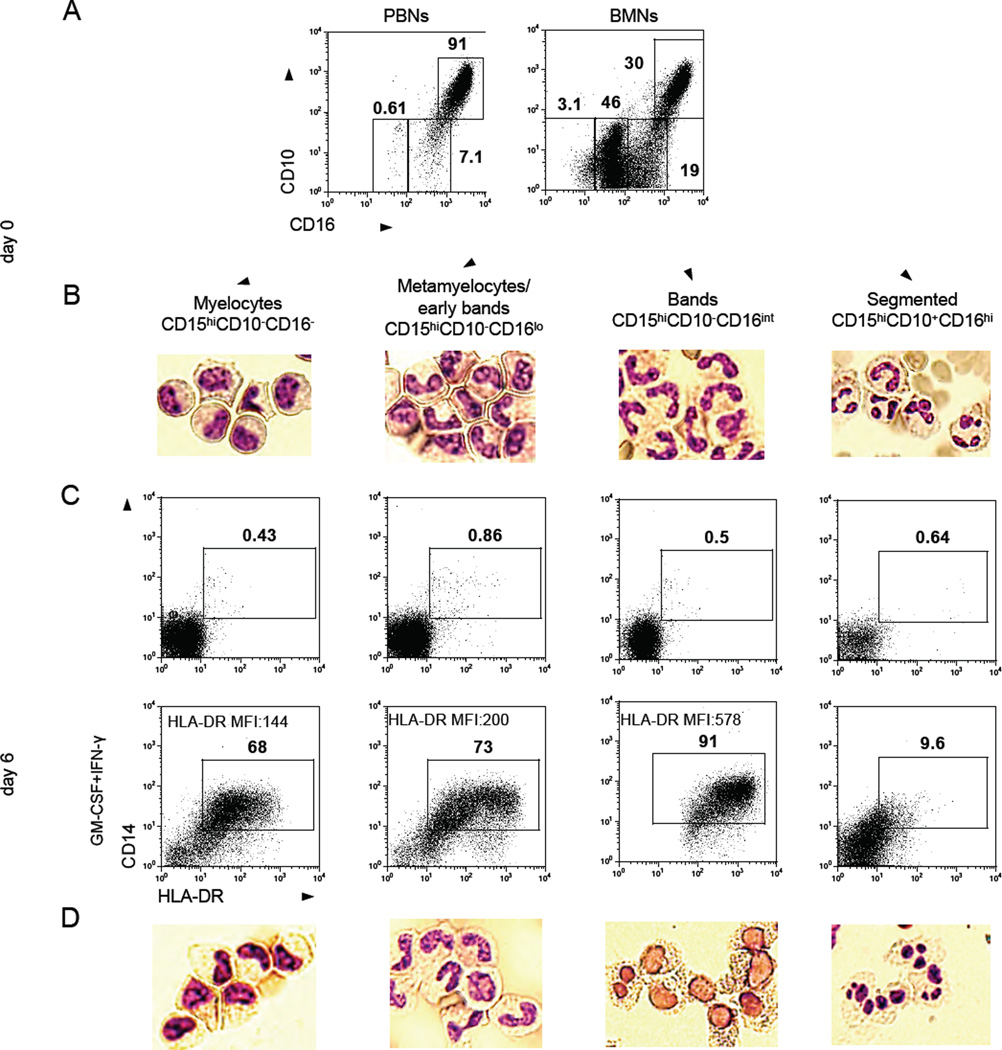
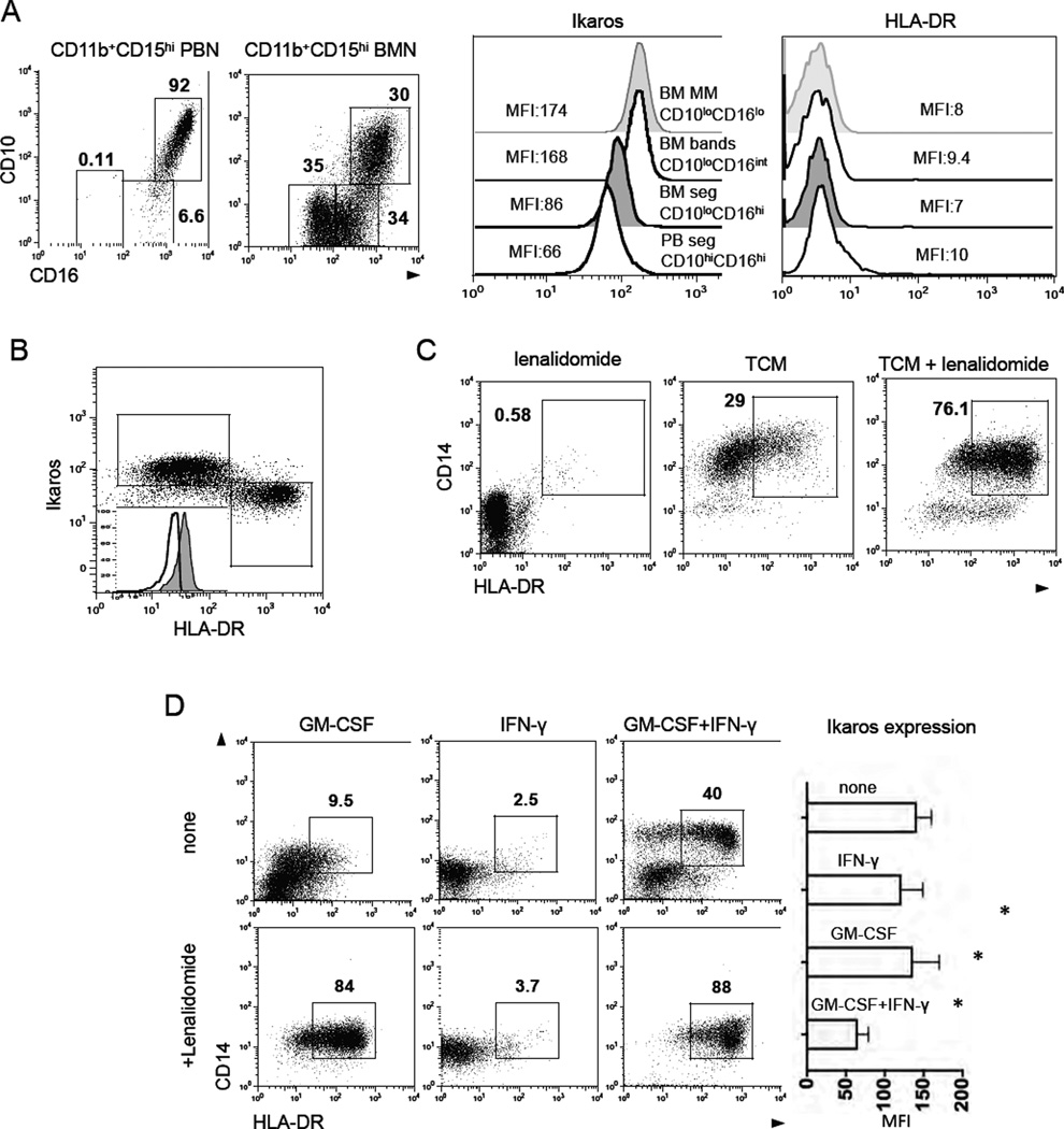
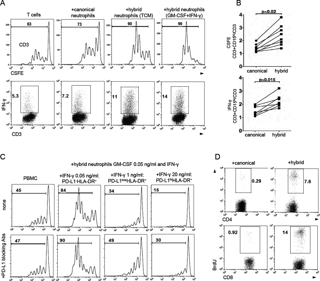
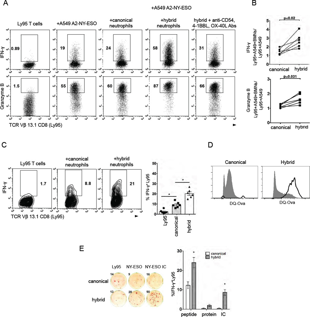
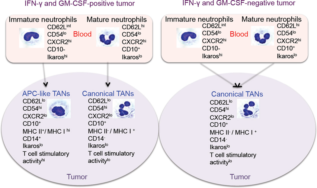
Based on studies in mouse tumor models, granulocytes appear to play a tumor-promoting role. However, there are limited data about the phenotype and function of tumor-associated neutrophils (TANs) in humans. Here, we identify a subset of TANs that exhibited characteristics of both neutrophils and antigen-presenting cells (APCs) in early-stage human lung cancer. These APC-like "hybrid neutrophils," which originate from CD11b(+)CD15(hi)CD10(-)CD16(low) immature progenitors, are able to cross-present antigens, as well as trigger and augment anti-tumor T cell responses. Interferon-γ and granulocyte-macrophage colony-stimulating factor are requisite factors in the tumor that, working through the Ikaros transcription factor, synergistically exert their APC-promoting effects on the progenitors. Overall, these data demonstrate the existence of a specialized TAN subset with anti-tumor capabilities in human cancer.


Conflict of interest statement

Conflict of interest:

No conflict of interest exists.

Figures

Figure 1
Figure 1. A subset of TANs with hybrid characteristics of neutrophils and APCs
(A) A single-cell suspension was obtained from fresh tumor and the expression of the indicated granulocytic markers was analyzed by flow cytometry on gated live CD11b cells. Total TANs are shown in blue boxes. (B) Flow cytometric analysis of the expression of APC markers on gated CD11b+CD15hiCD66b+ TANs. The representative cytomorphology of canonical (green boxes) and APC-like hybrid TANs (red boxes) in NSCLC. Scale bar, 10 µm. (C) The presence of APC-like hybrid TANs in tumor detected by IHC and IF double-staining. Scale bar: 50 µm (left image) and 10 µm (other images). (D) The frequency of APC-like hybrid neutrophils in tumors, distant lung tissue, peripheral blood (PB) (right graph) and in tumors of different sizes (left graph) (line represents mean ± SEM, n = 50, one-way ANOVA test and unpaired t test). APC-like hybrid TANs were defined as a live HLA-DR+CD11b+CD15hiCD66b+ cells. (E) Intracellular TNF-α and IL-12 production by HLA-DR+ hybrid or HLA-DR canonical TANs after stimulation with LPS. TANs were gated on CD11b+CD15hiCD66b+ cells. Representative results from 1 of 5 experiments are shown. (F) The proliferation of autologous CFSE-labeled PBMC stimulated with plate-bound anti-CD3 Abs in the presence of hybrid HLA-DR+ or canonical HLA-DR TANs. T cell stimulatory activity was defined as the ratio CFSElo (T cells+TANs) / CFSElo (T cells) (n=6, Wilcoxon matched-pairs rank test). (G) Autologous virus-specific memory T cell responses in the presence of APC-like hybrid HLA-DR+ or canonical HLA-DR TANs. IFN-γ-ELISPOT assay (mean ± SEM, n=3, *p ≤ 0.01 canonical vs. hybrid, Mann-Whitney test). See also Figure S1.
Figure 2
Figure 2. Tumor-derived factors differentiate long-lived immature BMNs into a hybrid subset with a partial phenotype of dendritic cells and macrophages
(A) Fixable Viability Dye eFluor® 660 (FVD 660) was used to discriminate viable neutrophils in cell culture. Representative dot plots from 1 of 6 experiments are shown. (B) Flow cytometric analysis of the expression of MPO, CD66b, and CD15 markers on freshly isolated BMNs (day 0) and BMNs cultured with (HLA-DR+ BMNs) or without hybrid-inducing TCM (HLA-DR BMNs) for 7 days. Cytospins show the cytomorphology of these BMNs. Scale bar, 10 µm. (C) Survival of BMNs in the cell culture in the presence or absence of TCM. Viability dye FVD 660 was used to discriminate viable BMNs in cell culture (mean ± SEM, n = 6, *p ≤ 0.01,Wilcoxon matched-pairs rank test). (D) Heat map comparing the phenotypes of BMNs, PBNs, canonical TANs (Can TAN), hybrid TANs (Hyb TAN) and BM-derived hybrid neutrophils (Hyb BM). (E–G) Flow cytometric analysis of the expression of indicated APC markers on BM-derived hybrid neutrophils (E) (red boxes), dendritic cells (F) and macrophages (G). Expression of APC markers was analyzed by flow cytometry on gated CD11b+CD15hiCD66b+ BMNs. See also Figure S2.
Figure 3
Figure 3. Tumor-derived IFN-γ and GM-SCF synergistically differentiate immature neutrophils into a subset of APC-like hybrid neutrophils
(A) Flow cytometric analysis of CD14 and HLA-DR expression on gated live CD11b+CD15hiCD66b+ BMNs cultured in the presence of hybrid-inducing TCM under normoxic and hypoxic cell culture conditions. (B) Flow cytometric analysis of CD14 and HLA-DR expression on gated live CD11b+CD15hiCD66b+ BMNs cultured in the presence of different TCMs (top panel) or with IFN-γ and/or GM-CSF (low panel). (C) The effect of IFN-γ and GM-CSF blocking Abs (5 µg/ml) in blunting the formation of HLA-DR+CD14+ hybrid neutrophils in vitro (red box). (D) The expression of CD14 and HLA-DR markers on live CD11b+CD15hiCD66b+ BMNs (top panel) and PD-L1 on gated HLA-DR+CD14+ hybrid neutrophils (low panel) differentiated with GM-CSF (50 pg/ml) and the increasing doses of IFN-γ in vitro. (E and F) Levels of IFN-γ and GM-CSF in supernatants collected from the cell culture of small size tumor digests where APC-like hybrid TANs were or were not previously detected (set-off was >10% among all TANs) (line represents mean ± SEM, n = 10, Mann-Whitney test for unpaired data). Lower panels represent the correlation between the absolute levels of IFN-γ and GM-CSF in the TCM and the frequency of hybrid neutrophils in each tumor showed in top graphs. Non-parametric Spearman test was used to determine the degree of correlation. Representative dot plots from 1 of 5 experiments are shown in (A–D). See also Figure S3.
Figure 4
Figure 4. APC-like hybrid neutrophils originate from CD11b+CD15hiCD66b+CD10CD16lo/int progenitors
(A) Flow cytometric analysis of the expression of CD10 and CD16 on gated live CD11b+CD15hiCD66b+ neutrophils isolated from peripheral blood (PBNs) and bone marrow (BMNs) of cancer patients. (B) Cytospins were made from sorted BMNs at different stages of maturation and stained with the Hema3 Stat Pack Kit (Wright-Giemsa-like stain). (C) Sorted BMNs at different stages of maturation were differentiated in the presence of IFN-γ (50 pg/ml) and GM-CSF (50 pg/ml) in vitro. Expression of HLA-DR and CD14 markers was analyzed by flow cytometry on CD11b+CD15hiCD66b+ BMNs. (D) Cytomorphology of APC-like HLA-DR+ hybrid neutrophils differentiated from the sorted populations of BMNs at different stages of maturation. Representative results from 1 of 4 experiments are shown in (A–D). Scale bar, 10 µm. See also Figure S4.
Figure 5
Figure 5. Transcription factor Ikaros negatively regulates the differentiation of hybrid neutrophils
(A) Flow cytometric analysis of the level of Ikaros and HLA-DR expression in PBNs and BMNs at different stages of maturation Results are shown as Mean Fluorescence Intensity (MFI). (B) Flow cytometric analysis of the level of Ikaros expression in the HLA-DR+ hybrid and HLA-DR canonical CD11b+CD15hiCD66b+ BMNs. (C) Flow cytometric analysis of CD14 and HLA-DR expression on gated live CD11b+CD15hiCD66b+ BMNs cultured in the presence of lenalidomide (10 µM) and hybrid-inducing TCM (30% v/v) for 6 days. (D) The effect of IFN-γ (50 pg/ml) and GM-CSF (50 pg/ml) on the formation of HLA-DR+ CD14+ hybrid neutrophils in the absence (top panel) or presence (bottom panel) of lenalidomide (10 µM) in vitro. The level of Ikaros expression (MFI) in BMNs treated with IFN-γ (50 pg/ml) and GM-CSF (50 pg/ml) for 5 days (mean ± SEM, n = 3, *p ≤ 0.01, Wilcoxon matched-pairs rank test). Representative dot plots from 1 of 6 experiments are shown in (A–D).
Figure 6
Figure 6. APC-like hybrid neutrophils stimulate antigen non-specific T cell responses
(A) The proliferation and IFN-γ production of anti-CD3 Abs stimulated autologous T cells in the presence of BM-derived canonical and hybrid neutrophils differentiated with hybrid-inducing TCM or IFN-γ (50 pg/ml) and GM-CSF (50 pg/ml). (B) Summary results of autologous T cell proliferation (top graph) and IFN-γ production (bottom graph) in the presence of canonical and hybrid neutrophils. Data are presented as a ratio (CD3 cells+CD15hi)/(CD3) (n = 8,Wilcoxon matched-pairs rank test). (C) The proliferation of CFSE-labeled autologous PBMCs cultured with hybrid BMNs with different level of PD-L1 expression in the presence (bottom panel) or absence PD-L1 blocking Abs (5 µg/ml) (top panel). PD-L1−/lo/hiHLA-DR+ hybrid neutrophils were differentiated with GM-CSF (50 pg/ml) and the increasing doses of IFN-γ. (D) The proliferation of allogeneic T cells from healthy donors in the presence of APC-like hybrid neutrophils in mixed lymphocyte reaction (MLR). Representative results from 1of 6 experiments are shown in (C) and (D). See also Figure S5.
Figure 7
Figure 7. APC-like hybrid neutrophils are able to trigger and stimulate NY-ESO specific effector T cell responses
(A) NY-ESO-specific Ly95 cells (TCR Vβ13.1+CD8+) were stimulated with A549 tumor cell line expressing NY-ESO-1 in the context of HLA-A*02 (A2/NY-ESO-1 A549) in the presence of BM-derived canonical and hybrid neutrophils. Intracellular IFN-γ and Granzyme B production was measured by flow cytometry. (B) Cumulative results showing the Ly95 cell stimulatory activity of canonical and hybrid neutrophils. Stimulatory activity was defined as a ratio (Ly95 cells+A549-NY-ESO+BMN)/(Ly95cells+A549-NY-ESO) (n=6,Wilcoxon matched-pairs rank test). (C) HLA-A02+ canonical or hybrid neutrophils were pulsed with synthetic NY-ESO-1 peptide and co-cultured with Ly95 cells for 24 hr. Intracellular IFN-γ was assessed by flow cytometry, (mean ± SEM, n = 6, *p ≤ 0.01,Wilcoxon matched-pairs rank test). (D) DQ-OVA uptake and processing by BM-derived canonical or hybrid neutrophils (open histograms). Cells incubated at 4°C served as controls (shaded histograms). (E) Cross-presentation of NY-ESO-1 epitopes to Ly95 cells by HLA-A02+ canonical or hybrid neutrophils preloaded with NY-ESO-1 protein, NY-ESO-1 peptide or NY-ESO-immune complex (IC). IFN-γ ELISpot (mean ± SEM, n = 6, *p ≤ 0.01 canonical vs. hybrid, Wilcoxon matched-pairs rank test).
Figure 8
Figure 8. Schematic model of neutrophil differentiation in early-stage human lung cancer

Comment in

References

    1. Abi Abdallah DS, Egan CE, Butcher BA, Denkers EY. Mouse neutrophils are professional antigen-presenting cells programmed to instruct Th1 and Th17 T-cell differentiation. Int. Immunol. 2011;5:317–326. - PMC - PubMed
    1. Araki H, Katayama N, Yamashita Y, Mano H, Fujieda A, Usui E, Mitani H, Ohishi K, Nishii K, Masuya M, et al. Reprogramming of human postmitotic neutrophils into macrophages by growth factors. Blood. 2004;8:2973–2980. - PubMed
    1. Ashtekar AR, Saha B. Poly's plea: membership to the club of APCs. Trends Immunol. 2003;9:485–490. - PubMed
    1. Brandau S. The dichotomy of neutrophil granulocytes in cancer. Semin. Cancer Biol. 2013;3:139–140. - PubMed
    1. Carus A, Ladekarl M, Hager H, Pilegaard H, Nielsen PS, Donskov F. Tumor-associated neutrophils and macrophages in non-small cell lung cancer: no immediate impact on patient outcome. Lung Cancer. 2013;1:130–137. - PubMed

Publication types

MeSH terms

Substances