. 2016 Jul 28;166(3):624-636.
doi: 10.1016/j.cell.2016.05.076. Epub 2016 Jun 30.

Hexokinase Is an Innate Immune Receptor for the Detection of Bacterial Peptidoglycan

Affiliations

Hexokinase Is an Innate Immune Receptor for the Detection of Bacterial Peptidoglycan

Andrea J Wolf et al. Cell. .

Abstract

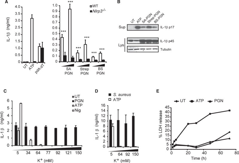
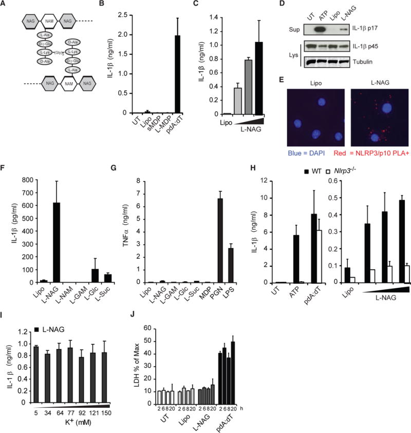
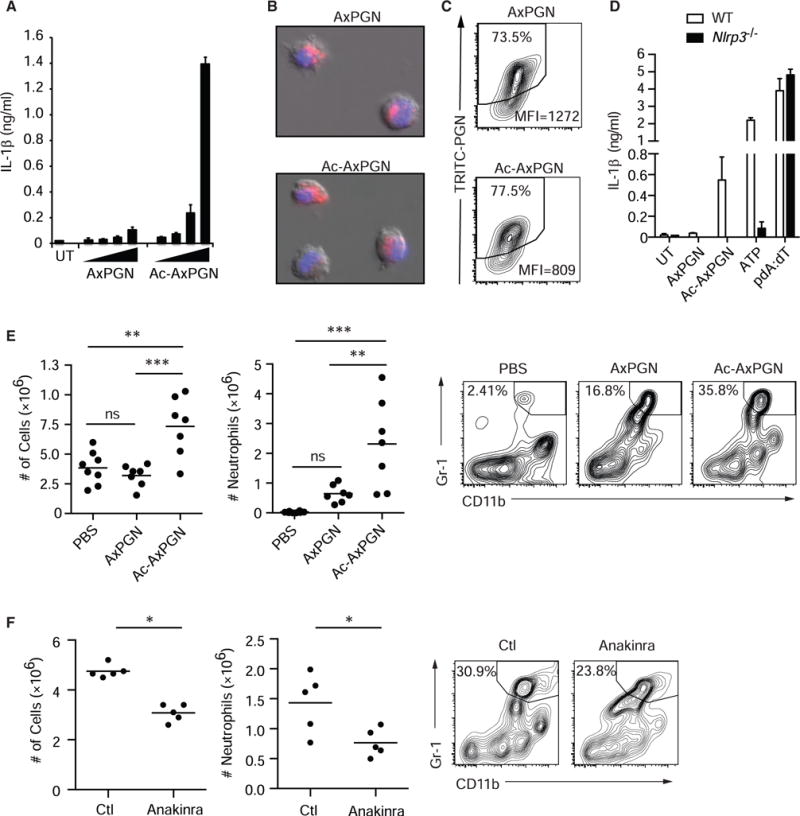
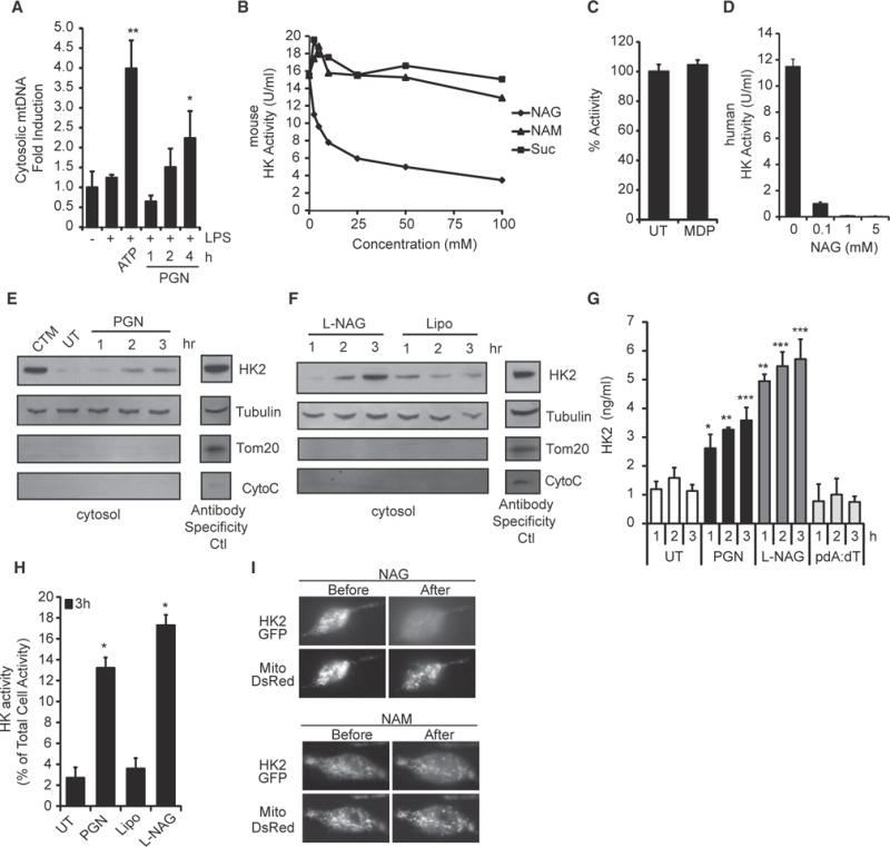
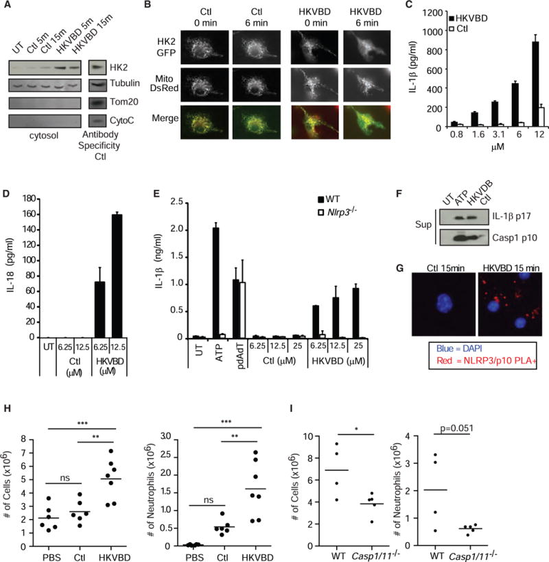
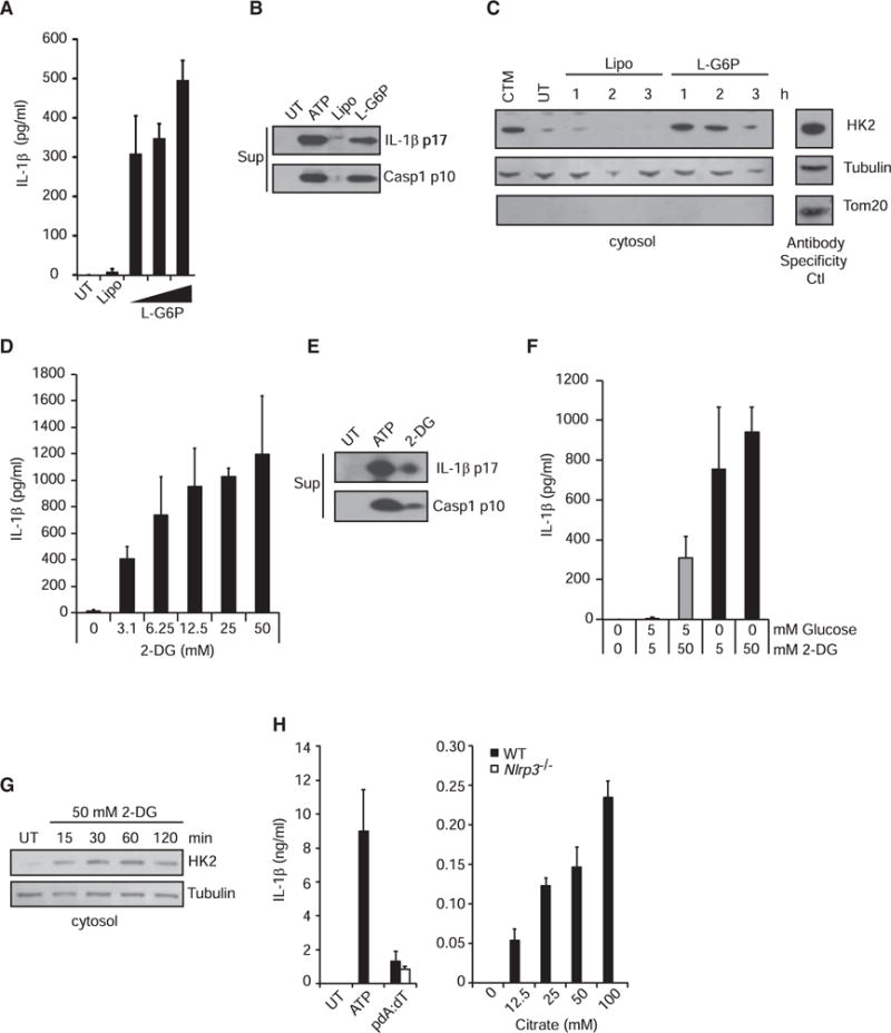
Degradation of Gram-positive bacterial cell wall peptidoglycan in macrophage and dendritic cell phagosomes leads to activation of the NLRP3 inflammasome, a cytosolic complex that regulates processing and secretion of interleukin (IL)-1β and IL-18. While many inflammatory responses to peptidoglycan are mediated by detection of its muramyl dipeptide component in the cytosol by NOD2, we report here that NLRP3 inflammasome activation is caused by release of N-acetylglucosamine that is detected in the cytosol by the glycolytic enzyme hexokinase. Inhibition of hexokinase by N-acetylglucosamine causes its dissociation from mitochondria outer membranes, and we found that this is sufficient to activate the NLRP3 inflammasome. In addition, we observed that glycolytic inhibitors and metabolic conditions affecting hexokinase function and localization induce inflammasome activation. While previous studies have demonstrated that signaling by pattern recognition receptors can regulate metabolic processes, this study shows that a metabolic enzyme can act as a pattern recognition receptor. PAPERCLIP.


Figures

Figure 1
Figure 1. PGN Activates the NLRP3 Inflammasome Independent of Potassium Efflux and Cell Death
(A) LPS-primed BMDMs from wild-type and Nlrp3−/−mice were untreated (UT) or stimulated with 5 mM ATP for 2 hr or pdA:dT or PGN from ΔoatA S. aureus (SA), Streptomyces (Strep), or B. subtilis (BS) at 20–40 μg/ml, and IL-1β was assayed in the supernatant after 6 hr. (B) Immunoblot of mature IL-1β in supernatants (Sup) or pro-IL-1β in cell lysates (Lys) of wild-type macrophages stimulated as in (A). (C and D) LPS-primed BMDMs were stimulated in the presence of increasing concentrations of extracellular KCl with (C) 20 μg/ml PGN 6 hr, 5 mM ATP 2 hr, 10 μg/ml nigericin (Nig) 2 hr, or (D), S. aureus (ΔoatA) 6 hr. (E) LPS-primed BMDMs were stimulated with PGN (20 μg/ml) or ATP (5 mM), and release of lactate dehydrogenase (LDH) into supernatants was measured at indicated times and shown as percentage of maximum at each time point. Error bars indicate SD. ***p < 0.001. See also Figure S1.
Figure 2
Figure 2. NAG Is the NLRP3 Inflammasome-Activating Component of PGN
(A) Schematic diagram of PGN structure. (B) LPS-primed BMDMs were stimulated with lipofectamine-complexed MDP (L-MDP), soluble MDP (sMDP, 10 μg/ml), pdA:dT, or lipofectamine alone (Lipo) for 6 hr. UT, untreated. (C) Cells were stimulated for 6 hr with lipofectamine complexes containing increasing amounts of NAG. (D) IL-1β processing was assessed by immunoblot as in (C). Sup, supernatant; Lys, lysate. (E) Proximity ligation assay (for association of NLRP3 with caspase-1) of LPS-primed BMDMs treated for 3 hr with lipofectamine alone (Lipo) or complexed with NAG (L-NAG) (experiment was performed 2×). (F) Cells were stimulated for 6 hr with different sugars complexed with lipofectamine (NAM, N-acetylmuramic acid; GAM, glucosamine; Gluc, glucose; and Suc, Sucrose). (G) Unprimed BMDMs were treated with lipofectamine-complexed sugars as in (F), and TNF-α was measured in the supernatant after 6 hr. (H) LPS-primed BMDMs from wild-type (WT) and Nlrp3−/−mice were stimulated as described above. (I) LPS-primed BMDMs were stimulated with lipofectamine-complexed NAG in the presence of increasing concentrations of extracellular KCl for 6 hr. (J) LPS-primed BMDMs, stimulated as described above, were assessed for LDH release at indicated times. Error bars indicate SD. ***p < 0.001, Student’s t test. See also Figure S2.
Figure 3
Figure 3. Acetylation of NAG Is Necessary for Inflammasome Activation by PGN
(A) LPS-primed BMDMs were treated with the increasing doses of native (AxPGN) or re-acetylated (Ac-AxPGN) anthrax PGN (20–160 μg/ml) for 6 hr, and IL-1β in the supernatant was measured by ELISA. UT, untreated. (B) TRITC-labeled native or re-acetylated anthrax PGN (40 μg/ml) was incubated with LPS-primed BMDMs for 1 hr, and internalization was confirmed by fluorescence microscopy. (C) TRITC-labeled native or re-acetylated anthrax PGN internalization by BMDMs was measured by flow cytometry after 6 hr. (D) LPS-primed BMDMs from wild-type (WT) and Nlrp3−/−mice were stimulated with 80 μg/ml AxPGN for 6 hr, 80 μg/ml Ac-AxPGN for 6 hr, 5 mM ATP for 2 hr, or pdA:dT for 6 hr, and IL-1β was measured by ELISA. (E) Mice were injected i.p. with PBS (n = 8) or 10 μg of AxPGN (n = 7) or Ac-AxPGN (n = 7). After 4 hr, cells in the peritoneal lavage were harvested and analyzed by flow cytometry. Total cells (left panel) and neutrophils (middle and right panels) were increased in response to re-acetylation of AxPGN. ns, not significant. (F) Mice were injected i.p. with 10 μg Ac-AxPGN without (Ctl) (n = 5) or with 25 mg/kg anakinra (n = 5) and assayed as in (E) (experiment was performed 1×). Error bars indicate SD. **p % 0.01; ***p < 0.001, one-way ANOVA and Newman-Keuls multiple comparison test (E) and unpaired Student’s t test (F). See also Figure S3.
Figure 4
Figure 4. Hexokinase Is the Receptor that Detects NAG for Inflammasome Activation
(A) LPS-primed BMDMs were stimulated with ATP (5 mM) for 2 hr or PGN (40 μg/ml) as indicated, and the presence of mtDNA in the cytosolic fraction was measured by RT-PCR. (B and C) Hexokinase (HK) activity in purified mouse macrophage mitochondria was assessed in the presence of increasing concentrations of (B) NAG, NAM, sucrose, or (C) MDP (25 mM). UT, untreated. (D) NAG inhibits the activity of purified human hexokinase. (E and F) Hexokinase in the cytosol fraction of LPS-primed BMDMs stimulated for the indicated times with PGN (40 μg/ml, from S. aureus) or lipofectamine-complexed NAG was detected by immunoblot. Clotrimazole (CTM) treatment was used as a positive control for hexokinase release from mitochondria, and mitochondrial markers Tom20 and cytochrome c (CytoC) were included to control for mitochondrial integrity. Control lysates were included (Antibody Specificity Ctl) to confirm antibody staining for each marker (non-continuous lane from the same gel). Lipo, lipofectamine. (G) LPS-primed BMDMs were stimulated as indicated PGN (40 μg/ml), and hexokinase 2 in the cytosol was determined by ELISA. (H) LPS-primed BMDMs were stimulated as indicated, and cytosolic hexokinase enzyme activity was measured. (I) LPS-primed BMDMs expressing hexokinase 2 fused to GFP (HK2-GFP) and DsRed targeted to mitochondria (Mito-DsRed) were microinjected with NAG or NAM as indicated. Association of hexokinase with mitochondria was visualized before and 1 min after injection (n = 10 cells assessed for each sugar). Error bars indicate SD. *p % 0.05; **p % 0.01; ***p % 0.001, Student’s t test. See also Figure S4.
Figure 5
Figure 5. Hexokinase Dissociation from Mitochondria Is Sufficient to Activate the NLRP3 Inflammasome
(A) LPS-primed BMDMs were treated with cell-permeable hexokinase dissociation peptide (HKVBD) or scrambled control peptides (Ctl) fused to TAT peptide (20 μM) for the indicated times, and the amount of hexokinase in the cytosolic fraction was determined by immunoblot. Control lysate was included on each gel (Antibody Specificy Ctl) to confirm antibody staining for each marker (non-continuous lane from the same gel). UT, untreated. (B) LPS-primed BMDMs expressing HK2-GFP and mitochondria-localized DsRed (Mito-DsRed) were imaged following treatment with HKVBD and Ctl peptides fused to TAT (20 μM) to assess hexokinase redistribution. (C and D) LPS-primed BMDMs were treated with HKVBD or control peptides fused to cell-permeable antennapedia peptide; IL-1β (C) and IL-18 (D) were measured by ELISA after 2 hr. (E) LPS-primed BMDMs from wild-type and Nlrp3−/−mice were stimulated with 5 mM ATP, pdA:dT, HKVBD or control peptides fused to antennapedia peptide, and IL-1β was measured in the supernatant after 2 hr. (F and G) LPS-primed BMDMs were treated with HKVBD or control peptides fused to TAT peptide; (F) cleaved IL-1β and caspase-1 were detected by immunoblot at 2 hr, and (G) inflammasome assembly was observed by NLRP3 and caspase-1 p10 proximity ligation (PLA,red). Nucleiwere stained withDAPI(blue). Sup, supernatant. (H and I) Indicated mice were injected i.p. with 500 μl of PBS (n = 6), 240 μM HKVBD (n = 7), or control peptide fused to TAT peptide (n = 6); peritoneal cavities were lavaged after 4 hr; total cells were counted; and neutrophil content was determined by flow cytometry (both experiments were each done 1×). ns, not significant. Error bars indicate SD. *p % 0.05; **p % 0.01; ***p % 0.001, Student’s t test (C–E and H) and one-way ANOVA and Newman-Keuls multiple comparison test (G). See also Figure S5.
Figure 6
Figure 6. Metabolic Perturbations Affecting Hexokinase Activate the NLRP3 Inflammasome
(A and B) LPS-primed BMDMs were treated with increasing concentrations of lipofectamine-complexed glucose-6-phosphate (G6P) for 6 hr. IL-1β was measured in the supernatant by ELISA (A), and cleaved IL-1β and caspase-1 were detected by immunoblot (B). UT, untreated; Lipo, lipofectamine. (C) Hexokinase was detected in the cytosolic fraction following treatment with lipofectamine-complexed G6P for the indicated times. Control lysate was included on each gel (Antibody Specificity Ctl) to confirm antibody staining for each marker (non-continuous lane from the same gel). CTM, clotrimazole. (D and E) LPS-primed BMDMs were treated with increasing concentrations of 2-deoxyglucose (2-DG) for 6 hr. IL-1β was measured in the supernatant (Sup) by ELISA (D), and cleaved IL-1β and caspase-1 were detected by immunoblot (E). (F) LPS-primed BMDMs were treated with 2-DG in the presence or absence of glucose for 6 hr, and IL-1β was measured in the supernatant by ELISA. (G) Hexokinase was measured in the cytosolic fraction following treatment with 2-DG in the absence of glucose. (H) LPS-primed wild-type or Nlrp3/BMDMs were treated with increasing concentrations of sodium citrate for 6 hr, and IL-1β was measured in the supernatant by ELISA. Error bars indicate SD. *p % 0.05; **p % 0.01; ***p % 0.001, Student’s t test. See also Figure S6.

Comment in

References

    1. Aleshin AE, Zeng C, Bartunik HD, Fromm HJ, Honzatko RB. Regulation of hexokinase I: crystal structure of recombinant human brain hexokinase complexed with glucose and phosphate. J Mol Biol. 1998a;282:345–357. - PubMed
    1. Aleshin AE, Zeng C, Bourenkov GP, Bartunik HD, Fromm HJ, Honzatko RB. The mechanism of regulation of hexokinase: new insights from the crystal structure of recombinant human brain hexokinase complexed with glucose and glucose-6-phosphate. Structure. 1998b;6:39–50. - PubMed
    1. Berg JM, Tymoczko JL, Stryer L, editors. Biochemistry. Fifth. New York: W. H. Freeman; 2002. The glycolytic pathway is tightly controlled. Section 16.2. http://www.ncbi.nlm.nih.gov/books/NBK21154/
    1. Chiara F, Castellaro D, Marin O, Petronilli V, Brusilow WS, Juhaszova M, Sollott SJ, Forte M, Bernardi P, Rasola A. Hexokinase II detachment from mitochondria triggers apoptosis through the permeability transition pore independent of voltage-dependent anion channels. PLoS ONE. 2008;3:e1852. - PMC - PubMed
    1. da-Silva WS, Gómez-Puyou A, de Gómez-Puyou MT, Moreno-Sanchez R, De Felice FG, de Meis L, Oliveira MF, Galina A. Mitochondrial bound hexokinase activity as a preventive antioxidant defense: steady-state ADP formation as a regulatory mechanism of membrane potential and reactive oxygen species generation in mitochondria. J Biol Chem. 2004;279:39846–39855. - PubMed

Publication types