. 2016 Aug 8;26(15):1998-2002.
doi: 10.1016/j.cub.2016.05.054. Epub 2016 Jun 30.

Changing Balance of Spinal Cord Excitability and Nociceptive Brain Activity in Early Human Development

Affiliations

Changing Balance of Spinal Cord Excitability and Nociceptive Brain Activity in Early Human Development

Caroline Hartley et al. Curr Biol. .

Abstract

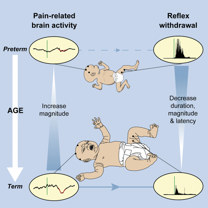
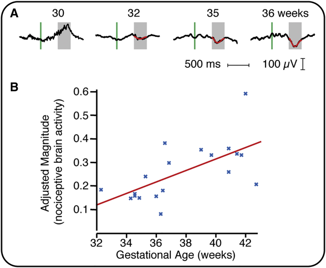
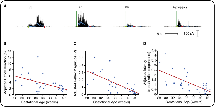
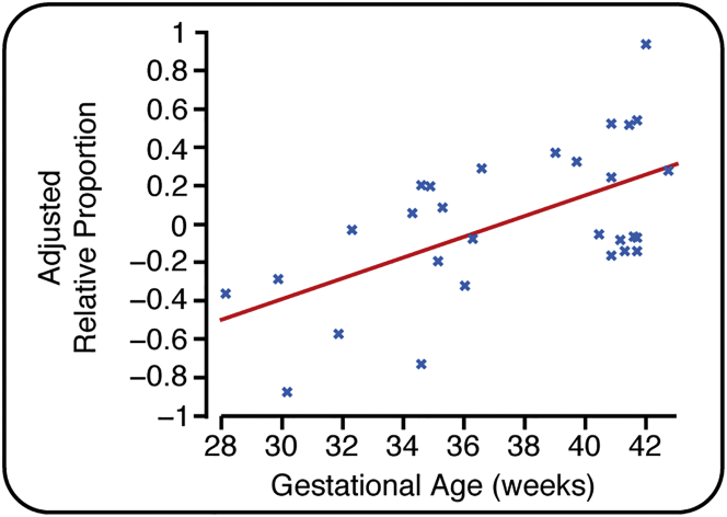
In adults, nociceptive reflexes and behavioral responses are modulated by a network of brain regions via descending projections to the spinal dorsal horn [1]. Coordinated responses to noxious inputs manifest from a balance of descending facilitation and inhibition. In contrast, young infants display exaggerated and uncoordinated limb reflexes [2]. Our understanding of nociceptive processing in the infant brain has been advanced by the use of electrophysiological and hemodynamic imaging [3-6]. From approximately 35 weeks' gestation, nociceptive-specific patterns of brain activity emerge [7], whereas prior to this, non-specific bursts of activity occur in response to noxious, tactile, visual, and auditory stimulation [7-10]. During the preterm period, refinement of spinal cord excitability is also observed: reflex duration shortens, response threshold increases, and improved discrimination between tactile and noxious events occurs [2, 11, 12]. However, the development of descending modulation in human infants remains relatively unexplored. In 40 infants aged 28-42 weeks' gestation, we examined the relationship between nociceptive brain activity and spinal reflex withdrawal activity in response to a clinically essential noxious procedure. Nociceptive-specific brain activity increases in magnitude with gestational age, whereas reflex withdrawal activity decreases in magnitude, duration, and latency across the same developmental period. By recording brain and spinal cord activity in the same infants, we demonstrate that the maturation of nociceptive brain activity is concomitant with the refinement of noxious-evoked limb reflexes. We postulate that, consistent with studies in animals, infant reflexes are influenced by the development of top-down inhibitory modulation from maturing subcortical and cortical brain networks.


Figures

None
Graphical abstract
Figure 1
Figure 1
Relationship between Nociceptive-Specific Brain Activity and Gestational Age (A) Four example EEG traces (black lines) from infants at different gestational ages. The green vertical line indicates the point of stimulation, and nociceptive-specific activity is overlaid in red. (B) The magnitude of the nociceptive-specific brain activity significantly increased with gestational age (n = 19). The red line indicates the regression. No significant effect was observed with postnatal age, estimated cumulative prior pain exposure, and previous diagnosis of postnatal infection (Table S1). The magnitude is shown adjusted for these variables and only includes data where nociceptive-specific brain activity has been identified. See also Figure S1.
Figure 2
Figure 2
Refinement of Reflex Withdrawal Activity (A) Four example EMG traces (black lines) from infants at different gestational ages. The green vertical line indicates the point of stimulation, and the blue line represents the smoothed reflex withdrawal activity. The start and end points are identified by the green and red dots, respectively (see also Figure S3). (B–D) The duration (B), magnitude (C), and latency to the peak of the reflex withdrawal activity (D) significantly decreased with gestational age (n = 32). No significant effect was observed with postnatal age, estimated cumulative prior pain exposure, and previous diagnosis of postnatal infection (Table S1). The data are shown adjusted for these variables.
Figure 3
Figure 3
The Relationship between Nociceptive-Specific Brain Activity and Reflex Withdrawal Activity with Gestational Age The relative proportion of brain and spinal cord activity for each infant plotted against gestational age (n = 29; where brain activity and reflex activity were both recorded without artifact). The values of the relative proportion are limited between −1 and 1, where −1 indicates maximal reflex withdrawal (within the population) with no concomitant nociceptive-specific brain activity and 1 indicates maximal nociceptive-specific brain activity with no concomitant reflex withdrawal (see the Experimental Procedures). See also Figure S2 and Table S1.

References

    1. Bingel U., Tracey I. Imaging CNS modulation of pain in humans. Physiology (Bethesda) 2008;23:371–380. - PubMed
    1. Cornelissen L., Fabrizi L., Patten D., Worley A., Meek J., Boyd S., Slater R., Fitzgerald M. Postnatal temporal, spatial and modality tuning of nociceptive cutaneous flexion reflexes in human infants. PLoS ONE. 2013;8:e76470. - PMC - PubMed
    1. Bartocci M., Bergqvist L.L., Lagercrantz H., Anand K.J. Pain activates cortical areas in the preterm newborn brain. Pain. 2006;122:109–117. - PubMed
    1. Goksan S., Hartley C., Emery F., Cockrill N., Poorun R., Moultrie F., Rogers R., Campbell J., Sanders M., Adams E. fMRI reveals neural activity overlap between adult and infant pain. eLife. 2015;4:e06356. - PMC - PubMed
    1. Slater R., Cantarella A., Gallella S., Worley A., Boyd S., Meek J., Fitzgerald M. Cortical pain responses in human infants. J. Neurosci. 2006;26:3662–3666. - PMC - PubMed

Publication types

LinkOut - more resources