Review
. 2017 Jun;14(3):460-469.
doi: 10.1111/iwj.12622. Epub 2016 Jul 4.

Vascular assessment of wound healing: a clinical review

Affiliations
Review

Vascular assessment of wound healing: a clinical review

William W Li et al. Int Wound J. 2017 Jun.

Abstract

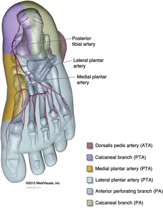
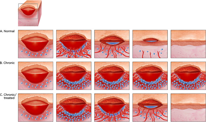
Although macrovascular screening of patients with chronic wounds, particularly in the lower extremities, is accepted as part of clinical practice guidelines, microvascular investigation is less commonly used for a variety of reasons. This can be an issue because most patients with macrovascular disease also develop concomitant microvascular dysfunction. Part of the reason for less comprehensive microvascular screening has been the lack of suitable imaging techniques that can quantify microvascular dysfunction in connection with non-healing chronic wounds. This is changing with the introduction of fluorescence microangiography. The objective of this review is to examine macro- and microvascular disease, the strengths and limitations of the approaches used and to highlight the importance of microvascular angiography in the context of wound healing.

Keywords: Chronic wounds; Fluorescence microangiography; Macrovascular disease; Microvascular dysfunction.


Figures

Figure 1
Figure 1
Anterior view of leg showing the anterior tibial artery, which feeds the dorsal pedis artery angiosome, primarily the dorsal portion of the foot.
Figure 2
Figure 2
Posterior view of the leg showing the posterior tibial artery, which feeds its calcaneal branch angiosome, primarily the inside of the heel, and the peroneal artery, which feeds its calcaneal branch angiosome, primarily the outside of the ankle, and the anterior perforating branch angiosome, the lateral and posterior area surrounding the ankle.
Figure 3
Figure 3
Plantar view of the foot, showing the posterior tibial artery branches (calcaneal and medial and lateral plantar arteries), which feed their corresponding angiosomes (inside bottom of the heel and most of the plantar foot.
Figure 4
Figure 4
Decision‐making algorithm for vascular consultation based on available vascular assessment methods in the wound care clinic.
Figure 5
Figure 5
(A) Progression of normal wound healing showing angiogenesis (blood vessels growing into the wound from bottom and sides of the wound) accompanied by oedema (fluid leakage around the new vessels and edges); granulation in the wound bed (granulation tissue filling the wound bed), less oedema and fewer blood vessels; migration of the epidermal tissue from the wound margins (keratinocytes) with no oedema and few blood vessels, and final closure of the wound with slight scarring. (B) A chronic wound stuck in the inflammatory phase. Note the extensive number of inflammatory cells around the margins of the wound and the copious amount of oedema resulting from continuous leaks of the few microvessels of chronic wound that are growing and which can be seen via indocyanine green angiography. (C) A chronic wound that is treated. Although the wound at the beginning is similar to (B), there is resultant vessel growth, a transient increase in microvessel leaks, followed by resolution with a similar pattern to (A).
Figure 6
Figure 6
Possible explanations behind ‘chronic capillary ischaemia’ in the diabetic foot: (1) Thermo‐regulating arteriovenous (AV) shunts are innervated by the sympathetic nerve system. In diabetes, autonomic neuropathy may lead to denervation of the AV shunts, which lose their normal contraction leading to blood passing through these shunts instead of the capillaries, (2) endothelial dysfunction with a disturbed balance between endogenous vasodilators and vasoconstrictors leading to precapillary vasoconstriction, (3) hemorheological alterations such as elevated levels of plasma fibrinogen (Reprinted with permission from reference 48 and Professor Bengt Fagrell).

References

    1. Guo S, DiPietro LA. Factors affecting wound healing. J Dent Res 2010;89:219–29. - PMC - PubMed
    1. Lazarus GS, Cooper DM, Knighton DR, Percoraro RE, Rodeheaver G, Robson MC. Definitions and guidelines for assessment of wounds and evaluation of healing. Wound Repair Regen 1994;2:165–70. - PubMed
    1. Shah JB. Correction of hypoxia, a critical element for wound bed preparation guidelines: TIMEO2 principle of wound bed preparation. J Am Col Certif Wound Spec 2011;3:26–32. - PMC - PubMed
    1. Sussman C. Management of wound healing with biophysical agent technologies. In: Sussman C, Bates‐Jensen B, editors. Wound care. A practice manual for health professionals, 3rd edn. Baltimore: Lippincott Williams & Wilkins, 2007:498.
    1. Snyder RJ, Driver V, Fife CE, Lantis J, Peirce B, Serena T, Weir D. Using a diagnostic tool to identify elevated protease activity levels in chronic and stalled wounds: a consensus panel discussion. Ostomy Wound Manage 2011;57:36–46. - PubMed

MeSH terms