. 2016 Jul 4:6:29159.
doi: 10.1038/srep29159.

Topical and Targeted Delivery of siRNAs to Melanoma Cells Using a Fusion Peptide Carrier

Affiliations

Topical and Targeted Delivery of siRNAs to Melanoma Cells Using a Fusion Peptide Carrier

Renquan Ruan et al. Sci Rep. .

Abstract

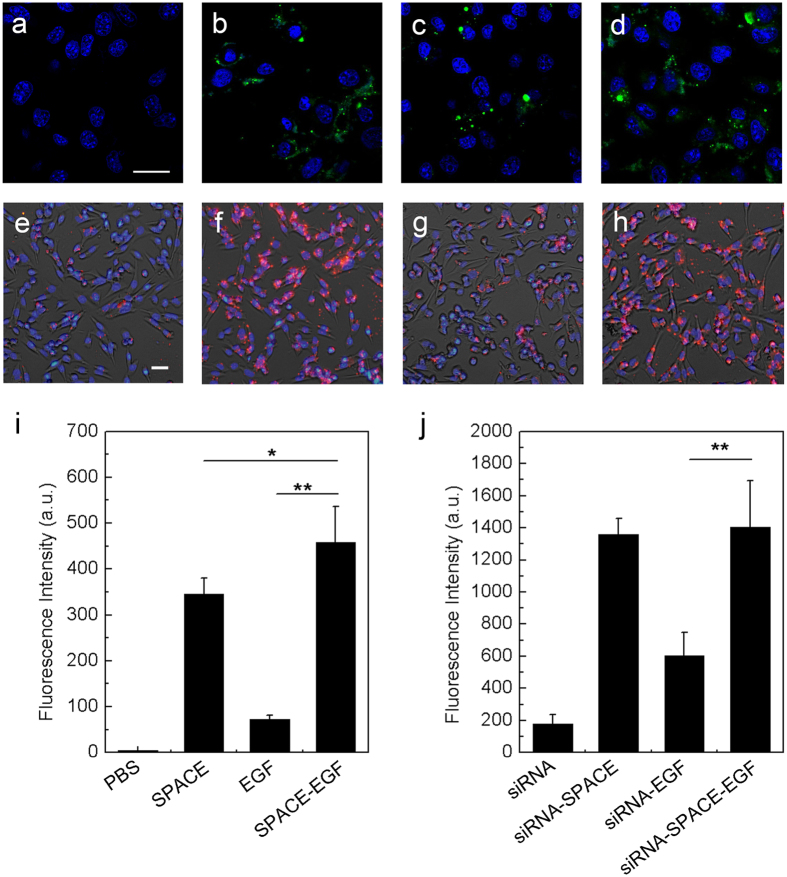
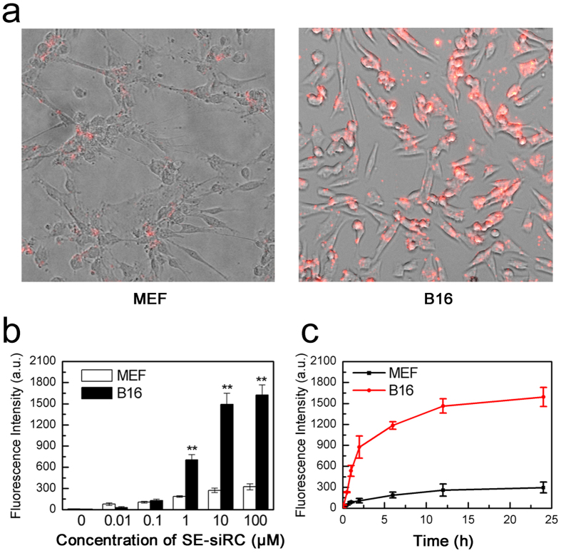
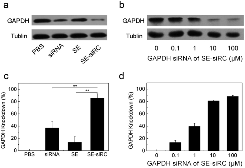
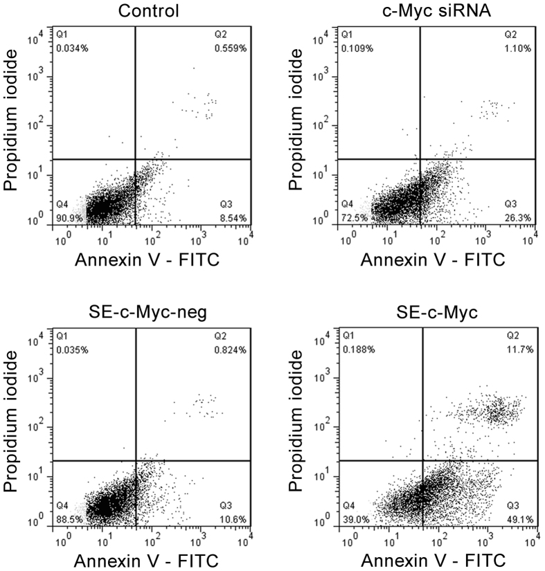
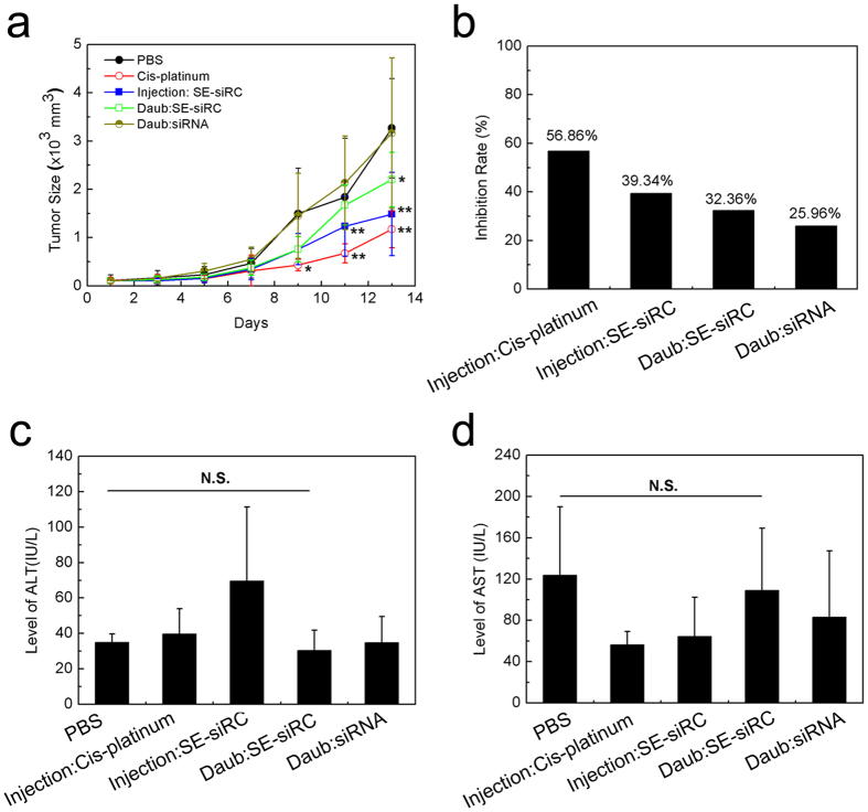
Topical application of siRNAs through the skin is a potentially effective strategy for the treatment of melanoma tumors. In this study, we designed a new and safe fusion peptide carrier SPACE-EGF to improve the skin and cell penetration function of the siRNAs and their targeting ability to B16 cells, such that the apoptosis of B16 cells can be induced. The results show that the carrier is stable and less toxic. The EGF motif does not affect the skin and cell penetration function of the SPACE. Because EGF can strongly bind EGFR, which is overexpressed in cancer cells, the targeting ability of the SPACE-EGF-siRNA complex is increased. In vitro experiments indicate that GAPDH siRNAs conjugated with SPACE-EGF can significantly reduce the GAPDH concentration in B16 cells, and c-Myc siRNAs can cause the gene silencing of c-Myc and thus the apoptosis of cells. In vivo experiments show that the topical application of c-Myc siRNAs delivered by SPACE-EGF through the skin can significantly inhibit the growth of melanoma tumors. This work may provide insight into the development of new transdermal drug carriers to treat a variety of skin disorders.


Figures

Figure 1
Figure 1. Determination of the characteristics of SPACE-EGF.
(a) The clone construction of SPACE-EGF in pGEX-6P-1. (b) The protein purification detected by SDS-PAGE. (c) The size distribution of SPACE-EGF. (d) The size distribution of the SPACE-EGF-siRNA complex (SE-siRC). The linked siRNA was GAPDH-siRNA.
Figure 2
Figure 2. Stability and cytotoxicity of SPACE-EGF.
(a) SPACE-EGF was incubated for 1, 3, 5 and 7 days at 37 °C and 55 °C. (b) The viabilities of MEF cells treated with 1 mg/mL of SPACE-EGF or 1 μM of Lipofectamine 2000 for 6, 12 and 24 hours. The experiments were repeated at least 3 times.
Figure 3
Figure 3. Skin penetration of SPACE-EGF and SE-siRC.
(a) The skin penetration of EGF (20 μg) after 4 hours. (b) The skin penetration of SPACE-EGF (20 μg) after 4 hours. (c) The skin penetration of EGF-siRNA after 8 hours. (d) The skin penetration of siRNA after 8 hours with co-administered Lipofectamine 2000. (e) The skin penetration of SPACE-siRNA after 8 hours. (f) The skin penetration of SE-siRC after 8 hours. (g) The permeated SPACE-EGF or EGF in the skin layer after 16 hours. (h) The permeated SPACE-EGF or EGF across the skin layer after 16 hours. The linked siRNA was GAPDH-siRNA. ELISA assay was used to quantify the permeated protein. Scale bar: 40 μm. The experiments were repeated at least 3 times.
Figure 4
Figure 4. Cell penetration of SPACE-EGF and SE-siRC.
In (a–d) cells were treated for 6 hours with PBS, SPACE, EGF, and SPACE-EGF, respectively. In (e–h) cells were incubated for 6 hours with siRNA, siRNA-SPACE, siRNA-EGF, and siRNA-SPACE-EGF, respectively. In (b) SPACE was also labeled fluorescently. In (i,j) the mean fluorescence intensities in melanoma cells after 6 hours were compared. Scale bar: 20 μm. The linked siRNA was GAPDH-siRNA. The experiments were repeated at least 5 times.
Figure 5
Figure 5. Targeting of SE-siRC to melanoma cells.
(a) Representative images of the targeting of SE-siRC to B16 cells with overexpressing EGFR and mouse embryo fibroblast cells (MEF) after treating for 1 hour with 10 μM of SE-siRC. (b) Fluorescence intensities of cells treated with various concentrations of SE-siRC for 12 hours. (c) Binding efficiencies at various times with 10 μM of SE-siRC. The linked siRNA was GAPDH-siRNA. The experiments were repeated at least 5 times.
Figure 6
Figure 6. GAPDH silencing by SE-siRC.
(a,c) The knockdown degree of the GAPDH protein in B16 cells treated for 48 hours with 10 μM GAPDH SE-siRC, PBS, siRNA, or SE. (b,d) The expression of GAPDH under various concentrations of GAPDH siRNA. The experiments were repeated at least 3 times.
Figure 7
Figure 7. Induction of apoptosis of melanoma cells by SPACE-EGF-mediated c-Myc siRNAs.
B16 cells were incubated with 10 μM of c-Myc siRNAs, negative c-Myc SE-siRC and c-Myc SE-siRC for 48 hours, and then the apoptosis was assessed by the flow cytometry analysis. In this work, early apoptotic (Annexin V+/PI−), late apoptotic/necrotic (Annexin V+/PI+), and live (Annexin V−/PI−) populations were clearly identified.
Figure 8
Figure 8. Growth inhibition of B16F10 tumors and hepatotoxicity of SE-siRC (n ≥ 6).
(a) The size of tumors under various formulations. (b) The growth inhibition rate of B16F10 tumors with SE-siRC. (c) The level of ALT in the mice after the treatment with SE-siRC. (d) The level of AST in the mice after the treatment with SE-siRC. The linked siRNA was c-Myc siRNA. N.S.: No Significance.

References

    1. Jerant A. F., Johnson J. T., Sheridan C. D. & Caffrey T. J. Early detection and treatment of skin cancer. Am Fam Physician 62, 357–368, 375–356, 381–352 (2000). - PubMed
    1. Hersey P. Apoptosis and melanoma: how new insights are effecting the development of new therapies for melanoma. Curr Opin Oncol 18, 189–196 (2006). - PubMed
    1. Huang Y. Y. et al. Melanoma resistance to photodynamic therapy: new insights. Biol Chem 394, 239–250 (2013). - PMC - PubMed
    1. Walther W. & Schlag P. M. Current status of gene therapy for cancer. Curr Opin Oncol 25, 659–664 (2013). - PubMed
    1. Liu Y. et al. Targeting hypoxia-inducible factor-1alpha with Tf-PEI-shRNA complex via transferrin receptor-mediated endocytosis inhibits melanoma growth. Mol Ther 17, 269–277 (2009). - PMC - PubMed

Publication types