. 2016 Oct;37(10):991-1003.
doi: 10.1002/humu.23042. Epub 2016 Aug 21.

Mutational Spectrum of MYO15A and the Molecular Mechanisms of DFNB3 Human Deafness

Affiliations

Mutational Spectrum of MYO15A and the Molecular Mechanisms of DFNB3 Human Deafness

Atteeq U Rehman et al. Hum Mutat. 2016 Oct.

Abstract

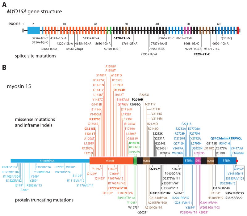
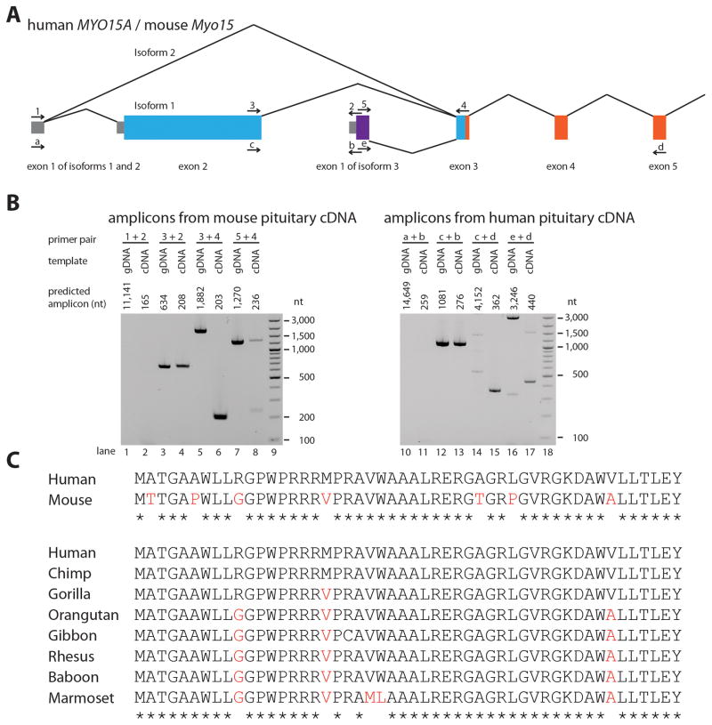
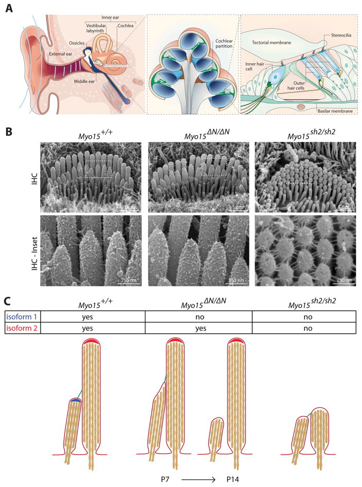
Deafness in humans is a common neurosensory disorder and is genetically heterogeneous. Across diverse ethnic groups, mutations of MYO15A at the DFNB3 locus appear to be the third or fourth most common cause of autosomal-recessive, nonsyndromic deafness. In 49 of the 67 exons of MYO15A, there are currently 192 recessive mutations identified, including 14 novel mutations reported here. These mutations are distributed uniformly across MYO15A with one enigmatic exception; the alternatively spliced giant exon 2, encoding 1,233 residues, has 17 truncating mutations but no convincing deafness-causing missense mutations. MYO15A encodes three distinct isoform classes, one of which is 395 kDa (3,530 residues), the largest member of the myosin superfamily of molecular motors. Studies of Myo15 mouse models that recapitulate DFNB3 revealed two different pathogenic mechanisms of hearing loss. In the inner ear, myosin 15 is necessary both for the development and the long-term maintenance of stereocilia, mechanosensory sound-transducing organelles that extend from the apical surface of hair cells. The goal of this Mutation Update is to provide a comprehensive review of mutations and functions of MYO15A.

Keywords: DFNB3; MYO15A; deafness; giant exon; micro exon; myosin 15; shaker 2.


Conflict of interest statement

statement: All authors declare no conflicts of interest

Figures

Figure 1
Figure 1
Human MYO15A gene structure, the location of mutations associated with nonsyndromic deafness DFNB3 and the domains of myosin 15. Only pathogenic or likely pathogenic mutations are shown here while the variants of unknown significance are listed in Table 2. The c.6487delG (p.Ala2153fs) allele is not included in this figure (see Supp. Table S1). New mutations reported in this study are in bold font (pedigrees in Supp. Figure S2). A: Each rectangle represents one of the 66 reported exons of MYO15A while the horizontal black line represents intronic sequence. The 5′ and 3′ untranslated regions (UTRs) are denoted by light grey rectangles (exon 1 and part of exon 66). Exons and the encoded protein domains of myosin 15 in panel B have the same color. B: Missense mutations and inframe indels are shown on top of the drawing whereas protein truncating mutations are shown below. The three vertical green disks represent two consensus IQ motifs and a third IQ-like motif. Black colored rectangles denote regions of myosin 15 with no predicted domains. The small red square at the C-terminus represents a class 1 PDZ binding motif that interacts with whirlin. MyTh4, myosin tail homology 4 domain; FERM (band 4.1, ezrin, radixin, moesin) domain; SH3, Src homology 3 domain.
Figure 2
Figure 2
Two alternative transcription start sites of MYO15A, three isoform classes and data supporting a novel exon 1 of isoform 3 identified in both human and mouse. A: Transcription for isoforms 1 and 2 starts from exon 1, whereas isoform 3 uses an alternate transcription start site of a second exon 1 located in intron 2. We identified this novel exon 1 of isoform 3 through 5′RACE from mouse pituitary cDNA and later confirmed it by PCR amplification and Sanger sequencing. Location of primers used to amplify and sequence novel exon 1 from mouse and human pituitary tissue cDNA are shown by arrows above and below exons, respectively. Lines indicate splicing of the primary transcript. Coloring of exons is the same as in Fig. 1A, except for the novel exon 1 (purple) of isoform 3 not illustrated in Fig 1. B: Images of 2% agarose gels used to size-separate amplicons. Each primer pair used to amplify cDNA was also used to amplify genomic DNA as a control template. Bands in lanes 4, 8 (upper band) and 13 show that the commercial cDNA libraries also contained some genomic DNA. C: Clustal Omega alignments of the 50 amino acid residues encoded by exon 1 of isoform 3. Non-identical amino acids are highlighted in red font whereas identical amino acids for all species examined are noted with a star. gDNA, genomic DNA; cDNA, complementary DNA; nt, nucleotide.
Figure 3
Figure 3
A: The cochlea is the sensory organ that detects sound within the inner ear. Sound waves enter the external auditory canal and are transmitted to the inner ear via the tympanic membrane and ossicles. Sound energy sets up oscillations within the fluid-filled ducts of the cochlea that displaces the cochlea partition and organ of Corti. Mechanosensory hair cells within the organ of Corti detect these displacements and release neurotransmitters to trigger afferent neural signaling. This panel reproduced from Frolenkov et al., (2004) with permission from Gregory Frolenkov and the publisher. B: Scanning electron microscopy (SEM) showing that mutations in mouse myosin 15 (Myo15) can have different effects upon development and maintenance of mechanosensory stereocilia. The missense (sh2) mutation in exon 20 blocks stereocilia elongation, while conversely, the nonsense (ΔN) mutation in exon 2 does not. Both mouse models exhibit profound deafness. Figure modified with permission from the publisher of Fang et al., (2015). C: Summary of myosin 15 isoform localization and their functions within stereocilia. In maturing stereocilia (P7 and older), isoforms 1 and 2 are concentrated at the tips of different stereocilia rows in inner hair cells. Isoform 2 is required to drive the developmental elongation of stereocilia, whilst isoform 1 is essential postnatally to stabilize shorter stereocilia rows that elicit mechano-electric transduction (MET) currents when the stereocilia bundle is defelected.

References

    1. Ahmed ZM, Morell RJ, Riazuddin S, Gropman A, Shaukat S, Ahmad MM, Mohiddin SA, Fananapazir L, Caruso RC, Husnain T, Khan SN, Riazuddin S, et al. Mutations of myo6 are associated with recessive deafness, dfnb37. Am J Hum Genet. 2003;72:1315–1322. - PMC - PubMed
    1. Ahmed ZM, Riazuddin S, Aye S, Ali RA, Venselaar H, Anwar S, Belyantseva PP, Qasim M, Riazuddin S, Friedman TB. Gene structure and mutant alleles of pcdh15: Nonsyndromic deafness dfnb23 and type 1 usher syndrome. Hum Genet. 2008;124:215–223. - PMC - PubMed
    1. Ammar-Khodja F, Bonnet C, Dahmani M, Ouhab S, Lefevre GM, Ibrahim H, Hardelin JP, Weil D, Louha M, Petit C. Diversity of the causal genes in hearing impaired algerian individuals identified by whole exome sequencing. Mol Genet Genomic Med. 2015;3:189–196. - PMC - PubMed
    1. Anderson DW, Probst FJ, Belyantseva IA, Fridell RA, Beyer L, Martin DM, Wu D, Kachar B, Friedman TB, Raphael Y, Camper SA. The motor and tail regions of myosin xv are critical for normal structure and function of auditory and vestibular hair cells. Hum Mol Genet. 2000;9:1729–1738. - PubMed
    1. Askew C, Rochat C, Pan B, Asai Y, Ahmed H, Child E, Schneider BL, Aebischer P, Holt JR. Tmc gene therapy restores auditory function in deaf mice. Sci Transl Med. 2015;7:295ra108. - PMC - PubMed

Publication types