Review
. 2016 Jun 7;6(9):1306-23.
doi: 10.7150/thno.14858. eCollection 2016.

The Smart Drug Delivery System and Its Clinical Potential

Affiliations
Review

The Smart Drug Delivery System and Its Clinical Potential

Dong Liu et al. Theranostics. .

Abstract

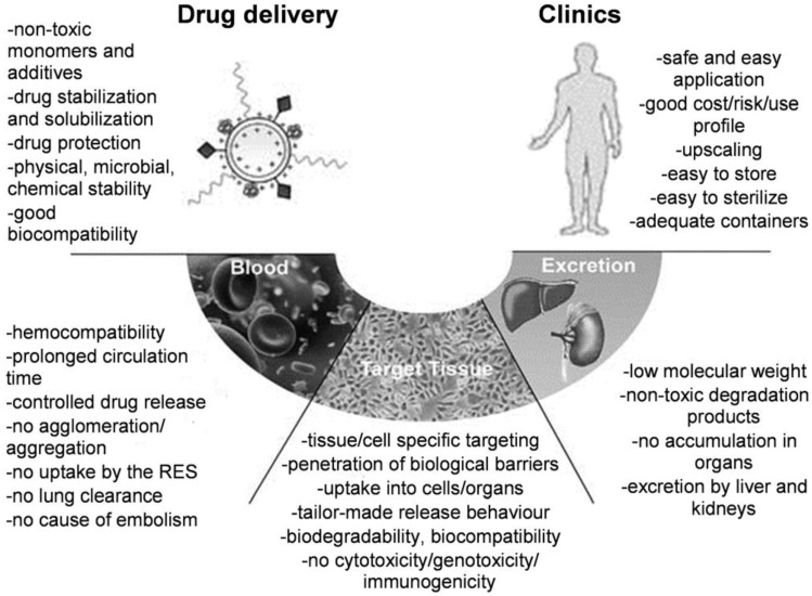
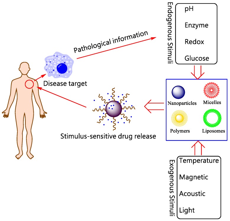
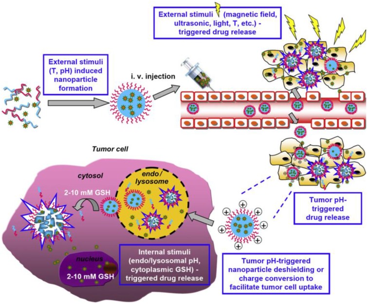
With the unprecedented progresses of biomedical nanotechnology during the past few decades, conventional drug delivery systems (DDSs) have been involved into smart DDSs with stimuli-responsive characteristics. Benefiting from the response to specific internal or external triggers, those well-defined nanoplatforms can increase the drug targeting efficacy, in the meantime, reduce side effects/toxicities of payloads, which are key factors for improving patient compliance. In academic field, variety of smart DDSs have been abundantly demonstrated for various intriguing systems, such as stimuli-responsive polymeric nanoparticles, liposomes, metals/metal oxides, and exosomes. However, these nanoplatforms are lack of standardized manufacturing method, toxicity assessment experience, and clear relevance between the pre-clinical and clinical studies, resulting in the huge difficulties to obtain regulatory and ethics approval. Therefore, such relatively complex stimulus-sensitive nano-DDSs are not currently approved for clinical use. In this review, we highlight the recent advances of smart nanoplatforms for targeting drug delivery. Furthermore, the clinical translation obstacles faced by these smart nanoplatforms have been reviewed and discussed. We also present the future directions and perspectives of stimuli-sensitive DDS in clinical applications.

Keywords: Biomaterials; Controlled release; Drug delivery system (DDS); Nanomedicine.; Smart nanoplatform.


Conflict of interest statement

Competing Interests: The authors have declared that no competing interest exists.

Figures

Figure 1
Figure 1
Essential components for design of polymeric drug delivery systems (DDSs). “RES” is the abbreviation of “Reticuloendothelial System”. Reproduced with permission from reference .
Figure 2
Figure 2
Schematic illustration for stimuli-responsive DDSs.
Figure 4
Figure 4
Schematic representation of the two strategies to control drug release from polymers in the tumor extracellular pH (pHe) environment. (A) The polymer carrier maintains its stealth function while circulating in the blood circulation. (B) Upon arrival at slightly acidic tumor sites through the EPR effect, the stealth polymer carrier is activated by pH to release its cargos. (C) Surface charge reversal to a positive charge when the stealth polymer carrier arrived at tumor vicinity, and drugs were released.
Figure 3
Figure 3
Dual and multi-stimuli (e.g., T (temperature), pH, magnetic field, US, and light) smart polymeric materials used for smart drugs in solid tumors. Reproduced with permission from reference .
Figure 5
Figure 5
(A) Schematic illustration of the fabrication of PIC NP/Pt@PPC-DA pH-responsive nanoparticles; (B) Schematic illustration of the procedure of drug release from polymer nanoparticles. (i) non-specific interactions are scarcely happened between NP/Pt@PPC-DA and serum components; (ii) nanoparticles accumulate at the tumor site owing to the enhanced permeability and retention effect; (iii) the positively charged NP/Pt nanoparticles are exposed again to enhance the Pt(IV) pro-drug internalization; (iv) the Pt(IV) pro-drug is rapidly released from NP/Pt nanoparticles. Reproduced with permission from reference .
Figure 6
Figure 6
Schematic illustration for the redox-responsive degradation process of PSSG, Reproduced with permission from reference .
Figure 7
Figure 7
Schematic illustration of thioketal nanocarriers formulated by ROS-sensitive polymer to release siRNA at sites of intestinal inflammation through oral administration. (a) Firstly, TNF-α-siRNA were prepared with the cationic lipid DOTAP (1, 2-dioleoyl-3-trimethylammonium-propane), and then TNF-α-TKNs were obtained through adding TNF-α-DOTAP complexes to an organic solution containing PPADT. Right image shows the scanning electron micrograph of TNF-α-TKNs with the scale bar of 1.5 μm. (b) TNF-α-TKNs remained to be stable in the gastrointestinal tract when delivered orally, and TNF-α-siRNA were protected and prevented its release unless at sites of intestinal inflammation, where infiltrating phagocytes produce unusually high levels of ROS. (c) PPADT 3 was synthesized by the acetal exchange reaction. PTSA represented p-toluenesulfonic acid. Reproduced with permission from reference .
Figure 8
Figure 8
Typical photochromic compounds used in photo-responsive polymer systems.
Figure 9
Figure 9
Schematic diagram depicting the construction of temperature-sensitive liposomes with thermo-sensitive poly(EOEOVE)-OD4 (Octadecyl vinyl ether), and heat-triggered release of DOX from liposomes. (a) poly(EOEOVE)-OD4, (b) EYPC (Egg yolk phosphatidylcholine), (c) cholesterol, (d) PEG-DSPE ((polyethylene glycol)- distearoyl phosphatidylethanolamine). Reproduced with permission from reference .
Figure 10
Figure 10
The various stimuli that induce the MSNP-hybridized DDSs to release the cargos. Reproduced with permission from reference .
Figure 11
Figure 11
Schematic illustration of microvesicles encapsulated magnetic nanoparticles and glucose oxidase for dual-stimuli responsive programmable delivery system. The encapsulated glucose-specific enzyme firstly catalyzes glucose into gluconic acid and H2O2. The subsequent alternating magnetic field increases the porosity of the polymer shell, leading to the reaction between H2O2 and L-arginine to produce nitric oxides. Reproduced with permission from reference .
Figure 12
Figure 12
(a) Schematic diagram to describe the synthesis of PEGylated Mn-Zn ferrite MNCs (C: chloroform; W: water). (b) TEM image of PEGylated MNCs with core-shell structure, and the white layer surrounding the magnetic cores indicating the DSPE-PEG2000 coating layer. (c) Schematic illustration of PEGylated MNCs with 8.27 nm magnetic core and 3.45 nm lipid shell. (d) DLS curve of PEGylated MNCs in water and serum. (e) Hysteresis loops at room temperature for PEGylated MNCs. Reproduced with permission from reference .

References

    1. Hrubý M, Filippov SK, Štěpánek P. Smart polymers in drug delivery systems on crossroads: Which way deserves following? European Polymer Journal. 2015;65:82–97.
    1. Kopeček J, Yang J. Hydrogels as smart biomaterials. Polymer International. 2007;56:1078–98.
    1. Lee BK, Yun YH, Park K. Smart nanoparticles for drug delivery: Boundaries and opportunities. Chemical Engineering Science. 2015;125:158–64. - PMC - PubMed
    1. Bamrungsap S, Zhao Z, Chen T, Wang L, Li C, Fu T. et al. Nanotechnology in therapeutics: a focus on nanoparticles as a drug delivery system. Nanomedicine. 2012;7:1253–71. - PubMed
    1. Allen TM, Cullis PR. Liposomal drug delivery systems: from concept to clinical applications. Advanced Drug Delivery Reviews. 2013;65:36–48. - PubMed

Publication types