. 2016 May 19;7(6):2269-84.
doi: 10.1364/BOE.7.002269. eCollection 2016 Jun 1.

In vivo assessment of optical properties of basal cell carcinoma and differentiation of BCC subtypes by high-definition optical coherence tomography

Affiliations

In vivo assessment of optical properties of basal cell carcinoma and differentiation of BCC subtypes by high-definition optical coherence tomography

Marc Boone et al. Biomed Opt Express. .

Abstract

High-definition optical coherence tomography (HD-OCT) features of basal cell carcinoma (BCC) have recently been defined. We assessed in vivo optical properties (IV-OP) of BCC, by HD-OCT. Moreover their critical values for BCC subtype differentiation were determined. The technique of semi-log plot whereby an exponential function becomes linear has been implemented on HD-OCT signals. The relative attenuation factor (µraf ) at different skin layers could be assessed.. IV-OP of superficial BCC with high diagnostic accuracy (DA) and high negative predictive values (NPV) were (i) decreased µraf in lower part of epidermis and (ii) increased epidermal thickness (E-T). IV-OP of nodular BCC with good to high DA and NPV were (i) less negative µraf in papillary dermis compared to normal adjacent skin and (ii) significantly decreased E-T and papillary dermal thickness (PD-T). In infiltrative BCC (i) high µraf in reticular dermis compared to normal adjacent skin and (ii) presence of peaks and falls in reticular dermis had good DA and high NPV. HD-OCT seems to enable the combination of in vivo morphological analysis of cellular and 3-D micro-architectural structures with IV-OP analysis of BCC. This permits BCC sub-differentiation with higher accuracy than in vivo HD-OCT analysis of morphology alone.

Keywords: (170.1870) Dermatology; (170.3660) Light propagation in tissues; (170.4500) Optical coherence tomography; (170.6935) Tissue characterization; (170.7050) Turbid media; (290.0290) Scattering.


Figures
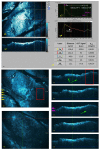
Fig. 1
Fig. 1
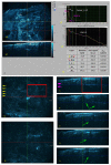
Superficial BCC. (a) A 3-D HD-OCT DICOM (digital imaging and communication in medicine) image of the lesion is selected: en face image (a1) and cross-sectional image (a2). This DICOM file is then opened using LabView software. A region of interest (ROI) (> 0.1 mm2) is chosen in the en face image (red square). In a first step, a plot z-axis profile of the scanned volume (> 0.05 mm3) is performed. The obtained graph (a3) displays the reflectance and attenuation of the ballistic photons (OCT-signal: measured on the y-axis with arbitral units (a.u.)) versus imaging depth which is indicated on the x-axis (µm). The distance from Z = 0 to the first peak is the thickness of the gel. The first peak at about 120 µm corresponds to the skin entrance signal (SE-S). The valley is noticed 352 µm deeper than the first peak. Consequently, the epidermis is approximately 232 µm thick in this ROI. The second peak is situated 50 µm deeper than the valley. Hence, the papillary dermis is approximately 50 µm thick in this ROI. In a second step, The HD-OCT signals were divided by SE-S in order to normalize them to the SE-S The natural logarithm ln(x) of those values is taken (a4). A semi-log plot is performed; an exponential function becomes linear described by y = ax + b. Successive layers with clear exponential decay are identified and plotted. A straight line is fitted in each of these layers resulting in equation of type yi = axi + b whereby coefficient a provides the relative attenuation factor for each of the identified layers “i” given by µrafi. Data are summarized in table (a5): OCT signal at different Z-values (a.u.), distance (µm) between different Z values (vertical coloured lines) and µraf in different layers (1/µm). (b) high-resolution en face images at different Z-values. Yellow arrows indicate the position of cross-sectional images (c). Magenta arrows indicate the position of the en face images. Green arrows indicate the sBCC hemi-lobules. Optical properties of sBCC with high diagnostic accuracy (>95%) and high negative predictive values (NPV) (>97.5%) are the presence of a nod in semi-log plot of layer-1, much lower relative attenuation factor in lower part of epidermal layer (µraf1b) and strongly increased E-T.
Fig. 2
Fig. 2
Nodular BCC. (a-c). For details about arrows see Fig. 1. Optical properties of nBCC with moderate to high diagnostic accuracy (91.7% - 95.8%) and high NPV (93.8% - 97.0%) were strongly decreased epidermal - and papillary dermal thickness and less negative µraf2 compared to normal skin.
Fig. 3
Fig. 3
Infiltrative BCC. (a-c) For details see Fig. 1. Regarding the optical properties of iBCC an increase of µraf3b of >0.0051 cm-1 compared to normal skin and the presence of peaks and falls in reticular dermis had good diagnostic accuracy (79.1% - 93.8%) and high NPV (93.8% - 96.8%).

References

    1. Flohil S. C., de Vries E., Neumann H. A., Coebergh J. W., Nijsten T., “Incidence, prevalence and future trends of primary basal cell carcinoma in the Netherlands,” Acta Derm. Venereol. 91(1), 24–30 (2011).10.2340/00015555-1009 - DOI - PubMed
    1. Roewert-Huber J., Lange-Asschenfeldt B., Stockfleth E., Kerl H., “Epidemiology and aetiology of basal cell carcinoma,” Br. J. Dermatol. 157(Suppl 2), 47–51 (2007).10.1111/j.1365-2133.2007.08273.x - DOI - PubMed
    1. Carsin A. E., Sharp L., Comber H., “Geographical, urban/rural and socioeconomic variations in nonmelanoma skin cancer incidence: a population-based study in Ireland,” Br. J. Dermatol. 164(4), 822–829 (2011).10.1111/j.1365-2133.2011.10238.x - DOI - PubMed
    1. Sellheyer K., “Basal cell carcinoma: cell of origin, cancer stem cell hypothesis and stem cell markers,” Br. J. Dermatol. 164(4), 696–711 (2011).10.1111/j.1365-2133.2010.10158.x - DOI - PubMed
    1. Trakatelli M., Morton C., Nagore E., Ulrich C., Del Marmol V., Peris K., Basset-Seguin N., BCC subcommittee of the Guidelines Committee of the European Dermatology Forum , “Update of the European guidelines for basal cell carcinoma management,” Eur. J. Dermatol. 24(3), 312–329 (2014). - PubMed

LinkOut - more resources