Review
. 2016 Jun 8:4:53.
doi: 10.3389/fcell.2016.00053. eCollection 2016.

ERK1 and ERK2 Map Kinases: Specific Roles or Functional Redundancy?

Affiliations
Review

ERK1 and ERK2 Map Kinases: Specific Roles or Functional Redundancy?

Roser Buscà et al. Front Cell Dev Biol. .

Abstract

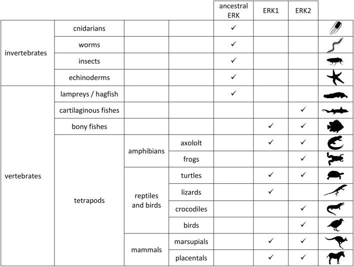
The MAP kinase signaling cascade Ras/Raf/MEK/ERK has been involved in a large variety of cellular and physiological processes that are crucial for life. Many pathological situations have been associated to this pathway. More than one isoform has been described at each level of the cascade. In this review we devoted our attention to ERK1 and ERK2, which are the effector kinases of the pathway. Whether ERK1 and ERK2 specify functional differences or are in contrast functionally redundant, constitutes an ongoing debate despite the huge amount of studies performed to date. In this review we compiled data on ERK1 vs. ERK2 gene structures, protein sequences, expression levels, structural and molecular mechanisms of activation and substrate recognition. We have also attempted to perform a rigorous analysis of studies regarding the individual roles of ERK1 and ERK2 by the means of morpholinos, siRNA, and shRNA silencing as well as gene disruption or gene replacement in mice. Finally, we comment on a recent study of gene and protein evolution of ERK isoforms as a distinct approach to address the same question. Our review permits the evaluation of the relevance of published studies in the field especially when measurements of global ERK activation are taken into account. Our analysis favors the hypothesis of ERK1 and ERK2 exhibiting functional redundancy and points to the concept of the global ERK quantity, and not isoform specificity, as being the essential determinant to achieve ERK function.

Keywords: ERK1 and ERK2 isoforms; MAP kinases; expression of isoforms in vertebrates; gene disruption; gene silencing; intracellular signaling; protein sequence evolution.


Figures

Figure 1
Figure 1
Expression of ERK proteins in animals. ERK1 and ERK2 proteins were classified as such upon phylogenic study of amino-acid coding sequences (same conclusions were reached with nucleotide sequences; Busca et al., 2015). In invertebrates only one erk gene was identified so far. Ancestral ERK corresponds to ERK protein sequence that cannot be classified into ERK1 or ERK2 group. Protein expression in vertebrate brains was described in the same study. Animal silhouettes are from phylopic (http://phylopic.org/).
Figure 2
Figure 2
erk1b RNAs from related species drive synthesis of unrelated proteins domains. Genomic sequences corresponding to exon7-intron7-exon8 of MAPK3 (erk1) were retrieved from Ensembl release 83. First line: extension of the introns and exons, separated by vertical bars. “stop” indicates the final of ERK1 and ERK1b coding sequences. Sequences were aligned by multalin program with identity matrix (Corpet, 1988). Alignments were performed also with Tcoffee and Clustal Omega with the same results (not shown). Rat (Rattus norvegicus), guinea pig (Cavia porcellus), rabbit (Oryctolagus cuniculus), kangaroo-rat (Dipodomys ordii), squirrel (Ictidomys tridecemlineatus), mouse (Mus musculus), platypus (Ornithorhynchus anatinus). Letters in red, amino-acids highly conserved for a position among the protein sequences; letters in blue, amino-acids showing limited conservation; letters in black, amino-acids showing no conservation in the aligned sequences. (A) The C-terminal sequences of rodents' ERK1 proteins are highly conserved; platypus'ERK1 protein sequence is highly similar to rodents' ERK1s. (B) Rodents' ERK1b proteins display no significant conserved protein motifs after exon7.
Figure 3
Figure 3
Position of ERK “dimerization” domains on 3D representation of ERK1. The two domains of ERK implicated in “dimerization” are highlighted in yellow on the 3D structure of ERK1 (still-images of structure 4QTB from RCSB PDB, www.rcsb.org, Chaikuad et al., viewed with CN3D software). “4 leucines” of ERK1 are equivalent to leucines L333, L336, L341, and L344 of mouse ERK2 (Khokhlatchev et al., 1998). In the sequence PEHD (equivalent to P174DHD177 of mouse ERK2), the glutamic acid of mammalian ERK1s is highlighted in gold. On mammalian ERK2s this glutamic-acid is replaced by an aspartic acid that is less bulky.

References

    1. Adachi M., Fukuda M., Nishida E. (2000). Nuclear export of MAP kinase (ERK) involves a MAP kinase kinase (MEK)-dependent active transport mechanism. J. Cell Biol. 148, 849–856. 10.1083/jcb.148.5.849 - DOI - PMC - PubMed
    1. Aebersold D. M., Shaul Y. D., Yung Y., Yarom N., Yao Z., Hanoch T., et al. (2004). Extracellular signal-regulated kinase 1c (ERK1c), a novel 42-kilodalton ERK, demonstrates unique modes of regulation, localization, and function. Mol. Cell. Biol. 24, 10000–10015. 10.1128/MCB.24.22.10000-10015.2004 - DOI - PMC - PubMed
    1. Agrawal A., Dillon S., Denning T. L., Pulendran B. (2006). ERK1-/- mice exhibit Th1 cell polarization and increased susceptibility to experimental autoimmune encephalomyelitis. J. Immunol. 176, 5788–5796. 10.4049/jimmunol.176.10.5788 - DOI - PubMed
    1. Alessandrini A., Brott B. K., Erikson R. L. (1997). Differential expression of MEK1 and MEK2 during mouse development. Cell Growth Differ. 8, 505–511. - PubMed
    1. Alter B. J., Zhao C., Karim F., Landreth G. E., Gereau R. W. t.. (2010). Genetic targeting of ERK1 suggests a predominant role for ERK2 in murine pain models. J. Neurosci. 30, 11537–11547. 10.1523/JNEUROSCI.6103-09.2010 - DOI - PMC - PubMed