Review
. 2018 Jan;91(1081):20160363.
doi: 10.1259/bjr.20160363. Epub 2016 Jul 22.

Advances in PET/MR instrumentation and image reconstruction

Affiliations
Review

Advances in PET/MR instrumentation and image reconstruction

Jorge Cabello et al. Br J Radiol. 2018 Jan.

Abstract

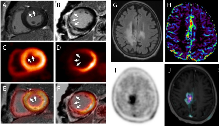
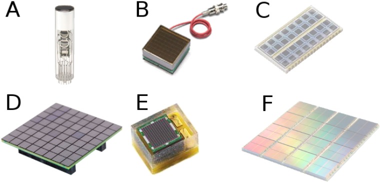
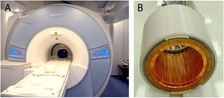
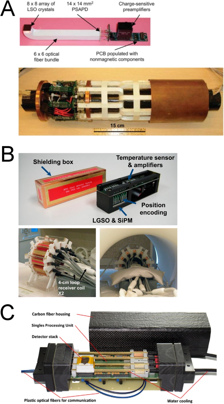
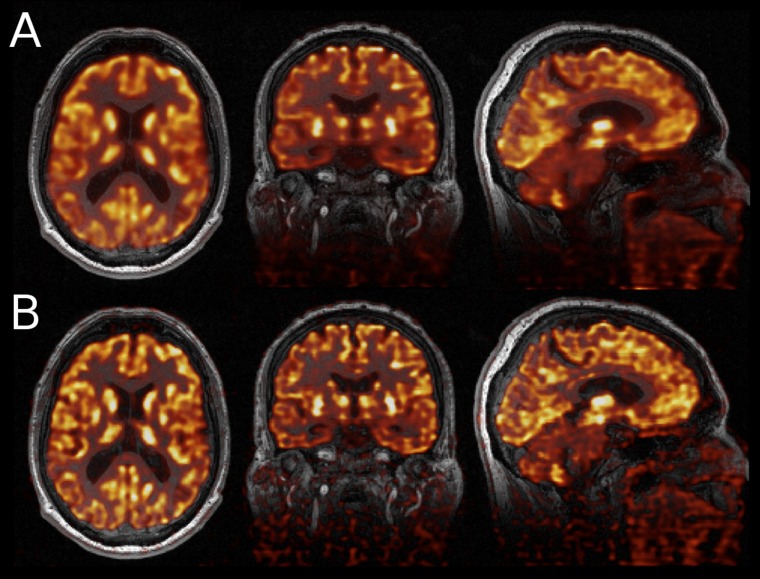
The combination of positron emission tomography (PET) and MRI has attracted the attention of researchers in the past approximately 20 years in small-animal imaging and more recently in clinical research. The combination of PET/MRI allows researchers to explore clinical and research questions in a wide number of fields, some of which are briefly mentioned here. An important number of groups have developed different concepts to tackle the problems that PET instrumentation poses to the exposition of electromagnetic fields. We have described most of these research developments in preclinical and clinical experiments, including the few commercial scanners available. From the software perspective, an important number of algorithms have been developed to address the attenuation correction issue and to exploit the possibility that MRI provides for motion correction and quantitative image reconstruction, especially parametric modelling of radiopharmaceutical kinetics. In this work, we give an overview of some exemplar applications of simultaneous PET/MRI, together with technological hardware and software developments.


Figures

Figure 1.
Figure 1.
Cardiology example: two cases with myocardial infarction of late gadolinium-enhanced (LGE) MRI (a,b), fluorine-18 fludeoxyglucose-positron emission tomography (18F-FDG-PET) uptake (c,d) and fused images (e,f) showing the diagnostic agreement between LGE transmurality and 18F-FDG uptake. Neuro-oncology example: fluid-attenuated inversion recovery (g), diffusion-weighted imaging (h), fluorine-18 fluoroethyl-L-tyrosine (18F-FET)-PET (i) and fusion of 18F-FET-PET with T1 weighted imaging (j) of a patient with grade III anaplastic astrocytoma after radiotherapy are demonstrating tumour recurrence. Reproduced from Rischpler et al with permission from Oxford University Press.
Figure 2.
Figure 2.
Photodetectors used for positron emission tomography detectors: a photomultiplier tube (PMT) for block detector (Hamamatsu Photonics) (a), flat-panel position-sensitive PMT (Hamamatsu Photonics) (b), avalanche photodiode array (Hamamatsu Photonics) (c), silicon photomultipliers (SiPM) array (SensL Technologies) (d), SiPM detector (Ketek) (e) and digital SiPM array (Philips) (f).
Figure 3.
Figure 3.
A 3.0-T MRI scanner MAGNETOM Tim-Trio with BrainPET insert within the MR bore (a) and MR-compatible BrainPET insert (b). Courtesy of Hans Herzog.
Figure 4.
Figure 4.
A positron emission tomography (PET) detector module based on position-sensitive avalanche photodiode (PSAPD) technology, including crystals, optical fibers and electronics on the printed circuit board (PCB), and complete PET insert (originally published in JNM reproduced with permission from the Society of Nuclear Medicine and Molecular Imaging, Inc.) (a). Silicon photomultipliers (SiPM) detector cassette with shielding box and complete PET insert outside and inside the MRI scanner (originally published in JNM reproduced with permission from Society of Nuclear Medicine and Molecular Imaging, Inc.) (b). Digital SiPM detector cassette with shielding box from Hyperion IID (Reproduced from Wehner et al published under terms of the Creative Commons Attribution License CC-BY, http://creativecommons.org/licences/by/4.0/ (c).
Figure 5.
Figure 5.
Gated MRI reconstructions with shortened scan times (10, 7, 5, 3 and 1 min) and different levels of k-space undersampling (a). Sagittal slice of fluorine-18 fludeoxyglucose uptake in the heart and in a lesion, as a result of an ungated reconstruction using all the positron emission tomography (PET) data (b), gated reconstruction using 40% of the PET data (c) and gated reconstruction using post-reconstruction registration (d). Reproduced from Grimm et al with permission from Elsevier.
Figure 6.
Figure 6.
Sagittal, coronal and axial exemplar µ-maps obtained from CT (a), Dixon (b), dual ultrashort time echo (UTE) (c), Artificial neural network-based atlas (d), dual UTE-based R2 (e), magnetization-prepared rapid acquisition gradient echo (MPRAGE)-based atlas (f) and MPRAGE-based template (g). Reproduced from Cabello et al with permission from Springer.
Figure 7.
Figure 7.
Sample transaxial slices for fluorine-18 fludeoxyglucose simulations, corrected for partial volume using various approaches: no correction (a), reblurred van Cittert (b), anisotropic diffusion prior (c), modified Bowsher prior (d), semi-parametric joint entropy (e), Gaussian mixture deconvolution (f), iterative projection (g), iterative Yang (h) and region-based voxelwise (i). Reproduced from Hutton et al published under the Creative Commons Attribution License CC-BY, http://creativecommons.org/licenses/by/4.0.
Figure 8.
Figure 8.
Axial, sagittal and coronal slices of a patient with glioblastoma injected with fluorine-18 fluoroethyl-L-tyrosine, showing the reconstructed map of the k3 microparameter obtained from indirect reconstruction (maximum a posteriori-expectation maximization using Green's one-step-late approach) (a), direct methods using distance weighting in the activity images (b) and Bowsher weighting with simultaneous regularization of activity images and kinetic parameters (c). Reproduced from Loeb et al with permission from the IEEE.
Figure 9.
Figure 9.
Axial, coronal and sagittal images of fluorine-18 fludeoxyglucose-positron emission tomography (PET) acquired with the Siemens mMR Biograph, reconstructed with maximum likelihood-expectation maximization (a) and multichannel MR-PET total generalized variation reconstruction (b) overlaid on MR images (conjugate gradient sensitivity encoding) as reference. Reproduced from Knoll et al from Springer. under the terms of Creative Commons Attribution License CC-BY, http://creativecommons.org/by/4.0/.

Similar articles

Cited by

  • Performance study of a radio-frequency field-penetrable PET insert for simultaneous PET/MRI.
    Chang CM, Lee BJ, Grant AM, Groll AN, Levin CS. Chang CM, et al. IEEE Trans Radiat Plasma Med Sci. 2018 Sep;2(5):422-431. doi: 10.1109/TRPMS.2018.2852686. Epub 2018 Jul 3. IEEE Trans Radiat Plasma Med Sci. 2018. PMID: 30911706 Free PMC article.
  • Induced volatolomics of pathologies.
    Djago F, Lange J, Poinot P. Djago F, et al. Nat Rev Chem. 2021 Mar;5(3):183-196. doi: 10.1038/s41570-020-00248-z. Epub 2021 Feb 2. Nat Rev Chem. 2021. PMID: 37117532 Review.
  • Brain Vascular Imaging Techniques.
    Laviña B. Laviña B. Int J Mol Sci. 2016 Dec 30;18(1):70. doi: 10.3390/ijms18010070. Int J Mol Sci. 2016. PMID: 28042833 Free PMC article. Review.
  • Advances in Molecular Imaging for Neuroendocrine Neoplasms.
    Girod B, Prasad V. Girod B, et al. Cancers (Basel). 2025 Jun 17;17(12):2013. doi: 10.3390/cancers17122013. Cancers (Basel). 2025. PMID: 40563663 Free PMC article. Review.
  • Advanced Monte Carlo simulations of emission tomography imaging systems with GATE.
    Sarrut D, Bała M, Bardiès M, Bert J, Chauvin M, Chatzipapas K, Dupont M, Etxebeste A, M Fanchon L, Jan S, Kayal G, S Kirov A, Kowalski P, Krzemien W, Labour J, Lenz M, Loudos G, Mehadji B, Ménard L, Morel C, Papadimitroulas P, Rafecas M, Salvadori J, Seiter D, Stockhoff M, Testa E, Trigila C, Pietrzyk U, Vandenberghe S, Verdier MA, Visvikis D, Ziemons K, Zvolský M, Roncali E. Sarrut D, et al. Phys Med Biol. 2021 May 14;66(10):10.1088/1361-6560/abf276. doi: 10.1088/1361-6560/abf276. Phys Med Biol. 2021. PMID: 33770774 Free PMC article. Review.

References

    1. Del Guerra A, Belcari N, Bisogni M. Positron emission tomography: its 65 years. Riv Nuovo Cimento 2016; 39: 155–223.
    1. Zaidi H, Del Guerra A. An outlook on future design of hybrid PET/MRI systems. Med Phys 2011; 38: 5667–89. doi: 10.1118/1.3633909 - DOI - PubMed
    1. Zaidi H, Becker M. The Promise of hybrid PET/MRI: technical advances and clinical applications. IEEE Signal Process Mag 2016; 33: 67–85. doi: 10.1109/MSP.2015.2482225 - DOI
    1. Zaidi H. PET/MRI: advances in instrumentation and quantitative procedures. PET Clin 2016; 11: 95–202. - PubMed
    1. Zaidi H, Ojha N, Morich M, Griesmer J, Hu Z, Maniawski P, et al. Design and performance evaluation of a whole-body ingenuity TF PET-MRI system. Phys Med Biol 2011; 56: 3091–106. doi: 10.1088/0031-9155/56/10/013 - DOI - PMC - PubMed

MeSH terms