. 2016 Sep;34(9):987-92.
doi: 10.1038/nbt.3625. Epub 2016 Jul 4.

Protein-retention expansion microscopy of cells and tissues labeled using standard fluorescent proteins and antibodies

Affiliations

Protein-retention expansion microscopy of cells and tissues labeled using standard fluorescent proteins and antibodies

Paul W Tillberg et al. Nat Biotechnol. 2016 Sep.

Abstract

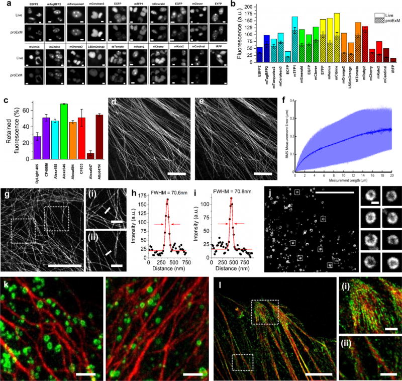
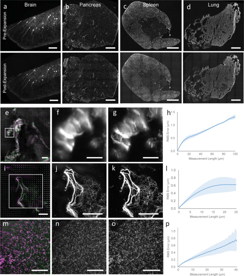
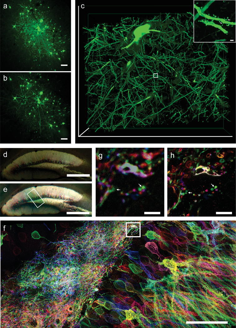
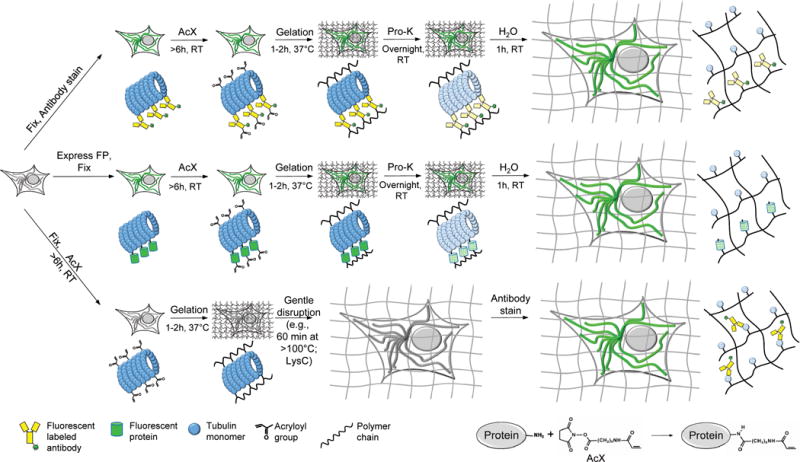
Expansion microscopy (ExM) enables imaging of preserved specimens with nanoscale precision on diffraction-limited instead of specialized super-resolution microscopes. ExM works by physically separating fluorescent probes after anchoring them to a swellable gel. The first ExM method did not result in the retention of native proteins in the gel and relied on custom-made reagents that are not widely available. Here we describe protein retention ExM (proExM), a variant of ExM in which proteins are anchored to the swellable gel, allowing the use of conventional fluorescently labeled antibodies and streptavidin, and fluorescent proteins. We validated and demonstrated the utility of proExM for multicolor super-resolution (∼70 nm) imaging of cells and mammalian tissues on conventional microscopes.


Conflict of interest statement

PWT, FC, ESB, and CCY have filed for patent protection on a subset of the technologies here described. ESB has helped co-found a company to help disseminate kits to the community.

Figures

Figure 1
Figure 1
Retention of fluorescent protein (FP) and antibody fluorescence signals in proExM and proExM of FP fusions. (a) Representative images of selected FP-histone fusion proteins in live HEK293FT cells (upper row) and in the same cells after proExM treatment (lower row); iRFP was expressed as N-terminal fusion with nuclear localization sequence (NLS). (b) Quantified fluorescence of experiments as in panel a, after proExM treatment (crosshatched bars; mean ± standard deviation; n = 4 transfection replicates each). Open bars, literature values of the brightnesses of these fluorophores, normalized to the brightness of EGFP. (c) Retention of fluorescence for selected dyes conjugated with antibodies, after proExM treatment (mean ± standard deviation, n = 3 samples each), in mouse brain slice. (d) Super-resolution structured illumination microscopy (SR-SIM) image of immunostained microtubules after the anchoring step vs. (e) post-expansion image of the same sample acquired with a spinning disk confocal microscope. (f) Root mean square (RMS) length measurement error as a function of measurement length for proExM vs SIM images (blue line, mean; shaded area, standard deviation; n = 4 samples). (g) Confocal image of mClover-α-tubulin fusion. HeLa cells are used throughout the rest of this figure. Panels (i and ii) are magnified views of boxed regions in (g). Linecuts are quantified in panels h, i. Solid red lines in (h, i) indicate the Gaussian fit used to determine the full width at half maximum (FWHM; illustrated with red arrows). (j) Confocal image of mEmerald-clathrin fusion (left) and magnified views of single CCPs in the boxed regions (right). (k) Dual color proExM of clathrin (fused to mEmerald, green) and keratin (mRuby2, red). (l) Dual color proExM image of actin (mRuby2, red) and paxillin (mEmerald, green) fusions. Panels (i and ii) are magnified views of boxed regions in (f). Scale bars: (a) 5 μm, (d) 5 μm, (e) 5 μm (physical size post-expansion, 20.5 μm), (g) 5 μm (21.5 μm), (i–ii) 1 μm; (j) 10 μm (42.6 μm), insets 200 nm; (k) 1 μm (4.3 μm), (l) 5 μm (21.5 μm), (i–ii) 1 μm.
Figure 2
Figure 2
Validation of proExM in different mammalian tissue types. (a–d) Low magnification, wide-field images of pre-expansion (top) and post-expansion (bottom) samples of Thy1-YFP mouse brain (a) and vimentin-immunostained mouse pancreas (b), spleen (c), and lung (d). (e) Composite fluorescence image of Tom20 in Thy1-YFP mouse brain imaged with super-resolution structured illumination microscopy (SR-SIM) (green) and proExM (purple) with conventional confocal microscopy with distortion vector field overlaid (white arrows). (f) Pre-expansion SR-SIM image showing boxed region in (a). (g) Post-expansion confocal image of (f). (h) RMS length measurement error as a function of measurement length for proExM vs SR-SIM pre-expansion for Tom20 staining in Thy1-YFP mouse brain (blue line, mean; shaded area, standard deviation; n = 3 mouse brain cortex samples). (i) High magnification, wide-field fluorescence composite image of vimentin in mouse pancreas before (green) and after (purple) expansion with distortion vector field overlaid (white arrows, see methods). (j) Pre-expansion wide-field image showing boxed region in (i). (k) Post-expansion image of (j). (l) Root mean square (RMS) length measurement error as a function of measurement length for proExM vs widefield pre-expansion images for the different tissue types in (b–d) (blue line, mean; shaded area, standard deviation; n = 3 samples from pancreas, spleen, and lung). (m) Composite fluorescence image of vimentin in mouse pancreas imaged with super-resolution structured illumination microscopy (SR-SIM) (green) and proExM (purple) with conventional confocal microscopy with distortion vector field overlaid (white arrows). (n) Pre-expansion SR-SIM image showing boxed region in (m). (o) Post-expansion confocal image of (n). (p) RMS length measurement error as a function of measurement length for proExM vs SR-SIM pre-expansion for vimentin staining in pancreas (blue line, mean; shaded area, standard deviation; n = 4 fields of view from 2 samples). Scale bars: (a) top 200 μm, bottom 200 μm (physical size post-expansion, 800 μm), (b–d) top 500 μm, bottom 500 μm (2.21 mm, 2.06 mm, 2.04 mm, respectively), (e, f) 10 μm, (g) 10 μm (40 μm), (i) 10 μm, (j) 5 μm, (k) 5 μm (20.4 μm), (m) 5 μm, (n) 5 μm, (o) 5 μm (20.65 μm).
Figure 3
Figure 3
proExM of mammalian brain circuitry. (a) Wide-field image of GFP fluorescence in virally injected rhesus macaque cortex. (b) Post-expansion wide-field fluorescence image of (a). (c) Volume rendering of confocal microscopy images of subregion of (b). Inset shows a zoom-in of boxed region in (c) showing dendritic spines. (d) Low magnification widefield fluorescence imaging showing immunostained mouse hippocampus expressing virally delivered Brainbow3.0. (e) Post-expansion wide-field image of sample from (e). (f) MIP high resolution confocal microscopy image following expansion of membrane labeled Brainbow3.0 neurons from boxed region in (e). (g) Pre-expansion confocal image showing one optical section of boxed region in (f). (h) Post-expansion image of (g). Scale bars: (a) 100 μm, (b) 100 μm (physical size post-expansion, 413 μm); (c) 300 μm × 254 μm × 25 μm, (c) (i) 1 μm, (d) 500 μm, (e) 500 μm (1980 μm); (f) 5 μm, (g) 5 μm (19.8 μm); (h) 50 μm (198 μm).
Figure 4
Figure 4
Workflows for expansion microscopy with protein retention. Three basic sample processing workflows were explored in this paper. Top, samples are chemically fixed and stained with antibodies, using conventional immunostaining protocols, before AcX treatment at room temperature and subsequent ExM processing (gelation, proteinase K treatment, and expansion in water). Middle, samples expressing fluorescent proteins (FPs) are chemically fixed (and optionally permeabilized) before AcX treatment, and subsequent ExM processing. Bottom, samples treated with AcX, followed by gelation, are then processed with a gentle homogenization procedure (e.g., alkaline hydrolysis and denaturation, or digestion with LysC), and finally antibody staining in the expanded state.

Comment in

  • Super-resolution microscopy writ large.
    Engerer P, Fecher C, Misgeld T. Engerer P, et al. Nat Biotechnol. 2016 Sep 8;34(9):928-30. doi: 10.1038/nbt.3669. Nat Biotechnol. 2016. PMID: 27606457 No abstract available.

References

    1. Chen F, Tillberg PW, Boyden ES. Expansion microscopy. Science (80-) 2015;347:543–548. - PMC - PubMed
    1. Laemmli UK. Cleavage of structural proteins during the assembly of the head of bacteriophage T4. Nature. 1970;227:680–685. - PubMed
    1. Hunt NCA, Jasani B, A R. High temperature antigen retrieval and loss of nuclear morphology: a comparison of microwave\rand autoclave techniques. J Clin Pathol. 1996;49:767–770. - PMC - PubMed
    1. Jekel PA, Weijer WJ, Beintema JJ. Use of endoproteinase Lys-C from Lysobacter enzymogenes in protein sequence analysis. Anal Biochem. 1983;134:347–354. - PubMed
    1. Wu CC, MacCoss MJ, Howell KE, Yates JR. A method for the comprehensive proteomic analysis of membrane proteins. Nat Biotechnol. 2003;21:532–8. - PubMed

Publication types

Substances