. 2016 Aug;23(8):738-43.
doi: 10.1038/nsmb.3257. Epub 2016 Jul 4.

Free backbone carbonyls mediate rhodopsin activation

Affiliations

Free backbone carbonyls mediate rhodopsin activation

Naoki Kimata et al. Nat Struct Mol Biol. 2016 Aug.

Abstract

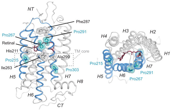
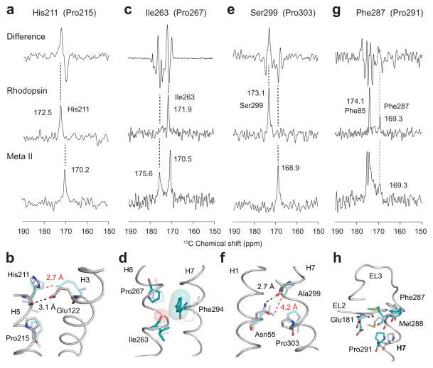
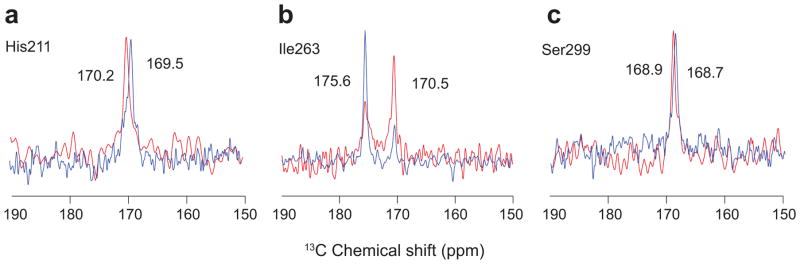
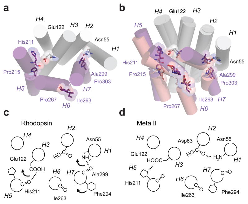
Conserved prolines in the transmembrane helices of G-protein-coupled receptors (GPCRs) are often considered to function as hinges that divide the helix into two segments capable of independent motion. Depending on their potential to hydrogen-bond, the free C=O groups associated with these prolines can facilitate conformational flexibility, conformational switching or stabilization of the receptor structure. To address the role of conserved prolines in family A GPCRs through solid-state NMR spectroscopy, we focus on bovine rhodopsin, a GPCR in the visual receptor subfamily. The free backbone C=O groups on helices H5 and H7 stabilize the inactive rhodopsin structure through hydrogen-bonds to residues on adjacent helices. In response to light-induced isomerization of the retinal chromophore, hydrogen-bonding interactions involving these C=O groups are released, thus facilitating repacking of H5 and H7 onto the transmembrane core of the receptor. These results provide insights into the multiple structural and functional roles of prolines in membrane proteins.


Conflict of interest statement

Competing Financial Interests

The authors declare no competing financial interests.

Figures

Figure 1
Figure 1
Crystal structure of the visual receptor rhodopsin (PDB ID 1U19 ) showing the positions of Pro2155.50, Pro2676.50, Pro2917.38 and Pro3037.50. These prolines are located on helices H5, H6 and H7. The lack of an NH group results in a carbonyl group at the i-4 position from these prolines that is free to form interhelical hydrogen-bonds. The residues with the free carbonyl (His2115.46, Ile2636.46, Phe2877.34, and Ala2997.46) are shown in grey. The three free C=O groups that are conserved across the family A GPCRs (His2115.46, Ile2636.46, and Ala2997.46) lie within or near the TM core of the receptor.
Figure 2
Figure 2
REDOR NMR as a probe of hydrogen bonding changes of carbonyl residues at the i-4 positions of Pro2155.50, Pro2676.50, Pro2917.38 and Pro3037.50. (a, c, e, g) One-dimensional MAS NMR difference spectra between rhodopsin and Meta II are shown in the top panel. REDOR filtered spectra of rhodopsin and Meta II are shown in the middle and lower panels, respectively. The spectra highlight the i-4 for carbonyls associated with His2115.46 using rhodopsin labeled with 1-13C His, 15N Phe (a), Ile2636.46 using rhodopsin labeled with 1-13C Ile, 15N Cys (c), Ser2997.46 using rhodopsin labeled with 1-13C Ser, 15N Val (e), and Phe2877.34 using rhodopsin labeled with 1-13C Phe, 15N Met (g). (b, d, f, h) Crystal structures of rhodopsin (gray, PDB ID 1U19) and Meta II (cyan, PDB ID 3PQR) are shown in the region of interest.
Figure 3
Figure 3
13C…15N REDOR NMR experiments of Meta II in the presence and absence of the Gα peptide of transducin. REDOR filtered spectra are shown for His2115.46 (a), Ile2636.46 (b) and Ser2997.46 (c). The experiments on rhodopsin without (red) and with (blue) added Gα peptide were carried out in DDM micelles and mixed DDM/DOPS micelles, respectively.
Figure 4
Figure 4
Receptor activation leads to repacking of helices H5–H7 on the TM core of rhodopsin. (a) Inactive structure of rhodopsin. Helices H1–H4 form a scaffold onto which helices H5–H7 pack. The key packing contacts on H5 and H7 are associated with Pro2155.50 and Pro3037.50 and their corresponding i-4 carbonyls. Retinal isomerization disrupts both interactions. For H5, the β-ionone ring has a steric clash at the position of the His2115.46-Glu1223.37 hydrogen bond upon conversion the all-trans configuration. For H7, the retinal is covalently attached to Lys2967.43. Upon isomerization, Trp2656.48 on H6 rotates away from H7 and disrupts a water mediated hydrogen bond with Asn3027.49, which is part of a hydrogen bonding network stretching from Asn551.50 and Asp832.50 to the Ala2997.46 C=O. (b) Overlap of the crystal structures of rhodopsin (1GZM, purple) and Meta II (3PQR, light purple) showing the positions of the TM helices. The disruption of the interactions of the His2115.46 C=O and Ala2997.46 C=O with the H1–H4 scaffold allows helices H5–H7 to reorient. (c,d) Schematic of the hydrogen bonding changes occurring between inactive rhodopsin and active Meta II rhodopsin.

References

    1. Sansom MSP, Weinstein H. Hinges, swivels and switches: The role of prolines in signalling via transmembrane α-helices. Trends Pharmacol Sci. 2000;21:445–451. - PubMed
    1. Cordes FS, Bright JN, Sansom MSP. Proline-induced distortions of transmembrane helices. J Mol Biol. 2002;323:951–960. - PubMed
    1. Williams KA, Deber CM. Proline residues in transmembrane helices: structural or dynamic role? Biochemistry. 1991;30:8919–8923. - PubMed
    1. von Heijne G. Proline kinks in transmembrane α-helices. J Mol Biol. 1991;218:499–503. - PubMed
    1. Fu Q, et al. Structural basis and functional role of intramembrane trimerization of the Fas/CD95 death receptor. Mol Cell. 2016;61:602–613. - PMC - PubMed

Publication types

LinkOut - more resources