. 2016 Oct;240(2):173-83.
doi: 10.1002/path.4766. Epub 2016 Sep 19.

Monomeric C-reactive protein and inflammation in age-related macular degeneration

Affiliations

Monomeric C-reactive protein and inflammation in age-related macular degeneration

Kathleen R Chirco et al. J Pathol. 2016 Oct.

Abstract

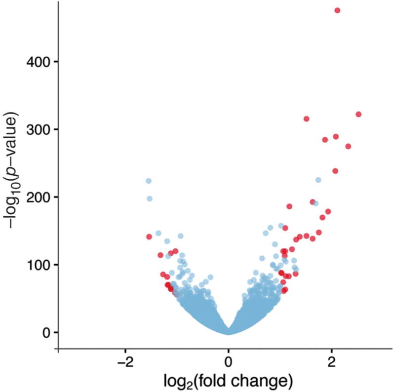
Age-related macular degeneration (AMD) is a devastating disease characterized by central vision loss in elderly individuals. Previous studies have suggested a link between elevated levels of total C-reactive protein (CRP) in the choroid, CFH genotype, and AMD status; however, the structural form of CRP present in the choroid, its relationship to CFH genotype, and its functional consequences have not been assessed. In this report, we studied genotyped human donor eyes (n = 60) and found that eyes homozygous for the high-risk CFH (Y402H) allele had elevated monomeric CRP (mCRP) within the choriocapillaris and Bruch's membrane, compared to those with the low-risk genotype. Treatment of choroidal endothelial cells in vitro with mCRP increased migration rate and monolayer permeability compared to treatment with pentameric CRP (pCRP) or medium alone. Organ cultures treated with mCRP exhibited dramatically altered expression of inflammatory genes as assessed by RNA sequencing, including ICAM-1 and CA4, both of which were confirmed at the protein level. Our data indicate that mCRP is the more abundant form of CRP in human choroid, and that mCRP levels are elevated in individuals with the high-risk CFH genotype. Moreover, pro-inflammatory mCRP significantly affects endothelial cell phenotypes in vitro and ex vivo, suggesting a role for mCRP in choroidal vascular dysfunction in AMD. Copyright © 2016 Pathological Society of Great Britain and Ireland. Published by John Wiley & Sons, Ltd.

Keywords: C-reactive protein; ICAM-1; age-related macular degeneration; choroid; inflammation; microvasculature.


Conflict of interest statement

The authors have declared that no conflict of interest exists.

Figures

Figure 1
Figure 1
Choroids from CFH high-risk donors have increased mCRP immunoreactivity. Donors were homozygous at the CFH allele for either the high-risk (A–C) or the low-risk (D–F) CFH genotype. Labelling with anti-mCRP antibody (B, E; purple, arrowheads) and anti-pCRP antibody (A, D; purple) was compared with labelling with secondary antibody only (C, F). A total of 25 eyes were evaluated and scored for each genotype (G). Scores were given based on the intensity of mCRP immunolabelling (H). RPE = retinal pigment epithelium; CC = choriocapillaris. Scale bar = 100 μm.
Figure 2
Figure 2
Choroids with the high-risk genotype have elevated mCRP. RPE/choroid tissue samples from heterozygous donors (HY) and those homozygous at the CFH allele for either the low-risk (YY) or the high-risk (HH) CFH genotype were analysed. (A) Total CRP ELISA was performed to compare total CRP levels between genotypes. Bars 1–4 = YY (n = 4); bars 5–10 = HY (n = 5); bars 11–15 = HH (n = 5). (B) Semi-native western blotting was performed using antibodies against pCRP (green, upper band) and mCRP (green, lower band). Lanes 2–5 = YY (n = 4); lanes 6–10 = HY (n = 5); lanes 11–15 = HH (n = 5). Relative band densities from the semi-native western blot were quantified for both pCRP (C) and mCRP (D). *p < 0.05.
Figure 3
Figure 3
mCRP localizes to sub-RPE deposits. Labelling with anti-mCRP antibody (A, C, E; purple) from three different donors compared with secondary antibody control (B, D, F). The black arrowhead indicates small druse (A); the asterisk indicates large hyaline druse (C); and the white arrowhead indicates large amorphous druse (E). Scale bar = 50 μm.
Figure 4
Figure 4
mCRP accelerates scratch closure and decreases TER in cultured endothelial cells. (A) Per cent scratch closure for media control (blue; n = 12), pCRP (20 μg/ml; green; n = 24), and mCRP (20 μg/ml; red; n = 24). (B) TER assay with media control (blue; n = 9), pCRP (20 μg/ml; green; n = 9), and mCRP (20 μg/ml; red; n = 9). *p < 0.05; **p < 0.01; ***p < 0.005.
Figure 5
Figure 5
Volcano plot summarizing RNA-Seq data for mCRP versus control RPE/choroid organ culture treatment. RNA-Seq analysis identified a list of 43 genes as differentially expressed (red points). The log2 fold change values and p-values were calculated using DESeq2. Red points indicate genes that were called as significant (adjusted p-value < 0.05; at least 2-fold difference between groups) by both DESeq2 and Cuffdiff (see supplementary material, Table S2).
Figure 6
Figure 6
Exogenous mCRP increases ICAM-1 after 12 h, while it decreases CA4 protein levels after 24 h in human choroid organ cultures. Representative western blots of human donor eye tissue after 12-h (A, C) or 24-h (B, D) treatment with mCRP or medium alone using anti-ICAM-1 (A, B; green, ∼80 kDa) and anti-CA4 (C, D; red, 35 kDa) antibodies. Lanes representing mCRP-treated tissue are indicated with a ‘+’. Quantification of relative band densities is shown for ICAM-1 at 12 h (A, n = 18), ICAM-1 at 24 h (B, n = 8), CA4 at 12 h (C, n = 6), and CA4 at 24 h (D, n = 8). *p < 0.05.

References

    1. Friedman DS, O’Colmain BJ, Muñoz B, et al. Prevalence of age-related macular degeneration in the United States. Arch Ophthalmol. 2004;122:564–572. - PubMed
    1. Bird AC, Bressler NM, Bressler SB, et al. An international classification and grading system for age-related maculopathy and age-related macular degeneration. Surv Ophthalmol. 1995;39:367–374. - PubMed
    1. Age-Related Eye Disease Study Research Group. The Age-Related Eye Disease Study (AREDS): design implications. AREDS report no. 1. Control Clin Trials. 1999;20:573–600. - PMC - PubMed
    1. Fritsche LG, Igl W, Bailey JNC, et al. A large genome-wide association study of age-related macular degeneration highlights contributions of rare and common variants. Nature Genet. 2016;48:134–143. - PMC - PubMed
    1. Edwards AO, Ritter R, Abel KJ, et al. Complement factor H polymorphism and age-related macular degeneration. Science. 2005;308:421–424. - PubMed

Publication types

MeSH terms

Substances