. 2016 Jul 5:7:11997.
doi: 10.1038/ncomms11997.

A spliceosome intermediate with loosely associated tri-snRNP accumulates in the absence of Prp28 ATPase activity

Affiliations

A spliceosome intermediate with loosely associated tri-snRNP accumulates in the absence of Prp28 ATPase activity

Carsten Boesler et al. Nat Commun. .

Abstract

The precise role of the spliceosomal DEAD-box protein Prp28 in higher eukaryotes remains unclear. We show that stable tri-snRNP association during pre-catalytic spliceosomal B complex formation is blocked by a dominant-negative hPrp28 mutant lacking ATPase activity. Complexes formed in the presence of ATPase-deficient hPrp28 represent a novel assembly intermediate, the pre-B complex, that contains U1, U2 and loosely associated tri-snRNP and is stalled before disruption of the U1/5'ss base pairing interaction, consistent with a role for hPrp28 in the latter. Pre-B and B complexes differ structurally, indicating that stable tri-snRNP integration is accompanied by substantial rearrangements in the spliceosome. Disruption of the U1/5'ss interaction alone is not sufficient to bypass the block by ATPase-deficient hPrp28, suggesting hPrp28 has an additional function at this stage of splicing. Our data provide new insights into the function of Prp28 in higher eukaryotes, and the requirements for stable tri-snRNP binding during B complex formation.


Figures
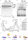
Figure 1
Figure 1. An excess of ATPase-deficient hPrp28 stalls splicing before spliceosomal B complex formation.
(a) In vitro splicing of 32P-labelled MINX-MS2 pre-mRNA in HeLa nuclear extract in the presence of increasing amounts (10–100 ng μl−1) of recombinant hPrp28 or an AAAD mutant (hPrp28AAAD) thereof, as indicated above each lane. RNA was analysed by denaturing PAGE and visualized by autoradiography. The positions of the pre-mRNA, and splicing intermediates and products are indicated on the right. The nucleotide length of the pre-mRNA, mRNA and 5′ exon is indicated on the left. The % spliced mRNA is indicated below each lane. The average % spliced mRNA±s.d. (from three independent experiments) observed in the presence of 50 and 100 ng μl−1 hPrp28AAAD was 2.65±0.80 and 2.05±0.77, respectively. (b) Analysis of splicing complexes formed in nuclear extract in the presence of 50 ng μl−1 hPrp28 or hPrp28AAAD by agarose gel electrophoresis in the presence of heparin. The positions of H, A, B and Bact/C complexes are indicated. (c) Splicing complexes were assembled on 32P-labelled MINX-MS2 pre-mRNA in HeLa nuclear extract for 6 min either in the absence or presence of an inhibitory concentration (50 ng μl−1) of the recombinant hPrp28AAAD protein, and were analysed on a 10–30% glycerol gradient containing G-150 buffer. The percent of total radioactivity in each gradient fraction is plotted. Sedimentation values were determined using prokaryotic ribosomal subunits run in parallel. (d) Spliceosomal complexes in peak fractions (37S pre-B, fractions 16–18; B complex, fractions 17–19) were subjected to MS2 affinity selection and RNA was analysed by denaturing PAGE followed by silver staining. RNA identities are indicated on the right. Nucleotide length (nt) markers are derived from the snRNAs from purified human 37S cross-exon complexes run in parallel. (e) Schematic of the spliceosome assembly stages leading to a pre-catalytic B complex.
Figure 2
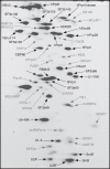
Figure 2. Identification of abundant proteins in purified 37S pre-B complexes.
Proteins (>25 kDa) associated with affinity-purified 37S pre-B complexes were separated by 2D gel electrophoresis, stained with RuBPS, and the identities of single protein spots were determined by mass spectrometry. Abundant proteins were identified by visual inspection and are indicated in black, and less abundant ones in grey.
Figure 3
Figure 3. Identification of RNA–RNA interactions in the 37S pre-B complex via psoralen crosslinking.
(a) Affinity-purified pre-B and B complexes were UV-irradiated ± psoralen (AMT) as indicated. Total psoralen-crosslinked RNA was incubated with RNase H and an oligonucleotide complementary to exon 2 of the MINX pre-mRNA as indicated above each lane. RNA–RNA crosslinks were identified by northern blot analyses, incubating sequentially with 32P-labelled probes against the pre-mRNA, and U1, U2, U4, U5 and U6 snRNAs. The blot was stripped of each 32P-probe before incubation with a subsequent probe. 32P-labelled MINX pre-mRNA, on which the pre-B and B complexes were formed, is visible in all panels. The lower intensity of the 32P-pre-mRNA in the U5 panel is due to decay of the original signal. The positions of crosslinked RNA species are indicated. Bands appearing after RNase H digestion are indicated with a diamond (◊). *A potential U1/U4 crosslink and **Internally crosslinked MINX pre-mRNA. (b) Schematic representation of the RNA–RNA networks in the pre-B and B complexes. The 5′ss base pairing interaction with U1 followed by U6 is highlighted purple, the U2-branch site base pairing interaction is shown in light blue and U2/U6 helix II is shown in dark blue.
Figure 4
Figure 4. Purified 37S pre-B complexes can be chased into catalytically active spliceosomes.
Affinity-purified pre-B or B complexes, or MINX-MS2 pre-mRNA (as indicated above) were incubated at 30 °C for the indicated times (0–90 min) under splicing conditions in the presence of buffer alone (lanes 1–3 and 10–12) or micrococcal nuclease-treated HeLa nuclear extract (MNxt; lanes 6–9 and 13–17). To outcompete the hPrp28AAAD mutant present in the pre-B complexes, 50 ng μl−1 of recombinant hPrp28wt protein was added before incubation at 30 °C (lanes 4–5 and 8–9). RNA was analysed by denaturing PAGE and visualized with a Phosphorimager. The positions of the pre-mRNA, splicing intermediates and products are indicated on the right. Nucleotide length (nt) markers are derived from the snRNAs from purified human 37S cross-exon complexes run in parallel.
Figure 5
Figure 5. Electron microscopy of the 37S pre-B complex.
Overviews of negatively stained, affinity-purified 37S pre-B (a) and B complexes (c) or affinity-purified 37S pre-B complexes incubated solely with the 5′ss RNA oligo (e). Representative class averages from 9,000 to 11,000 single-particle images of each complex are shown in the galleries in b,d and f. Scale bars, 50 nm. (g) Schematic representation of the 37S pre-B complex and B complex, with main structural features labelled according to Boehringer et al.
Figure 6
Figure 6. Addition of a 5′ss RNA oligonucleotide to 37S pre-B complexes induces stable tri-snRNP binding.
(a) Schematic of the effect of the addition of an excess of a 5′ss containing oligonucleotide on RNA–RNA interaction within the pre-B complex. Colouring as in Fig. 3. (b) 37S pre-B complexes were assembled in nuclear extract on 32P-labelled MINX-MS2 pre-mRNA in the presence of an inhibitory concentration of hPrp28AAAD. After 3 min, wild type or 2′Ome 5′ss oligo was added at an 100-fold excess. Splicing complex formation was analysed on an agarose gel in the presence of heparin and visualized by autoradiography. The positions of H, A, B and Bact/C complexes are indicated. (c) Glycerol gradient centrifugation (150 mM KCl) of affinity-purified 37S pre-B complexes alone or after incubating with either a wild-type 5′ss RNA oligonucleotide or a 2′Ome version thereof. Affinity-purified B complexes were run in parallel. The percent of total radioactivity (32P-MINX pre-mRNA) is plotted for each gradient fraction. (d) RNA was recovered from the indicated peak fractions, separated by denaturing PAGE and visualized by silver staining. Identities of snRNAs are indicated. Nucleotide length (nt) markers are derived from the spliceosomal snRNAs from purified human 37S cross-exon complexes run in parallel.
Figure 7
Figure 7. Summary of the cross-intron spliceosome assembly pathway.
The A, pre-B, B and Bact complexes and their RNA–RNA networks are shown schematically. The 5′ss base pairing interaction with U1 followed by U6 is highlighted purple, the U2-branch site base pairing interaction is shown in light blue, U2/U6 helix II is shown in dark blue and U2/U6 helix I is shown in yellow.

References

    1. Will C. L. & Lührmann R. Spliceosome structure and function. Cold Spring Harb. Perspect. Biol. 3, doi:10.1101/cshperspect.a003707 (2011). - PMC - PubMed
    1. Robberson B. L., Cote G. J. & Berget S. M. Exon definition may facilitate splice site selection in RNAs with multiple exons. Mol. Cell Biol. 10, 84–94 (1990). - PMC - PubMed
    1. Wahl M. C., Will C. L. & Lührmann R. The spliceosome: design principles of a dynamic RNP machine. Cell 136, 701–718 (2009). - PubMed
    1. Nilsen T. W. RNA Structure and Function 279–307Cold Spring Harbor Laboratory Press (1998).
    1. Valadkhan S. snRNAs as the catalysts of pre-mRNA splicing. Curr. Opin. Chem. Biol. 9, 603–608 (2005). - PubMed

Publication types

MeSH terms

LinkOut - more resources