

On the distribution of intranuclear and cytoplasmic aggregates in the brainstem of patients with spinocerebellar ataxia type 2 and 3
- PMID: 27377427
- PMCID: PMC8028910
- DOI: 10.1111/bpa.12412
On the distribution of intranuclear and cytoplasmic aggregates in the brainstem of patients with spinocerebellar ataxia type 2 and 3
Abstract
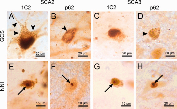
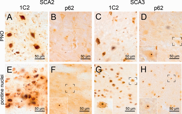
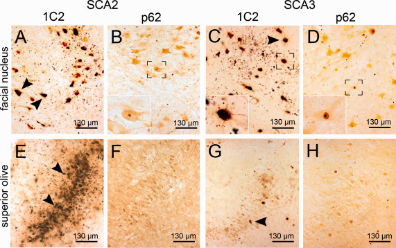
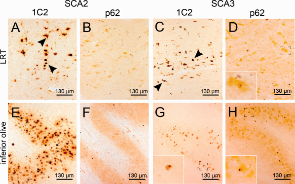
The polyglutamine (polyQ) diseases are a group of genetically and clinically heterogeneous neurodegenerative diseases, characterized by the expansion of polyQ sequences in unrelated disease proteins, which form different types of neuronal aggregates. The aim of this study was to characterize the aggregation pathology in the brainstem of spinocerebellar ataxia type 2 (SCA2) and 3 (SCA3) patients. For good recognition of neurodegeneration and rare aggregates, we employed 100 µm PEG embedded brainstem sections, which were immunostained with the 1C2 antibody, targeted at polyQ expansions, or with an antibody against p62, a reliable marker of protein aggregates. Brainstem areas were scored semiquantitatively for neurodegeneration, severity of granular cytoplasmic staining (GCS) and frequency of neuronal nuclear inclusions (NNI). SCA2 and SCA3 tissue exhibited the same aggregate types and similar staining patterns. Several brainstem areas showed statistically significant differences between disease groups, whereby SCA2 showed more severe GCS and SCA3 showed more numerous NNI. We observed a positive correlation between GCS severity and neurodegeneration in SCA2 and SCA3 and an inverse correlation between the frequency of NNI and neurodegeneration in SCA3. Although their respective disease proteins are unrelated, SCA2 and SCA3 showed the same aggregate types. Apparently, the polyQ sequence alone is sufficient as a driver of protein aggregation. This is then modified by protein context and intrinsic properties of neuronal populations. The severity of GCS was the best predictor of neurodegeneration in both disorders, while the inverse correlation of neurodegeneration and NNI in SCA3 tissue implies a protective role of these aggregates.
Keywords: neurodegeneration; p62; polyglutamine; protein aggregation disease; spinocerebellar ataxia.
© 2016 International Society of Neuropathology.
Figures
References
-
- Ansorge O, Giunti P, Michalik A, Van BC, Harding B, Wood N et al (2004) Ataxin‐7 aggregation and ubiquitination in infantile SCA7 with 180 CAG repeats. Ann Neurol 56:448–452. - PubMed
-
- Arrasate M, Mitra S, Schweitzer ES, Segal MR, Finkbeiner S (2004) Inclusion body formation reduces levels of mutant huntingtin and the risk of neuronal death. Nature 431:805–810. - PubMed
-
- Bjorkoy G, Lamark T, Johansen T (2006) p62/SQSTM1: a missing link between protein aggregates and the autophagy machinery. Autophagy 2:138–139. - PubMed
Publication types
MeSH terms
LinkOut - more resources
Full Text Sources
Other Literature Sources
Research Materials
