. 2016 Aug 5;371(1700):20150420.
doi: 10.1098/rstb.2015.0420.

The zinc sensing receptor, ZnR/GPR39, triggers metabotropic calcium signalling in colonocytes and regulates occludin recovery in experimental colitis

Affiliations

The zinc sensing receptor, ZnR/GPR39, triggers metabotropic calcium signalling in colonocytes and regulates occludin recovery in experimental colitis

Laxmi Sunuwar et al. Philos Trans R Soc Lond B Biol Sci. .

Erratum in

Abstract

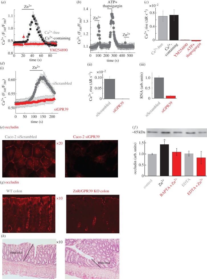
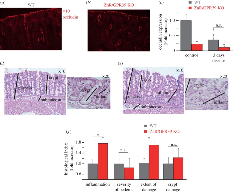
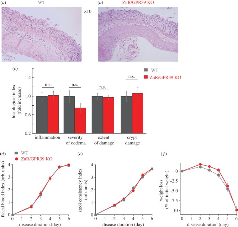
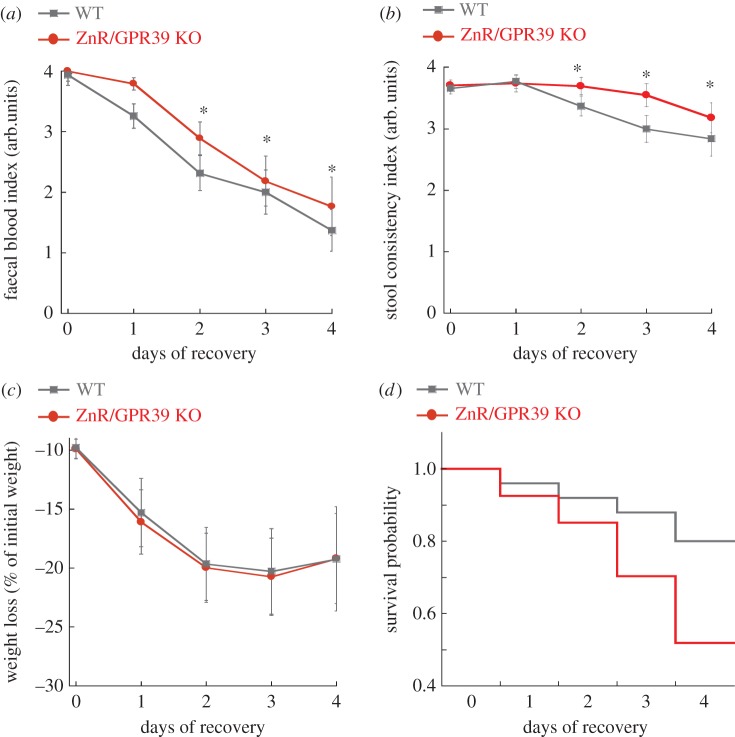
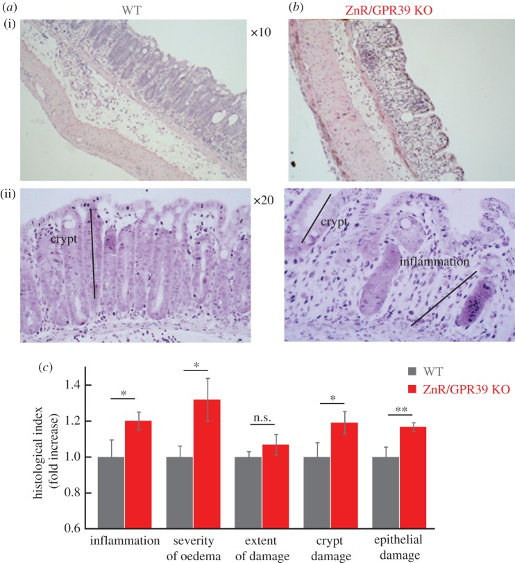
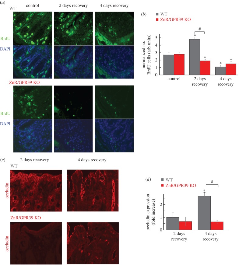
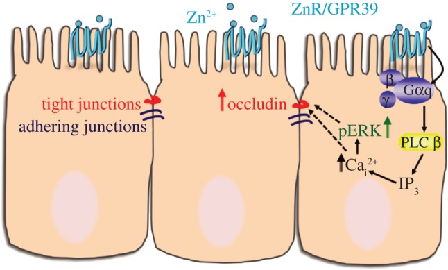
Impaired epithelial barrier function is a hallmark of inflammatory bowel diseases, such as colitis, contributing to diarrhoea and perpetuating inflammation. We show that the zinc sensing receptor, ZnR/GPR39, triggers intracellular Ca(2+) signalling in colonocytes thereby inducing occludin expression. Moreover, ZnR/GPR39 is essential for epithelial barrier recovery in the dextran sodium sulfate (DSS) ulcerative colitis model. Loss of ZnR/GPR39 results in increased susceptibility to DSS-induced inflammation, owing to low expression of the tight junction protein occludin and impaired epithelial barrier. Recovery of wild-type (WT) mice from the DSS insult was faster than that of ZnR/GPR39 knockout (KO) mice. Enhanced recovery of the epithelial layer and increased crypt regeneration were observed in WT mice compared with ZnR/GPR39 KO, suggesting that ZnR/GPR39 is promoting epithelial barrier integrity following DSS insult. Indeed, cell proliferation and apical expression of occludin, following the DSS-induced epithelial erosion, were increased in WT tissue but not in ZnR/GPR39 KO tissue. Importantly, survival following DSS treatment was higher in WT mice compared with ZnR/GPR39 KO mice. Our results support a direct role for ZnR/GPR39 in promoting epithelial renewal and barrier function following DSS treatment, thereby affecting the severity of the disease. We suggest ZnR/GPR39 as a novel therapeutic target that can improve epithelial barrier function in colitis.This article is part of the themed issue 'Evolution brings Ca(2+) and ATP together to control life and death'.

Keywords: Ca2+ signalling; ZnR/GPR39; colitis; intestinal epithelium; tight junction; zinc.


Figures

Figure 1.
Figure 1.
ZnR/GPR39 triggers Ca2+ signalling in Caco-2 colonocytes and regulates occludin expression. (a) Caco-2 colonocytes were loaded with Fura-2 and responses were monitored in Ringer's solution with (grey) and without (black) Ca2+-free following the addition of Zn2+ (200 µM) to control cell or cells treated with the Gq inhibitor YM254890 (1 µM). Averaged response from seven cells in one slide is shown. (b) The response to Zn2+ (200 µM, as in a) was monitored before or after Ca2+ store depletion (ATP 100 µM+thapsigargin 200 nM). (c) The average initial rate of the Ca2+ response as monitored in (a,b) (n = 10). (d) The response to Zn2+ (200 µM, as in a) was monitored in Caco-2 cells treated with an siRNA construct aimed to silence ZnR/GPR39 (siGPR39) or a scrambled control (siScrambled). Panel (ii) shows the average rate of initial Ca2+ response (n = 20), panel (iii) shows mRNA level in control or siGPR39 silenced cells (n = 3). (e) Representative immunofluorescence analysis of the expression of the tight junction protein occludin in siScrambled or siGPR39 Caco-2 cells. (f) Analysis of occludin expression level after Ca2+ depletion (see Material and methods) in differentiated Caco-2 cells treated with Zn2+ (200 µM) or without it (control) in the presence of the intracellular Ca2+ chelator BAPTA-AM (5 µM) or the non-permeable Ca2+ chelator EDTA (100 µM) (n = 2, *p < 0.05). (g) Representative immunofluorescence analysis of the expression of the tight junction protein occludin in colon tissue from WT or ZnR/GPR39 KO mice. (h) Histochemical (H&E) analysis of colon tissue sections from WT and ZnR/GPR39 KO mice in sections of colon. The mucosa layer is marked, showing similar crypt formation and low extent of inflammation in both genotypes.
Figure 2.
Figure 2.
Clinical and histological analysis of colon tissue at the acute phase of DSS treatment. (a,b) Representative immunofluorescence analysis of the expression of the tight junction protein occludin in colon tissue from WT or ZnR/GPR39 KO mice following 3 days DSS treatment. (c) Quantification of occludin expression levels during basal (figure 1f) or disease condition (figure 2a) using a threshold analysis (see Material and methods). Results were normalized to the average count in the WT group at baseline/control conditions (n = 15–35 slices from six to eight mice for each treatment, *p < 0.05 compared to control WT). (d,e) H&E analysis of colon tissue sections from WT and ZnR/GPR39 KO mice in sections of colon following 3 days DSS treatment. The black lines mark the mucosal layer, intact crypts in the mucosa or the inflamed mucosal layer (inflam). (f) Histological evaluation along the colon was done to obtain indices of: the inflammation level, severity of oedema, extent of inflamed tissue within the wall (extent of damage) and crypt damage (see Material and methods). Each of the indices is normalized to the level in WT tissues. (n = 22–23 per genotype, *p < 0.05 compared to WT).
Figure 3.
Figure 3.
Clinical and histological analysis of colon tissue at the acute phase of DSS treatment. (a,b) Representative HE-stained colon tissue sections obtained following 6 days of DSS treatment from WT (a) and ZnR/GPR39 KO (b). (c) Histological evaluation along the colon, performed as described in figure 1, shows no significant differences in the severity of the injury to WT and ZnR/GPR39 KO mice. (d) Faecal blood and (e) faecal consistency indices were monitored during the 6 days of DSS treatment (n = 22–23, per genotype). The scores for blood index and stool consistency were between 0 and 4, when 0 indicates the healthy stage (see Material and methods). (f) Weight loss during DSS treatment is shown. During the first 2 days of DSS treatment, the weight of treated mice increased by 0.6 ± 0.4% in WT mice and 1.5 ± 0.4% in ZnR/GPR39 KO mice (n = 22–23, per genotype).
Figure 4.
Figure 4.
ZnR/GPR39 ameliorates clinical symptoms of the disease and enhances survival during recovery from DSS treatment. Clinical scores were monitored daily during the recovery period, following removal of DSS treatment, while on a regular drinking regime. Day 0 is the 6th day of DSS and shows the data as figure 3. (a) Faecal blood and (b) stool consistency indices as well as (c) weight loss were determined in WT versus ZnR/GPR39 KO mice (as in figure 3 and Material and methods). (d) Survival probability during the recovery period is presented using the Kaplan–Maier plot, p < 0.05 between WT and ZnR/GPR39 KO mice.
Figure 5.
Figure 5.
ZnR/GPR39 promotes crypts regeneration during the recovery period. (a,b) Representative HE-stained colon tissues from WT (a) and ZnR/GPR39 KO (b) mice obtained on the second day of the recovery phase. (c) Histological evaluation along the colon was done to obtain indices of: the inflammation level, severity of oedema, extent of inflamed tissue within the wall (extent of damage), crypt damage and extent of epithelial damage (regions of destructed tissue along the epithelial layer). For clarity of presentation in a bar graph, each of the indices is normalized to the level in WT tissues. (n = 28–30 per genotype, *p < 0.05 compared to WT).
Figure 6.
Figure 6.
ZnR/GPR39 promotes cell proliferation. (a) Representative BrdU-stained tissues of WT or ZnR/GPR39 KO colon from control (non-treated) mice or following DSS treatment at 2 days or 4 days of the recovery period (×20). DAPI staining was added as a control. Note that DAPI staining is seen in epithelial as well as in inflammatory cells nuclei within the mucosa. (b) Analysis of epithelial proliferation, using BrdU-stained tissue, comparing the number of stained cells to the number of crypts observed in a field. (n = 37 per genotype, *p < 0.05 compared to control WT, #p < 0.05, between WT and ZnR/GPR39 KO on day 2 of recovery). (c) Representative images of occludin expression levels during the recovery period, on the second and fourth days following removal of DSS, recovery phase. (d) Quantitative analysis was performed as in figure 1 (n = 10–30 slices from 6 to 10 mice from each treatment and each genotype, *p < 0.05 compared to 2 days of recovery in WT, #p < 0.05 compared to 4 days of recovery in WT).
Figure 7.
Figure 7.
Scheme of ZnR/GPR39 signalling activated by Zn2+ to enhance tight junction formation. Our results indicate that ZnR/GPR39 signalling activates Ca2+ release from thapsigargin-dependent endoplasmic reticulum stores in colonocytes, thereby upregulating occludin expression. In vivo, ZnR/GPR39 expression is essential to enhance the recovery of occludin expression and colonocytes proliferation following barrier disruption by DSS.

References

    1. Danese S, Fiocchi C. 2011. Ulcerative colitis. N. Engl. J. Med. 365, 1713–1725. (10.1056/NEJMra1102942) - DOI - PubMed
    1. Hering NA, Fromm M, Schulzke JD. 2012. Determinants of colonic barrier function in inflammatory bowel disease and potential therapeutics. J. Physiol. 590, 1035–1044. (10.1113/jphysiol.2011.224568) - DOI - PMC - PubMed
    1. Dorfel MJ, Huber O. 2012. Modulation of tight junction structure and function by kinases and phosphatases targeting occludin. J. Biomed. Biotechnol. 2012, 807356 (10.1155/2012/807356) - DOI - PMC - PubMed
    1. Bertiaux-Vandaele N, et al. 2011. The expression and the cellular distribution of the tight junction proteins are altered in irritable bowel syndrome patients with differences according to the disease subtype. Am. J. Gastroenterol. 106, 2165–2173. (10.1038/ajg.2011.257) - DOI - PubMed
    1. Su L, et al. 2013. TNFR2 activates MLCK-dependent tight junction dysregulation to cause apoptosis-mediated barrier loss and experimental colitis. Gastroenterology 145, 407–415. (10.1053/j.gastro.2013.04.011) - DOI - PMC - PubMed

Publication types