. 2016 Aug 15;25(16):3467-3475.
doi: 10.1093/hmg/ddw188. Epub 2016 Jul 4.

Identification of plexin A4 as a novel clusterin receptor links two Alzheimer's disease risk genes

Affiliations

Identification of plexin A4 as a novel clusterin receptor links two Alzheimer's disease risk genes

Silvia S Kang et al. Hum Mol Genet. .

Abstract

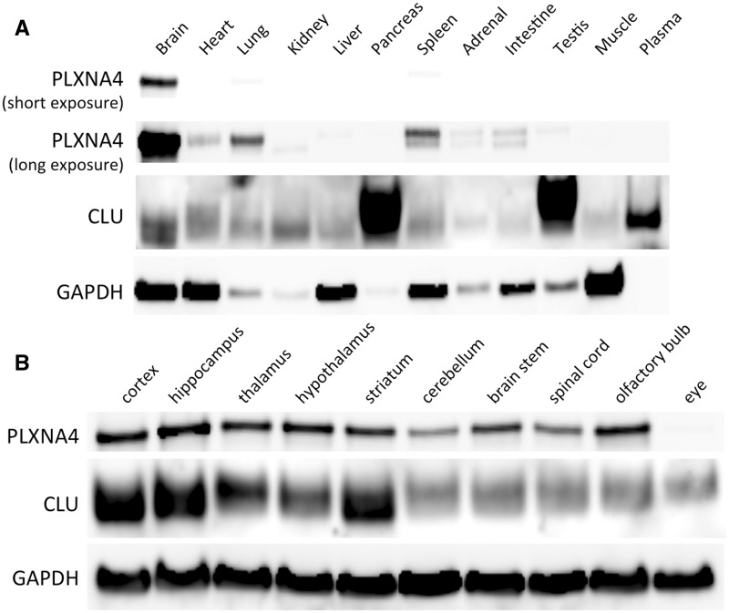
Although abundant genetic and biochemical evidence strongly links Clusterin (CLU) to Alzheimer disease (AD) pathogenesis, the receptor for CLU within the adult brain is currently unknown. Using unbiased approaches, we identified Plexin A4 (PLXNA4) as a novel, high-affinity receptor for CLU in the adult brain. PLXNA4 protein expression was high in brain with much lower levels in peripheral organs. CLU protein levels were significantly elevated in the cerebrospinal fluid (CSF) of Plxna4-/- mice and, in humans, CSF levels of CLU were also associated with PLXNA4 genotype. Human AD brains had significantly increased the levels of CLU protein but decreased levels of PLXNA4 by ∼50%. To determine whether PLXNA4 levels influenced cognition, we analyzed the behaviour of Plxna4+/+, Plxna4+/-, and Plxna4-/- mice. In comparison to WT controls, both Plxna4+/- and Plxna4-/- mice were hyperactive in the open field assay while Plxna4-/- mice displayed a hyper-exploratory (low-anxiety phenotype) in the elevated plus maze. Importantly, both Plxna4+/- and Plxna4-/- mice displayed prominent deficits in learning and memory in the contextual fear-conditioning paradigm. Thus, even a 50% reduction in the level of PLXNA4 is sufficient to cause memory impairments, raising the possibility that memory problems seen in AD patients could be due to reductions in the level of PLXNA4. Both CLU and PLXNA4 have been genetically associated with AD risk and our data thus provide a direct relationship between two AD risk genes. Our data suggest that increasing the levels of PLXNA4 or targeting CLU-PLXNA4 interactions may have therapeutic value in AD.


Figures

Figure 1.
Figure 1.
CLU binds directly to PLXNA4 in vivo and in vitro. (A) Silver stained gel showing a specific band (arrow) immunoprecipitated with anti-CLU antibody in WT but not Clu-/- brain lysates (* indicates non-specific band). The specific CLU-interacting band was cut out from the gel and identified as PLXNA4 by mass spectrometry. Image shown is representative of > 3 experimental replicates. (B) Immunoprecipitation for CLU from brain lysates followed by Western blotting for PLXNA4 demonstrates specific interaction between CLU and PLXNA4 in WT mice but not Clu-/- mice or when using non-specific goat IgG as control. Image shown is representative of > 3 experimental replicates. (C) Cell-free, solid phase binding assay with immobilized PLXNA4 incubated with increasing amounts of CLU showing high affinity (Kd =10.8 nM), saturable binding (average of three independent experiments). (D) Time-course of CLU binding to immobilized PLXNA4 demonstrates that the CLU-PLXNA4 interaction occurs very rapidly with half-maximal binding at 10.03 min. A.U. = arbitrary units.
Figure 2.
Figure 2.
Expression of PLXNA4 and CLU in various tissues by Western blotting. (A) PLXNA4 is highly expressed in brain tissue. Longer exposures show expression of PLXNA4 in lung and spleen, with low but detectable levels in heart, kidney, liver, adrenal, intestine, and testis. No detectable levels were found in pancreas, muscle, or plasma. Slight differences in molecular weights of PLXNA4 are likely due to post-translational modifications. CLU is broadly expressed throughout the body, with highest expression in pancreas, testis, and plasma. CLU is easily detectable in all tissues surveyed, including whole brain tissue, with some tissues also showing differences in molecular weights due to the known heavily glycosylated nature of CLU. (B) Both PLXNA4 and CLU are highly expressed throughout all regions of the central nervous system (with the exception of lack of PLXNA4 in the eye). Highest expression of both PLXNA4 and CLU is seen in the hippocampus.
Figure 3.
Figure 3.
PLXNA4 is associated with levels of CLU in the cerebrospinal fluid (CSF) in mice and humans. (A) CSF isolated from WT (N = 14) and Plxna4-/- (N = 10) littermate mice was assayed for CLU protein levels by a sensitive sandwich ELISA. CLU was significantly elevated in Plxna4-/- mice by ∼34%. Data analyzed by two-tailed t-test, ** indicates P < 0.01. (B) CLU protein is significantly elevated by ∼27% in the CSF of individuals with the T/T allele (n = 23) compared to C/T allele (n = 631) at rs117713945 of PLXNA4. ** indicates P < 0.01 (Bonferroni corrected).
Figure 4.
Figure 4.
Alterations in CLU and PLXNA4 protein levels in Alzheimer’s disease brain tissue. (A) Aβ40 and Aβ42 are significantly elevated in the brains of AD cases compared to controls. (B) ApoE is significantly decreased in TBS and TBS-X fractions in AD cases but increased in guanidine fractions. In contrast, CLU is significantly elevated in TBS, TBS-X and guanidine fractions. (C) Representative Western blot of control vs AD showing substantial decrease of PLXNA4 and increase in CLU in AD cases. (D) PLXNA4 levels were significantly decreased in AD cases vs controls by ∼50%. N = 20 cases and N = 20 controls for A, B, and D, analyzed by two-tailed t-test.
Figure 5.
Figure 5.
Behavioural alterations in the open field assay (OFA) in mice due to reduction or loss of PLXNA4. Plxna4+/- and Plxna4-/- mice were significantly more active than their WT littermates in the OFA by total distance travelled (A) and time spent mobile (B). However, only Plxna4-/- mice had alterations in rearing (C) and anxiety-like behaviour as measured by centre:total distance ratio (D) compared to WT littermates while and Plxna4+/- mice were apparently normal. Data analyzed by one-way ANOVA with post-hoc Fisher’s LSD t-test. N = 22/genotype of males and females combined. * P < 0.05, *** P < 0.001, **** P < 0.0001.
Figure 6.
Figure 6.
Loss of PLXNA4 in mice results in hyper-exploratory behaviour in the elevated plus maze (EPM). Compared to WT littermate controls, Plxna4-/- mice displayed significantly greater exploratory behaviour in the EPM as measured by time spent in the open arms (A) or as a ratio of the time spent in the open arms to the time spent in the closed arms (B). However, Plxna4+/- mice were apparently normal and were not significantly different that their WT littermate controls. Data analyzed by one-way ANOVA with post-hoc Fisher’s LSD t-test. N = 22/genotype of males and females combined. **** P < 0.0001.
Figure 7.
Figure 7.
Reduction or loss of PLXNA4 in mice results severe memory impairments in the contextual fear conditioning (CFC) paradigm. Compared to WT littermate controls, both Plxna4+/- and Plxna4-/- mice had significant memory impairments in the hippocampus-dependent contextual phase of the CFC (A) as well as the hippocampus/amygdala-dependent cued phase of the CFC (B). Data analyzed by one-way ANOVA with post-hoc Fisher’s LSD t-test. N = 22/genotype of males and females combined. * P < 0.05, ** P < 0.01, **** P < 0.0001.

References

    1. DeMattos R.B., Cirrito J.R., Parsadanian M., May P.C., O’Dell M.A., Taylor J.W., Harmony J.A.K., Aronow B.J., Bales K.R., Paul S.M., et al. (2004) ApoE and clusterin cooperatively suppress Abeta levels and deposition: evidence that ApoE regulates extracellular Abeta metabolism in vivo. Neuron, 41, 193–202. - PubMed
    1. DeMattos R.B., O’dell M.A., Parsadanian M., Taylor J.W., Harmony J.A.K., Bales K.R., Paul S.M., Aronow B.J., Holtzman D.M. (2002) Clusterin promotes amyloid plaque formation and is critical for neuritic toxicity in a mouse model of Alzheimer’s disease. Proc. Natl. Acad. Sci. U. S. A, 99, 10843–10848. - PMC - PubMed
    1. Lambert J.C., Heath S., Even G., Campion D., Sleegers K., Hiltunen M., Combarros O., Zelenika D., Bullido M.J., Tavernier B., et al. (2009) Genome-wide association study identifies variants at CLU and CR1 associated with Alzheimer’s disease. Nat. Genet., 41, 1094–1099. - PubMed
    1. Harold D., Abraham R., Hollingworth P., Sims R., Gerrish A., Hamshere M.L., Pahwa J.S., Moskvina V., Dowzell K., Williams A., et al. (2009) Genome-wide association study identifies variants at CLU and PICALM associated with Alzheimer’s disease. Nat. Genet., 41, 1088–1093. - PMC - PubMed
    1. Gajera C.R., Emich H., Lioubinski O., Christ A., Beckervordersandforth-Bonk R., Yoshikawa K., Bachmann S., Christensen E.I., Götz M., Kempermann G., et al. (2010) LRP2 in ependymal cells regulates BMP signaling in the adult neurogenic niche. J. Cell Sci., 123, 1922–1930. - PubMed

Publication types