. 2016 Aug 15;25(16):3564-3577.
doi: 10.1093/hmg/ddw202. Epub 2016 Jul 4.

Ectodysplasin signalling deficiency in mouse models of hypohidrotic ectodermal dysplasia leads to middle ear and nasal pathology

Affiliations

Ectodysplasin signalling deficiency in mouse models of hypohidrotic ectodermal dysplasia leads to middle ear and nasal pathology

Ali Azar et al. Hum Mol Genet. .

Abstract

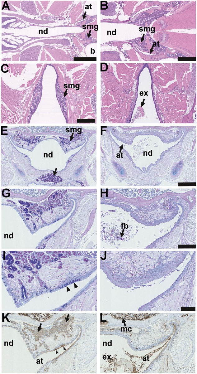
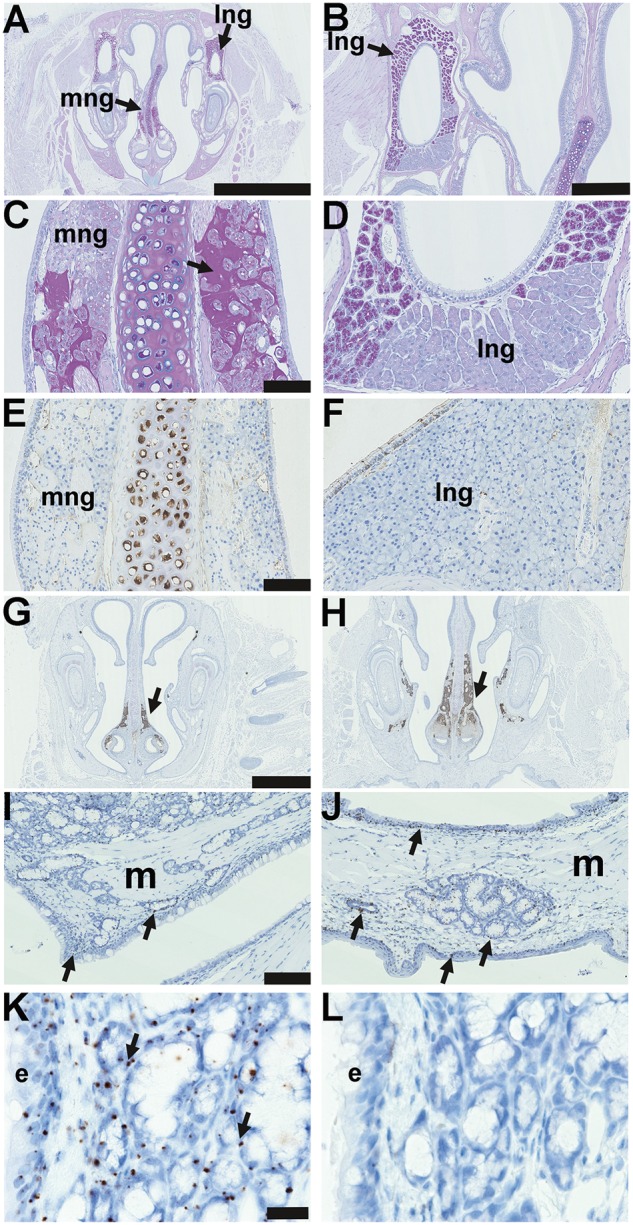
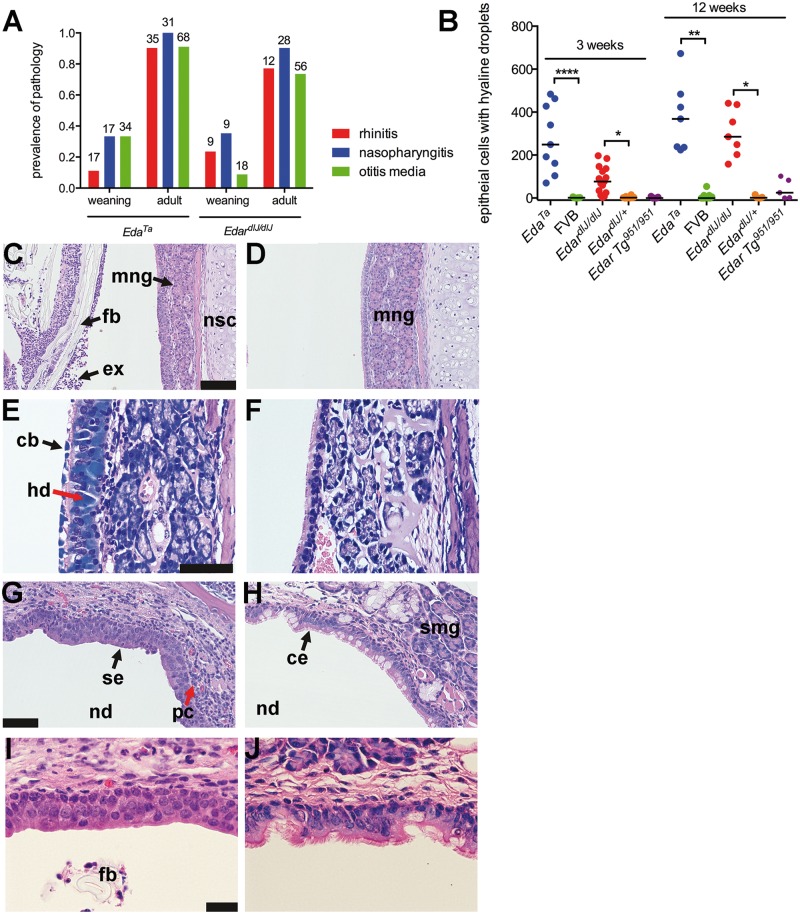
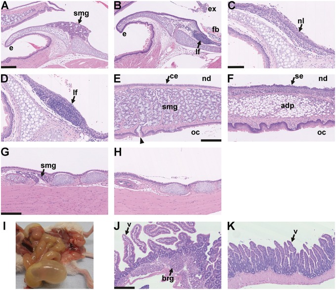
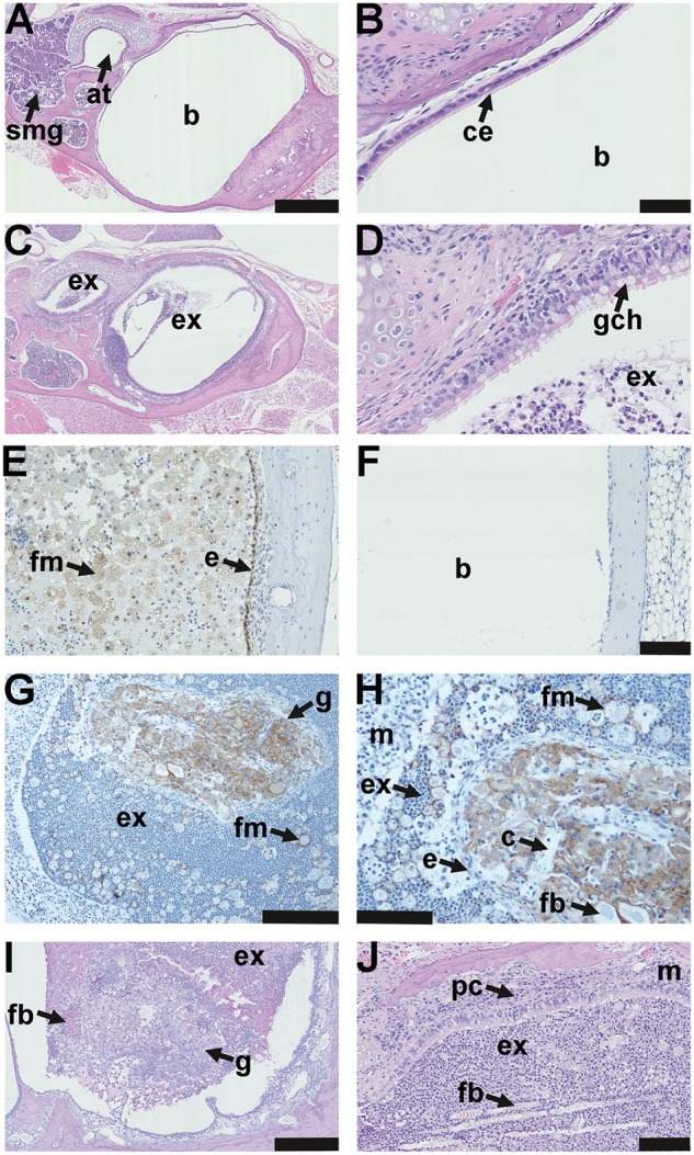
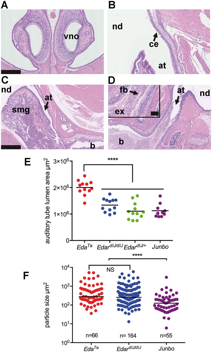
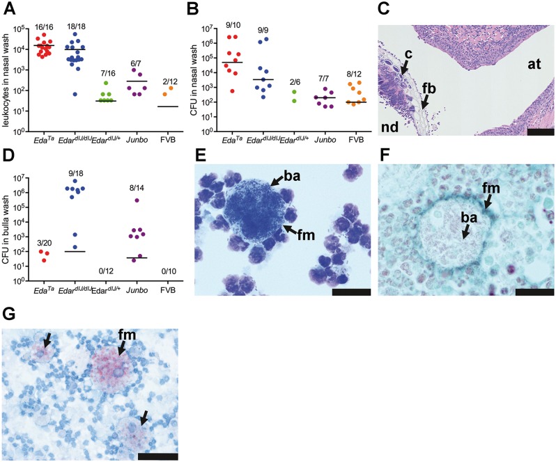
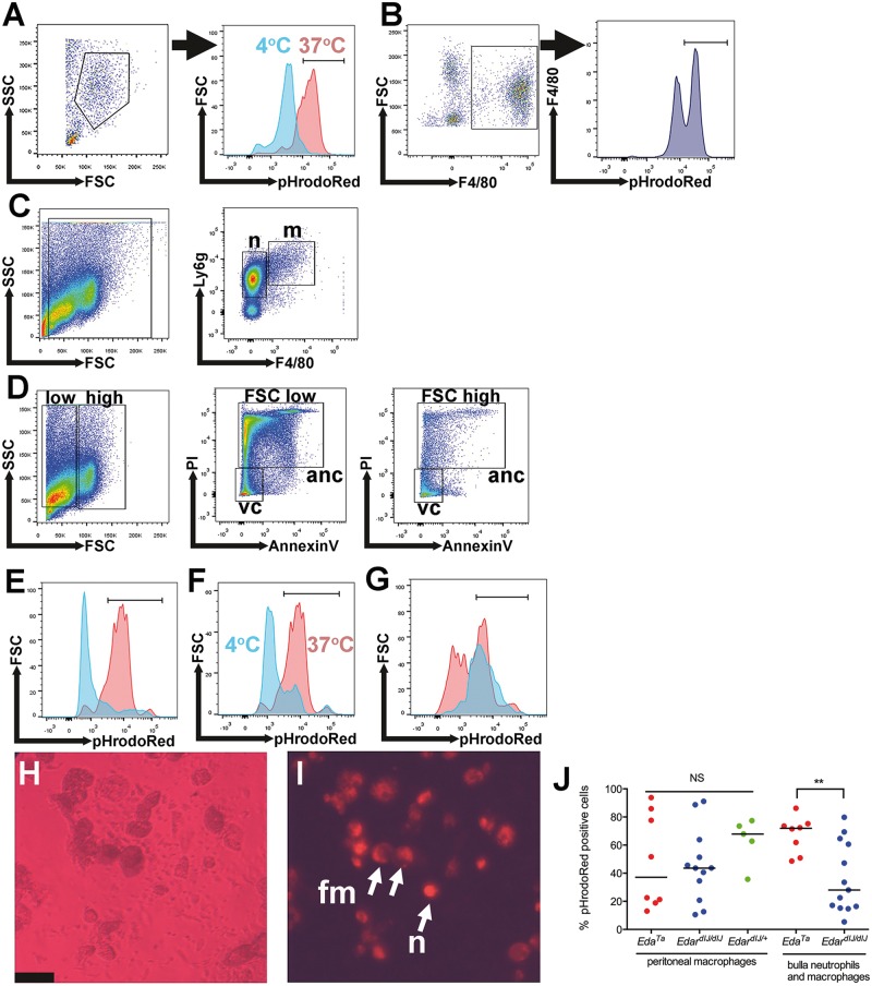
Hypohidrotic ectodermal dysplasia (HED) results from mutation of the EDA, EDAR or EDARADD genes and is characterized by reduced or absent eccrine sweat glands, hair follicles and teeth, and defective formation of salivary, mammary and craniofacial glands. Mouse models with HED also carry Eda, Edar or Edaradd mutations and have defects that map to the same structures. Patients with HED have ear, nose and throat disease, but this has not been investigated in mice bearing comparable genetic mutations. We report that otitis media, rhinitis and nasopharyngitis occur at high frequency in Eda and Edar mutant mice and explore the pathogenic mechanisms related to glandular function, microbial and immune parameters in these lines. Nasopharynx auditory tube glands fail to develop in HED mutant mice and the functional implications include loss of lysozyme secretion, reduced mucociliary clearance and overgrowth of nasal commensal bacteria accompanied by neutrophil exudation. Heavy nasopharynx foreign body load and loss of gland protection alters the auditory tube gating function and the auditory tubes can become pathologically dilated. Accumulation of large foreign body particles in the bulla stimulates granuloma formation. Analysis of immune cell populations and myeloid cell function shows no evidence of overt immune deficiency in HED mutant mice. Our findings using HED mutant mice as a model for the human condition support the idea that ear and nose pathology in HED patients arises as a result of nasal and nasopharyngeal gland deficits, reduced mucociliary clearance and impaired auditory tube gating function underlies the pathological sequelae in the bulla.


Figures

Figure 1.
Figure 1.
Anatomy and histology of the nasopharynx in EDA pathway deleted mice. (A–D) Dorsal plane sections, (E-L) coronal sections. (A) FVB wild-type controls, EdardlJ/+ and Edar Tg951/951 mice have submucosal glands (smg) located in caudal region of the nasopharynx duct (nd) associated with the auditory tubes (at) that connect the nasopharynx duct to the bulla (b); (B) is at higher magnification of this region. (C) Small nasopharynx submucosal glands (smg) are located caudal to the opening of the auditory tubes, the example shown is a 16-week-old EdardlJ/+ mouse; (D) caudal nasopharynx glands are absent EdardlJ/dlJ and EdaTa mice; the example shown is a 12-week-old EdaTa mouse. (E) Coronal section of the nasopharynx in a 37-week-old FVB mouse shows auditory tube submucosal glands (smg) and submucosal glands in the soft palate (arrow); (G,I) auditory tube submucosal glands shown at higher magnifications have serous cells and mucous cell populations, and the auditory tubes have Alcian Blue positive goblet cells (arrowheads); stained with PAS Alcian Blue. (F,H,J) Comparable sections in a 27-week-old EdaTa mouse lacking submucosal glands and auditory tube goblet cells: note foreign body (fb) in the nasopharynx duct. (K) In the FVB mouse the submucosal gland serous gland cell population (arrows), and the auditory tube epithelium secretory cells (arrowheads) stain positively for lysozyme. (L) In the EdaTa mouse, lysozyme positive myeloid cells (mc) in the bone marrow cavity and auditory tube lumen. Scale bars: 2.5 mm (A); 1.0 mm (B); 500 µm (C,D,E,F); 250 µm (G,H,K,L); 100 µm (I,J).
Figure 2.
Figure 2.
Nasal glands in EDA pathway deleted mice and Edar expression in adult glands. (A–L) are coronal sections. (A) Submucosal glands in the mid region of the nose of a 37-week-old FVB mouse, medial nasal glands (mng) are located in the mucosa either side of the nasal septum; and lateral nasal glands (lng) are located in the lateral walls of the nasal chambers; PAS Alcian Blue. (B) Higher magnification of the lateral nasal gland. (C) Higher magnification of the medial nasal gland and both nasal glands are comprised solely of serous cells; note that PAS-positive amorphous amyloid material (arrow) between submucosal gland acini is an incidental degenerative change in the medial nasal glands of older mice: see reference [13]. (D) Higher magnification of the lateral nasal glands shows a ventral area stains weakly with PAS. (E) Medial nasal gland and (F) lateral nasal gland in the FVB mouse do not stain for lysozyme; note that the nasal septum cartilage chondrocytes are lysozyme positive. Medial nasal glands in the rostral (G) and (H) mid nose regions in FVB and EdardlJ/dlJmice stain positively for cleaved caspase 3 (arrows), example shown in 3-week-old FVB mouse. (I-K) In situ hybridization detecting Edar mRNA in 3-week-old Edar Tg951/951. (I) Nasopharynx and (J) soft palate gives punctate signals (arrows) in the nasopharynx and soft palate submucosal glands and duct, nasopharynx and auditory tube ciliated epithelium, and squamous epithelium of the oral cavity. There are no hybridization signals in muscle (m). (K) Higher magnification of the Edar signals in auditory tube submucosal gland and epithelium (e). (L) There are no hybridization signals using the negative control probe DapB. Scale bars: 2.5 mm (A); 500 µm (B); 500 µm (C,D,E,F); 100 µm (I,J); 20 µm (K,L).
Figure 3.
Figure 3.
Pathology of the nose and nasopharynx in EDA pathway deleted mice. (A) Rhinitis, nasopharyngitis and otitis media increases in prevalence between weaning age (3 weeks) and adulthood in EdardlJ/dlJ(7–17-weeks old) and EdaTa (12–43-weeks old) mice: there was no evidence of this pathology in EdardlJ/+ (n = 45), Edar Tg951/951 (n = 23) or FVB (n = 36) mice. The number above each histogram bar indicates the number of tissues examined. (B) The number of nasal epithelial cells with hyaline droplets in rostral, mid and nasopharynx duct sections is higher in EdardlJ/dlJ and EdaTa mice compared to FVB and EdardlJ/+ respectively at 3-weeks (weaning) and 12-weeks of age. The graph represents data points and the median (that incudes zero values) as a bar; two-tailed Kruskal Wallis test; *P <0.05, **P <0.01, ****P< 0.001 Dunn’s multiple comparison test. (C) The rostral nasal passages of a 21-day-old EdaTa mouse with rhinitis contain foreign bodies (fb hair shaft fragments and plant material) mixed with neutrophil rich exudate (ex); medial submucosal gland (mng) and nasal septum cartilage (nsc). (D) Comparable image of 22-day-old FVB mouse shows no evidence of intraluminal exudate. (E) Ciliated epithelial cells with intracytoplasmic hyaline droplets (hd) and cytoplasmic blebbing (cb) in the nasal septum of a 12-week-old EdaTa mouse; Giemsa stain. (F) Comparable section in a 12-week-old FVB mouse shows normal respiratory epithelium without hyaline droplets. (G) Nasopharyngitis in 21-day-old EdardlJ/dlJ mouse with squamous epithelium (se) lining the roof of the nasopharynx (nd) and infiltration of the submucosa with lymphocytes and plasma cells (pc); (I) shows higher magnification of the squamous epithelium. (H) The nasopharynx of 21-day-old FVB mouse has a ciliated respiratory epithelium (ce) with goblet cells, and auditory tube submucosal glands (smg); (J) shows a higher magnification of the ciliated epithelium. Scale bars 100 µm (C,D,G,H); 50 µm (E,F); 20 µm (I,J).
Figure 8.
Figure 8.
Larynx histology and aerophagia in EDA pathway deleted mice A–D larynx mid-sagittal plane. (A) The epiglottis (e) of a 24-week-old FVB mouse has submucosal glands (smg) but these are absent in EdardlJ/dlJ and EdaTa mice. (B) The epiglottis of 14-week-old EdardlJ/dlJ mouse lacks glands and has a submucosal lymphoid follicle (lf). There is exudate (ex) and foreign body (fb) in the proximal trachea. (C,D) Two 25-week-old mice EdaTa mice. (C) One EdaTa mouse has neutrophil (nl) infiltration of the epiglottis epithelium. (D) a higher magnification image of a submucosal lymphoid follicle (lf) in a second EdaTa mouse. (E,F) Soft palate mid-sagittal plane. (E) The soft palate of a 24-week-old FVB mouse has submucosal gland comprising mucous cells (smg) that empty via ducts (arrow head) into the oral cavity (oc); nasopharynx (nd) is lined by ciliated epithelium (ce). (F) The soft palate of a 14-week-old EdardlJ/dlJ mouse lacks submucosal glands and this region is occupied by adipose tissue (adp); the nasopharynx is lined by squamous epithelium (se). (G,H) Trachea mid-sagittal plane. (G) The proximal trachea of a 14-week-old EdardlJ/+ mouse has submucosal glands (smg); (H) these glands are absent in a 25-week-old EdaTa mouse. (I) Post mortem appearance of an 18-week-old EdaTa mouse with aerophagia that has an air filled stomach, small intestines and caecum. (J,K) small intestine. (J) Duodenum of this EdaTa mouse has a normal submucosal Brunner’s gland (brg) and villi (v); (K) the jejunum has an empty lumen. Scale bars: 250 µm (A,B,E–H,J,K); 100 µm (C,D).
Figure 4.
Figure 4.
Pathology of the auditory bulla in EDA pathway deleted mice. (A) The healthy air filled bulla (b) and auditory tube (at) in a 37-week-old FVB mouse; note auditory tube submucosal glands (smg). (B) Higher magnification image (A) shows the bulla mucosa in the region adjacent to the auditory tube opening has ciliated epithelium (ce). (C) A 43-week-old EdaTa mouse with otitis media has exudate (ex) in the bulla lumen and auditory tube and (D) epithelium adjacent to the auditory tube opening, has goblet cell hyperplasia (gch). (E, F) In vivo labelling with the hypoxia tracer pimonidazole shows positive staining in epithelium (e) and bulla foamy macrophages (fm) in a representative 16-week-old EdaTa mouse. (F) Hypoxia signals are absent in the healthy air-filled bulla of a representative 8-week-old FVB control mouse. (G, H) The caudal bulla lumen of an 11-week-old EdardlJ/dlJ mouse has an organized vascular granuloma (g) surrounded by exudate (ex) containing neutrophils and foamy macrophages (fm); bulla mucosa (m). (H) Higher magnification of image (G) shows the granuloma has an epithelial margin (e), embedded foreign body (fb) and capillaries (c). Macrophages in the granuloma and foamy macrophages (fm) in bulla exudate stain positively for F4/80. (I) A granuloma located in the caudal bulla of a 14-week-old EdaTa mouse has PAS positive (plant-based) foreign body particle (fb) at the margin. (J) Hair shaft fragments (fb) in the neutrophil rich exudate (ex) of a 43-week-old EdaTa mouse; note plasma cell (pc) infiltration of the thickened bulla mucosa (m). Scale bars: 500 µm (A,C); 50 µm (B,D); 100 µm (E,F,H,J), 200 µm (G) 250 µm (I).
Figure 5.
Figure 5.
Auditory tube gating function is compromised in the absence of submucosal glands. (A) Coronal section of the rostral nose in a Junbo mouse has normal air filled nasal passages; note vomeronasal organ (vno). (B–D) Dorsal plane sections. (B) The opening of the auditory tube (at) into the nasopharynx (nd) in a Junbo mouse shows the normal mucosa of the nasopharynx is lined by ciliated epithelium (ce). (C,D) Nasopharynx (nd), auditory tubes (at) and bullae (b). (C) An auditory tube in a 45-week-old Junbo mouse with a slender profile and empty lumen; note submucosal glands (smg). (D) The auditory tube of a 13-week-old EdaTa mouse contains exudate (ex) and hair shaft foreign body material (fb) (inset shows higher magnification of the tube contents). (E) Auditory tube lumen area is significantly greater in EdaTa (n = 6 mice, 13-weeks-old) than in EdardlJ/dlJ(n = 6 mice, 16-weeks-old), EdardlJ/+ (n = 6 mice, 15-16 weeks-old) and Junbo (n = 5 mice, 24–45 weeks-old). Total area for each auditory tube was measured in 50 µm step sections, and data are represented as points with the average as a bar; one-way ANOVA and Tukey’s multiple comparison test; **** P <0.0001. (F) Bulla foreign body particle size in EdardlJ/dlJ and EdaTa mice is significantly larger than in Junbo mice. Particle sizes were measured in the bullae of EdardlJ/dlJ (n = 11 mice, 7-17 weeks old), EdaTa (n = 10, 13–30-weeks old) and Junbo (n = 9, 14–45-weeks old) mice. The graph represents data points and the median as a bar; 2-tailed Kruskal Wallis test; ****P <0.0001 and NS not significant in Dunn’s multiple comparison test. Scale bars: 250 µm (A,B); 500 µm (C,D); 100 µm (panel D inset).
Figure 6.
Figure 6.
Commensal bacteria populations and inflammatory response in the nose and auditory bulla of EDA pathway deleted mice. (A) Leukocyte numbers in the nasal wash of EdardlJ/dlJ and EdaTa mice are significantly elevated (see text for details) compared to FVB, EdardlJ/+ and Junbo mice. (B) Total bacterial load (CFU) in nasal samples are significantly elevated (see text for details) in EdardlJ/dlJ (predominately E. coli) and EdaTa (predominately Staphylococcus aureus) mice than FVB, EdardlJ/+ and Junbo mice. (C) Cocci colonies (c) associated with plant foreign body (fb) in the nasopharynx (nd) at the opening of the auditory tube (at) of a 43-week-old EdaTa mouse. (D) Total bacterial load in bulla samples was highest in EdardlJ/dlJ (predominately E. coli) and Junbo (mixed bacterial isolates, see Supplementary material, Table S1 for bacterial isolate list). (E) Giemsa stained cytology of bulla exudate from an E. coli culture positive 11-week-old EdardlJ/dlJmouse shows a foamy macrophage with bacilli (ba); (F) a histology section shows the Gram-negative bacilli (ba) are intracytoplasmic; F4/80 IHC (Vector SG detection kit has a blue-grey chromogen) and Neutral Red counterstain. (G) Bulla foamy macrophages from a 13-week-old EdaTa mouse contain lipid bodies; formalin fixed cytology preparation stained with Oil Red O. (A,B,D) The graphs represent data points, the median (that incudes zero values) as a bar, and the ratio above each represents the number of positive samples divided by the total number sampled. Scale bars: 100 µm (C); 20 µm (E,F); 50 µm (G).
Figure 7.
Figure 7.
Immune cell populations and function in EDA pathway deleted mice. (A,B) Peritoneal macrophages. (A) A Forward Scatter (FSC) Side Scatter (SCS) plot shows the selected peritoneal macrophage population from an EdardlJ/+ mouse. The overlay histogram shows pHrodoRed fluorescence in the macrophage gate for a control sample incubated at 4 °C to inhibit phagocytosis (blue) and sample incubated at 37 °C (red). (B) FSCHigh F4/80High gated peritoneal macrophages analysed for pHrodoRed fluorescent signals. (C–G) Bulla fluid leukocytes from EdardlJ/dlJ mice. (C) FSC SSC selected leukocytes were analysed in a F4/80 Ly6g plot and 85.6% of the leukocytes were Ly6gHigh F4/80Low neutrophils (n) (median 95% CI 71.7–94.6%) and 2.1% were Ly6gHigh F4/80High macrophages (m) (95% CI, 1.2–3.5%). (D) FSC SSC plots were used to select two leukocyte populations. The FSCLow SSCLow gated leukocytes had 23.9% PI- AnnexinV- viable cells (v) (median 95% CI 11.1–52.3) and 74% PI + AnnexinV+ apoptotic or necrotic cells (anc) (95% CI 41.6–76.3%). The FSCHigh SSCHigh gated leukocytes had 86.1% PI- AnnexinV- viable cells (median, 95% CI, 84.0–97.0) and 11.6% PI + AnnexinV+ apoptotic or necrotic cells (95% CI 1.8–11.9%). (E–G) The overlay histograms for pHrodoRed fluorescence. (E) FSCLow SSCLow selected leukocytes (blue) have a low population of viable cells capable of phagocytosis, whereas FSCHigh SSCHigh selected leukocytes (red) have high population of viable cells capable of phagocytosis. (F) A FSCHigh SSCHigh selected PI- AnnexinV- viable leukocytes incubated at 37 °C show a phagocytosis signal (red) but incubation at 4 °C (blue) inhibits phagocytosis. (G) 56% of Ly6gHighF4/80Low neutrophils (95% CI 50-71%) (red) were phagocytically active and 72% of Ly6gHighF4/80High macrophages (95% CI, 59–93%) (blue) were phagocytically active. (H,I) EdaTa mouse bulla leukocytes; (H) phase contrast image of cells; (I) pHrodoRed fluorescent image of the same field of view, the smaller cells are the same size as neutrophils (n), and larger cells are macrophages and foamy macrophages (fm). The untreated control had no detectable autofluorescent signal. Scale bar 50 µm. (J) The percentage of pHrodoRed positive peritoneal macrophages was not significantly different in EdaTa, EdardlJ/dlJ, EdardlJ/+ mice (Kruskal Wallis test); whereas the percentage of positive bulla neutrophils and macrophages was significantly higher in EdaTa than EdardlJ/dlJmice. The graph represents data points and the median as a bar. NS P >0.05; **P <0.01.

References

    1. Sadier A., Viriot L., Pantalacci S., Laudet V. (2014) The ectodysplasin pathway: from diseases to adaptations. Trends Genet, 30, 24–31. - PubMed
    1. Majumder K., Shawlot W., Schuster G., Harrison W., Elder F.F., Overbeek P.A. (1998) YAC rescue of downless locus mutations in mice. Mamm. Genome., 9, 863–868. - PubMed
    1. Mou C., Thomason H.A., Willan P.M., Clowes C., Harris W.E., Drew C.F., Dixon J., Dixon M.J., Headon D.J. (2008) Enhanced ectodysplasin-A receptor (EDAR) signaling alters multiple fiber characteristics to produce the East Asian hair form. Hum. Mutat., 29, 1405–1411. - PubMed
    1. Chang S.H., Jobling S., Brennan K., Headon D.J. (2009) Enhanced Edar signalling has pleiotropic effects on craniofacial and cutaneous glands. PLoS, One, 4, e7591.. - PMC - PubMed
    1. Mehta U., Brunworth J., Lewis R.A., Sindwani R. (2007) Rhinologic manifestations of ectodermal dysplasia. Am. J. Rhinol., 21, 55–58. - PubMed