. 2016 Sep 1;25(17):3647-3653.
doi: 10.1093/hmg/ddw210. Epub 2016 Jul 4.

Dystrophin contains multiple independent membrane-binding domains

Affiliations

Dystrophin contains multiple independent membrane-binding domains

Junling Zhao et al. Hum Mol Genet. .

Abstract

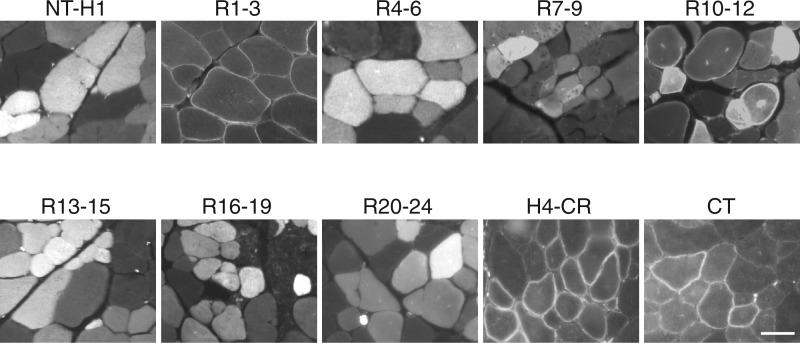
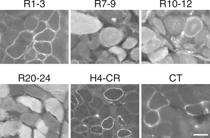
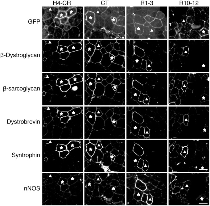
Dystrophin is a large sub-sarcolemmal protein. Its absence leads to Duchenne muscular dystrophy (DMD). Binding to the sarcolemma is essential for dystrophin to protect muscle from contraction-induced injury. It has long been thought that membrane binding of dystrophin depends on its cysteine-rich (CR) domain. Here, we provide in vivo evidence suggesting that dystrophin contains three additional membrane-binding domains including spectrin-like repeats (R)1-3, R10-12 and C-terminus (CT). To systematically study dystrophin membrane binding, we split full-length dystrophin into ten fragments and examined subcellular localizations of each fragment by adeno-associated virus-mediated gene transfer. In skeletal muscle, R1-3, CR domain and CT were exclusively localized at the sarcolemma. R10-12 showed both cytosolic and sarcolemmal localization. Importantly, the CR-independent membrane binding was conserved in murine and canine muscles. A critical function of the CR-mediated membrane interaction is the assembly of the dystrophin-associated glycoprotein complex (DGC). While R1-3 and R10-12 did not restore the DGC, surprisingly, CT alone was sufficient to establish the DGC at the sarcolemma. Additional studies suggest that R1-3 and CT also bind to the sarcolemma in the heart, though relatively weak. Taken together, our study provides the first conclusive in vivo evidence that dystrophin contains multiple independent membrane-binding domains. These structurally and functionally distinctive membrane-binding domains provide a molecular framework for dystrophin to function as a shock absorber and signaling hub. Our results not only shed critical light on dystrophin biology and DMD pathogenesis, but also provide a foundation for rationally engineering minimized dystrophins for DMD gene therapy.


Figures

Figure 1.
Figure 1.
Dystrophin R1-3, R10-12, CR and CT are independent membrane-binding domains. Full-length human dystrophin was split into ten subdomains and each subdomain fused with a GFP tag. The fusion proteins were individually expressed in mdx muscle by AAV gene transfer. Representative GFP photomicrographs of each indicated dystrophin subdomain are shown. Dystrophin R1-3, H4-CR and CT were exclusively localized at the sarcolemma. R10-12 was found at the sarcolemma and in the cytosol. NT-H1, R4-6, R7-9, R13-15, R16-19 and R20-24 were exclusively localized in the cytosol. Scale bar: 50 μm.
Figure 2.
Figure 2.
Microsomal western blot suggests the association of R1-3, R10-12, CR and CT with the sarcolemma. (A) Whole muscle lysate western blots revealing AAV-mediated expression of GFP-fused dystrophin subdomains in mdx muscle. (B) Detection of dystrophin R1-3, R10-12, H4-CR and CT in the membrane fraction by microsomal western blots. GAPDH marks the cytosolic fraction. (C) cytosolic fraction; M, membrane fraction.
Figure 3.
Figure 3.
Dystrophin R1-3, R10-12, H4-CR and CT bind to the sarcolemma in canine muscle. Indicated GFP fusion dystrophin subdomains were expressed in dystrophic dog muscle by AAV gene transfer. Representative GFP photomicrographs show the membrane binding of R1-3, R10-12, H4-CR and CT and cytosolic localization of R7-9 and R20-24. R10-12 is also seen in the cytosol. Scale bar: 50 μm.
Figure 4.
Figure 4.
Dystrophin CT restores the DGC at the sarcolemma. Representative serial section photomicrographs of GFP and immunostaining for β-dystroglycan, β-sarcoglycan, dystrobrevin and syntrophin in mdx muscle expressing the indicated GFP-dystrophin subdomain fusion proteins. Asterisk, the GFP-positive myofiber in serial sections; Triangle, the GFP-negative revertant fiber in serial sections. GFP signals co-localize with DGC components in myofibers transduced by the H4-CR and CT but not R1-3 and R10-12 subdomain AAV vectors. Scale bar: 50 μm.
Figure 5.
Figure 5.
Dystrophin R1-3, CR and CT bind to the sarcolemma in the heart. Indicated GFP fusion dystrophin subdomains were delivered to the mdx heart by systemic AAV injection. Uninjected BL10 and mdx hearts were used as negative controls. Subdomain H4-CR and CT showed membrane localization. Subdomain R1-3 was found in the intercalated disk and cytosol. Remaining subdomains were only seen in the cytosol. Scale bar: 50 μm.
Figure 6.
Figure 6.
A new model of dystrophin-sarcolemma interaction. (A) In muscle, dystrophin binds to the sarcolemma through four independent membrane-binding subdomains. (B) In the heart, dystrophin binds to the sarcolemma through three independent membrane-binding domains. These subdomains are marked by thick red lines.

References

    1. Straub V., Rafael J.A., Chamberlain J.S., Campbell K.P. (1997) Animal models for muscular dystrophy show different patterns of sarcolemmal disruption. J. Cell Biol., 139, 375–385. - PMC - PubMed
    1. Petrof B.J., Shrager J.B., Stedman H.H., Kelly A.M., Sweeney H.L. (1993) Dystrophin protects the sarcolemma from stresses developed during muscle contraction. Proc. Natl. Acad. Sci. U S A, 90, 3710–3714. - PMC - PubMed
    1. Hoffman E.P., Brown R.H.J., Kunkel L.M. (1987) Dystrophin: the protein product of the Duchenne muscular dystrophy locus. Cell, 51, 919–928. - PubMed
    1. Koenig M., Kunkel L.M. (1990) Detailed analysis of the repeat domain of dystrophin reveals four potential hinge segments that may confer flexibility. J. Biol. Chem., 265, 4560–4566. - PubMed
    1. Amann K.J., Renley B.A., Ervasti J.M. (1998) A cluster of basic repeats in the dystrophin rod domain binds F-actin through an electrostatic interaction. J Biol. Chem., 273, 28419–28423. - PubMed

Publication types