. 2016 Jun 16:9:44.
doi: 10.3389/fnmol.2016.00044. eCollection 2016.

Structural Studies of GABAA Receptor Binding Sites: Which Experimental Structure Tells us What?

Affiliations

Structural Studies of GABAA Receptor Binding Sites: Which Experimental Structure Tells us What?

Roshan Puthenkalam et al. Front Mol Neurosci. .

Abstract

Atomic resolution structures of cys-loop receptors, including one of a γ-aminobutyric acid type A receptor (GABAA receptor) subtype, allow amazing insights into the structural features and conformational changes that these pentameric ligand-gated ion channels (pLGICs) display. Here we present a comprehensive analysis of more than 30 cys-loop receptor structures of homologous proteins that revealed several allosteric binding sites not previously described in GABAA receptors. These novel binding sites were examined in GABAA receptor homology models and assessed as putative candidate sites for allosteric ligands. Four so far undescribed putative ligand binding sites were proposed for follow up studies based on their presence in the GABAA receptor homology models. A comprehensive analysis of conserved structural features in GABAA and glycine receptors (GlyRs), the glutamate gated ion channel, the bacterial homologs Erwinia chrysanthemi (ELIC) and Gloeobacter violaceus GLIC, and the serotonin type 3 (5-HT3) receptor was performed. The conserved features were integrated into a master alignment that led to improved homology models. The large fragment of the intracellular domain that is present in the structure of the 5-HT3 receptor was utilized to generate GABAA receptor models with a corresponding intracellular domain fragment. Results of mutational and photoaffinity ligand studies in GABAA receptors were analyzed in the light of the model structures. This led to an assignment of candidate ligands to two proposed novel pockets, candidate binding sites for furosemide and neurosteroids in the trans-membrane domain were identified. The homology models can serve as hypotheses generators, and some previously controversial structural interpretations of biochemical data can be resolved in the light of the presented multi-template approach to comparative modeling. Crystal and cryo-EM microscopic structures of the closest homologs that were solved in different conformational states provided important insights into structural rearrangements of binding sites during conformational transitions. The impact of structural variation and conformational motion on the shape of the investigated binding sites was analyzed. Rules for best template and alignment choice were obtained and can generally be applied to modeling of cys-loop receptors. Overall, we provide an updated structure based view of ligand binding sites present in GABAA receptors.

Keywords: GABAA receptors; allosteric modulatory sites; binding pockets; conformations; subtype selectivity.


Figures
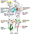
Figure 1
Figure 1
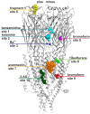
Ten small ligand binding sites are found in atomic structures of γ-aminobutyric acid type A receptor (GABAA receptor) homologs. The figure shows a side view of a superposition of the protein data bank (PDB) files shown in boldface in Table 1. Representative ligand positions were chosen for display of the 10 studied sites. Two subunits of 4COF are shown in ribbon representation (gray). The ligands are depicted in space-filling representation, and the orginal PDB files are specified in Table 1. More examples of ligands in these 10 site types are given in Table 1 and “Supplementary Figure 2”.
Figure 2
Figure 2
Conserved strand 8’ limits alignment choices in loop F region. The structural overlay of a single subunits’ extracellular domain (ECD) of all glycine receptor (GlyR) structures, all glutamate gated ion channel (GluCl) structures and the GABAA receptor structure 4COF reveals a strict conservation of the short strand 8’, which always starts with a hydrophobic residue. The image shows the sequence alignment of the loop F region which results from the 3D superposition, and the perfect overlay of the hydrophobic residues in the indicated representative structures. Thus, for aligning sequences of subunits with unknown structure to the resolved structures, the hydrophobic position alignment that is emphasized by the magenta box must be enforced. “Supplementary Figure 3A” (the master alignment) shows a possible alignment of the 19 GABAA receptor subunits in this region to the different structures to preserve strand 8’. Notably, Gloeobacter violaceus (GLIC) and Erwinia chrysanthemi (ELIC) also feature this conserved motif (see “Supplementary Figures 3D,E”).
Figure 3
Figure 3
3D superposition of 4PIR and 4COF and modeling of the GABAA receptor intact intracellular domain (ICD). (A) Superposition of 4PIR (cyan) and 4COF (red) shows high structural similarity between the distant homologs. (B) Magnified view of the trans-membrane domain (TMD)-ICD interface. Strictly conserved amino acids at the intracellular end of M3 and M4 superpose very well and are shown in stick representation, numbered according to 4COF (see also “Supplementary Figure 3B”). (C) Side view of a homology model of the β3+/α1− interface (α1: yellow, β3: red) of the GABAA receptor with the ICD based on the 4PIR structure. α1F385 localizes to the α minus side of the interface in the model and has contacts with the β3+ pre-MX loop, and with the M1-M2 linker of α1. This residue that has been implicated in propofol modulatory action thus likely localizes to the interface forming part of the ICD.
Figure 4
Figure 4
Multiple ligand binding sites at the extracellular domain (ECD)- interface. Left panel, 5AFJ: two different ligands occupy subsites 1 and 2 simultaneously in the nicotinic acetylcholine receptor (nAChR)- Acetylcholine binding protein (AChBP) chimera structure 5AFJ. Middle panel, 4F8H: subsite 2 is used by ketamine without a ligand in subsite 1 (which is a proton site in GLIC) in the GLIC structure 4F8H. Right panel, superposition: the color codes match the ligands for each structure in the overlay. Larger ligands like methyllycaconitine can use both sites simultaneously. However, most ligands are observed in subsite 1, as shown in this overlay of six structures that was generated with a secondary structure match superposition. Only fragment 1 in 5AFJ, ketamine in 4F8H and a part of the methyllycaconitine molecule in 2BYR occupy the site 2. Approximately 20 structures with ligands in the ECD interface were screened, and no other instances of site 2 usage were found in this sample (data not shown).
Figure 5
Figure 5
(A) Top and side views of the TMD of a GABAA receptor model (α6 subunit red, other subunits light pink) superposed with a single subunit of the bromoform bound GLIC F14’A 4HFD structure (green). The pocket volume in 4HFD is occupied by three bromoform molecules (dark cyan volume) in the GLIC structure at an intra-subunit site mainly formed by M1 and M2. The homologous pocket volume near α6I228 can accommodate a furosemide molecule (gray volume). The side view is rendered from the outside of the channel between M1 and M4. The viewing perspective is indicated in the left image with an arrow. (B) Localization of amino acids implicated in neurosteroid action relative to sites 9 and 10. The left panel displays a superposition of a single subunit’s TMD of the bromoform bound ELIC structure 3ZKR (gray) with a homology model of a GABAA receptor α1 subunit based on 4TNW (yellow). Only a single Br atom of the bromoform has been identified in the crystal structure, it is localized in a groove-like pocket between M1 and M4 on the minus side of the TMD and displayed here as a magenta space-filling atom. Among the binding site forming residues in GLIC is W220, the homologous residue in the GABAA receptor α1 subunit is Q241. The right panel depicts amino acids implicated in actions of neurosteroids at or near β+/α− interfaces along the whole length of the TMD (Hosie et al., ; Bracamontes et al., ; Chen et al., 2012).
Figure 6
Figure 6
Binding sites in GABAA receptor homology models based on structural evidence from homologous proteins. The image shows a homology model based on 4COF (ECD, TMD) and 4PIR (ICD) with one representative ligand per representative site: benzamidine in site 1 and ketamine in site 2 (cyan), Ba2+ in site 3 (dark blue), bromoform in site 5 (right subunit, magenta), fragment 1 in site 6 (left subunit, yellow), avermectin in site 7 (orange), desflurane in site 8 (green), bromoform in site 9 (right subunit, red), 2-arachidonglycerol (2-AG) in site 10 (left subunit, dark green). Note that each of the sites could occur in five subunits or at five interfaces, respectively, but may display ligand specificity. Some sites may not exist in all subunits or at all interfaces. Amino acids putatively forming these sites are color coded in the alignment shown in “Supplementary Figure 3A”.
Figure 7
Figure 7
Impact of protein motion on the ECD and TMD interface pockets. (A) ECD binding site forming segments in models based on the indicated six structures. The left panel shows the very close structural overlap that results from a superposition of the ECDs of single chains. The plus side forming segments A, B and C are shown from two different perspectives, the minus side is depicted once. The color codes match the PDB IDs. The right panel shows the distinctly interface geometries in the pentameric structures. The 4COF based interface (gray) is compared with models based on agonist (3JAE) and antagonist (3JAD) bound GlyR template structures (blue/ cyan/ gray), as well as with apo- (4TNV) and differently ligated GluCl structures (red/orange/brown). Every interface has a unique geometry due to the near rigid-body like movements which the ECD and TMD perform relative to one another during conformational changes, see also “Supplementary Figures 7, 8”. (B) The TMD interface in two different protein conformations contains different pocket forming residues. The left panel shows a superposition of 4COF with a β3 homopentamer model based on 4TNW, only the TMD is displayed. Two complete TMDs are displayed in different hues of red-brown, the plus side subunit is darker. Of the other three subunits, only M2 is displayed in gray for orientation relative to the pore. The four amino acids that were used to quantify differences of interface geometry (see “Supplementary Figure 9”) are shown in stick rendering. The other two panels display 4COF and the 4TNW based model viewed individually from the same perspective as in the superposition, but the far end of the TMD is clipped for more clarity. In the 4COF structure β3H267 is localized in a remote position to the subunit interface. A similar position is also observed in models based on the GluCl structure 3RIF. The 4TNW based β+/β− interface features β3H267 on the minus side of the interface vis a vis from β3N265, consistent with both residues being part of a single TMD-interface pocket. The position of β3H267 relative to the interface is strikingly different in the two protein conformations.

References

    1. Althoff T., Hibbs R. E., Banerjee S., Gouaux E. (2014). X-ray structures of GluCl in apo states reveal a gating mechanism of Cys-loop receptors. Nature 512, 333–337. 10.1038/nature13669 - DOI - PMC - PubMed
    1. Alvarez L. D., Estrin D. A. (2015). Exploring the molecular basis of neurosteroid binding to the β3 homopentameric GABAA receptor. J. Steroid Biochem. Mol. Biol. 154, 159–167. 10.1016/j.jsbmb.2015.07.012 - DOI - PubMed
    1. Antkowiak B. (2015). Closing the gap between the molecular and systemic actions of anesthetic agents. Adv. Pharmacol. 72, 229–262. 10.1016/bs.apha.2014.10.009 - DOI - PubMed
    1. Araujo F., Ruano D., Vitorica J. (1999). Native γ-aminobutyric acid type A receptors from rat hippocampus, containing both α 1 and α 5 subunits, exhibit a single benzodiazepine binding site with α 5 pharmacological properties. J. Pharmacol. Exp. Ther. 290, 989–997. Available online at: http://www.ncbi.nlm.nih.gov/pubmed/10454469 - PubMed
    1. Barrera N. P., Betts J., You H., Henderson R. M., Martin I. L., Dunn S. M. J., et al. . (2008). Atomic force microscopy reveals the stoichiometry and subunit arrangement of the α4β3δ GABAA receptor. Mol. Pharmacol. 73, 960–967. 10.1124/mol.107.042481 - DOI - PubMed

LinkOut - more resources