

Mild Nutrient Starvation Triggers the Development of a Small-Cell Survival Morphotype in Mycobacteria
- PMID: 27379076
- PMCID: PMC4909757
- DOI: 10.3389/fmicb.2016.00947
Mild Nutrient Starvation Triggers the Development of a Small-Cell Survival Morphotype in Mycobacteria
Abstract
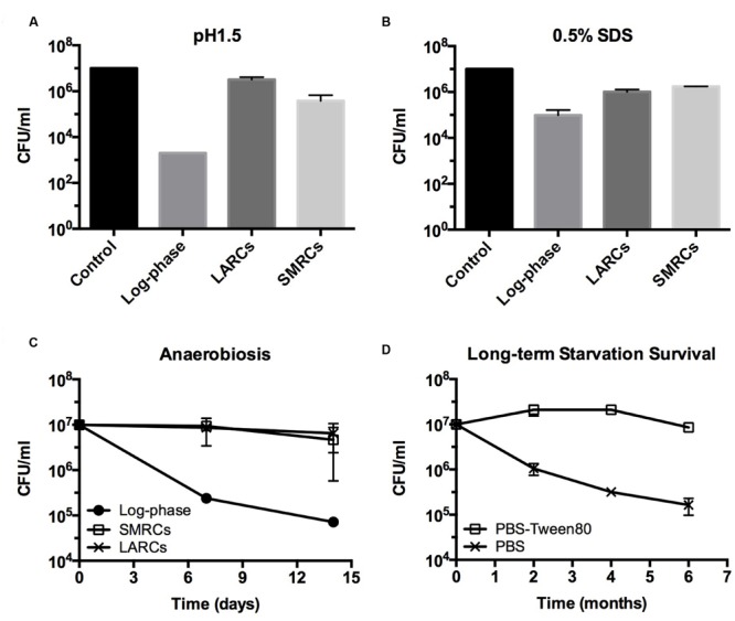
Mycobacteria, generally believed to be non-sporulating, are well known to survive shock starvation in saline for extended periods of time in a non-replicating state without any apparent morphological changes. Here, we uncover that mycobacteria can undergo cellular differentiation by exposing Mycobacterium smegmatis to mild starvation conditions. Traces of various carbon sources in saline triggered the development of a novel small resting cell (SMRC) morphotype. Development of SMRCs could also be observed for other mycobacteria, suggesting evolutionary conservation of this differentiation pathway. Fluorescence microscopic analyses showed that development of SMRCs progresses via septated, multi-nucleoided cell intermediates, which divide to generate mono-nucleoided SMRCs. Intriguingly, saline shock-starved large resting cells (LARCs), which did not show cell size or surface changes when observed by scanning electron microscopy, remodeled their internal structure to septated, multi-nucleoided cells, similar to the intermediates seen during differentiation to SMRCs. Our results suggest that mycobacteria harbor a starvation-induced differentiation program in which at first septated, multi-nucleoided cells are generated. Under zero-nutrient conditions bacteria terminate development at this stage as LARCs. In the presence of traces of a carbon source, these multi-nucleoided cells continue differentiation into mono-nucleoided SMRCs. Both SMRCs and LARCs exhibited extreme antibiotic tolerance. SMRCs showed increased long-term starvation survival, which was associated with the presence of lipid inclusion bodies.
Keywords: Mycobacterium smegmatis; bacterial differentiation; mycobacteria; non-replicating bacteria; quiescence; starvation.
Figures


Similar articles
-
Rel Is Required for Morphogenesis of Resting Cells in Mycobacterium smegmatis.Front Microbiol. 2016 Aug 31;7:1390. doi: 10.3389/fmicb.2016.01390. eCollection 2016. Front Microbiol. 2016. PMID: 27630635 Free PMC article.
-
Developmental transcriptome of resting cell formation in Mycobacterium smegmatis.BMC Genomics. 2016 Oct 26;17(1):837. doi: 10.1186/s12864-016-3190-4. BMC Genomics. 2016. PMID: 27784279 Free PMC article.
-
Novel Populations of Mycobacterium smegmatis Under Hypoxia and Starvation: Some Insights on Cell Viability and Morphological Changes.Microorganisms. 2024 Nov 10;12(11):2280. doi: 10.3390/microorganisms12112280. Microorganisms. 2024. PMID: 39597669 Free PMC article.
-
Alanine dehydrogenases in mycobacteria.J Microbiol. 2019 Feb;57(2):81-92. doi: 10.1007/s12275-019-8543-7. Epub 2019 Jan 31. J Microbiol. 2019. PMID: 30706339 Review.
-
Stable Regulation of Cell Cycle Events in Mycobacteria: Insights From Inherently Heterogeneous Bacterial Populations.Front Microbiol. 2018 Mar 21;9:514. doi: 10.3389/fmicb.2018.00514. eCollection 2018. Front Microbiol. 2018. PMID: 29619019 Free PMC article. Review.
Cited by
-
Measuring the Global Substrate Specificity of Mycobacterial Serine Hydrolases Using a Library of Fluorogenic Ester Substrates.ACS Infect Dis. 2018 Jun 8;4(6):904-911. doi: 10.1021/acsinfecdis.7b00263. Epub 2018 Apr 16. ACS Infect Dis. 2018. PMID: 29648787 Free PMC article.
-
Activity of combinations of bactericidal and bacteriostatic compounds in Mycobacterium abscessus-infected mice: an overview.Front Microbiol. 2025 Aug 1;16:1616149. doi: 10.3389/fmicb.2025.1616149. eCollection 2025. Front Microbiol. 2025. PMID: 40822395 Free PMC article. Review.
-
Pulmonary non-tuberculous mycobacterial infections: current state and future management.Eur J Clin Microbiol Infect Dis. 2020 May;39(5):799-826. doi: 10.1007/s10096-019-03771-0. Epub 2019 Dec 18. Eur J Clin Microbiol Infect Dis. 2020. PMID: 31853742 Free PMC article. Review.
-
Stress-Induced Reorganization of the Mycobacterial Membrane Domain.mBio. 2018 Jan 23;9(1):e01823-17. doi: 10.1128/mBio.01823-17. mBio. 2018. PMID: 29362232 Free PMC article.
-
Interfering With DNA Decondensation as a Strategy Against Mycobacteria.Front Microbiol. 2018 Sep 5;9:2034. doi: 10.3389/fmicb.2018.02034. eCollection 2018. Front Microbiol. 2018. PMID: 30233521 Free PMC article.
References
-
- Brennan P. J., Goren M. B. (1979). Structural studies on the type-specific antigens and lipids of the Mycobacterium avium. Mycobacterium intracellulare. Mycobacterium scrofulaceum serocomplex. Mycobacterium intracellulare serotype 9. J. Biol. Chem. 254 4205–4211. - PubMed
LinkOut - more resources
Full Text Sources
Other Literature Sources