Review
. 2016 Jun 14:7:234.
doi: 10.3389/fimmu.2016.00234. eCollection 2016.

Autoimmune Myopathies: Where Do We Stand?

Affiliations
Review

Autoimmune Myopathies: Where Do We Stand?

Jean-Philippe Simon et al. Front Immunol. .

Abstract

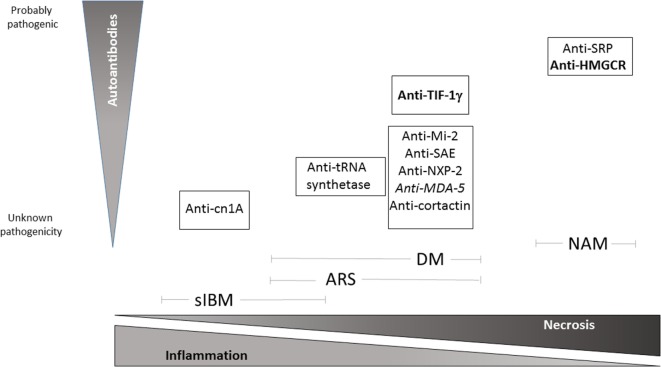
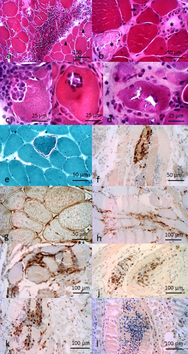
Autoimmune diseases (AIDs) as a whole represent a major health concern and remain a medical and scientific challenge. Some of them, such as multiple sclerosis or type 1 diabetes, have been actively investigated for many decades. Autoimmune myopathies (AIMs), also referred to as idiopathic inflammatory myopathies or myositis, represent a group of very severe AID for which we have a more limited pathophysiological knowledge. AIM encompass a group of, individually rare but collectively not so uncommon, diseases characterized by symmetrical proximal muscle weakness, increased serum muscle enzymes such as creatine kinase, myopathic changes on electromyography, and several typical histological patterns on muscle biopsy, including the presence of inflammatory cell infiltrates in muscle tissue. Importantly, some AIMs are strongly related to cancer. Here, we review the current knowledge on the most prevalent forms of AIM and, notably, the diagnostic contribution of autoantibodies.

Keywords: autoantibodies; dermatomyositis; inclusion-body myositis; myositis; necrotizing myopathy.


Figures

Figure 1
Figure 1
Schematic view of pathological pattern of inflammatory myopathies and proven degree of pathogenicity of specific autoantibodies relative to each IM subtypes. Bold: high risk of neoplasia, Italics: amyopathic DM. NAM, necrotizing autoimmune myopathy; DM, dermatomyositis; ARS, anti-t-rna synthetase syndrome; sIBM, sporadic inclusion-body myositis.
Figure 2
Figure 2
Pathological features of inflammatory myopathies. Pathological features of DM. (A) Perimysial and perivascular inflammatory cell infiltrate. Perifascicular myofiber atrophy (arrow heads). Pathological features of anti-HMGCR NAM. (B) Necrotic (asterisk) and regenerative (arrows) myofibers, sparsely infiltrate pathological features of sIBM. (C) Invaded fibers by inflammatory cells. (D) Rimmed vacuoles. (E) Ragged-red fiber. (F) P62 aggregates. (G) Immunohistochemichal evidence of MHC-I myofibers expression. (H) C5b–9 membrane attack complex in capillaries. Composition of the endomysial inflammatory infiltrate (I) CD4+ cells, (J) CD8+ cells, (K) CD68+ cells, and (L) rare CD20+ cells. All features provided by Dr. Jean-Philippe Simon.

References

    1. Bohan A, Peter JB. Polymyositis and dermatomyositis (first of two parts). N Engl J Med (1975) 292:344–7. 10.1056/NEJM197502132920706 - DOI - PubMed
    1. Griggs RC, Askanas V, DiMauro S, Engel A, Karpati G, Mendell JR, et al. Inclusion body myositis and myopathies. Ann Neurol (1995) 38:705–13. 10.1002/ana.410380504 - DOI - PubMed
    1. Benveniste O, Guiguet M, Freebody J, Dubourg O, Squier W, Maisonobe T, et al. Long-term observational study of sporadic inclusion body myositis. Brain (2011) 134:3176–84. 10.1093/brain/awr213 - DOI - PubMed
    1. Amato AA, Griggs RC. Unicorns, dragons, polymyositis, and other mythological beasts. Neurology (2003) 61:288–9. 10.1212/WNL.61.3.288 - DOI - PubMed
    1. van der Meulen MF, Bronner IM, Hoogendijk JE, Burger H, van Venrooij WJ, Voskuyl AE, et al. Polymyositis: an overdiagnosed entity. Neurology (2003) 61:316–21. 10.1212/WNL.61.3.316 - DOI - PubMed

LinkOut - more resources