. 2016 Jun 16:7:862.
doi: 10.3389/fpls.2016.00862. eCollection 2016.

The Aquaporin Splice Variant NbXIP1;1α Is Permeable to Boric Acid and Is Phosphorylated in the N-terminal Domain

Affiliations

The Aquaporin Splice Variant NbXIP1;1α Is Permeable to Boric Acid and Is Phosphorylated in the N-terminal Domain

Henry Ampah-Korsah et al. Front Plant Sci. .

Abstract

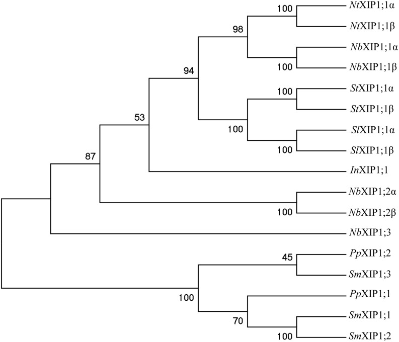
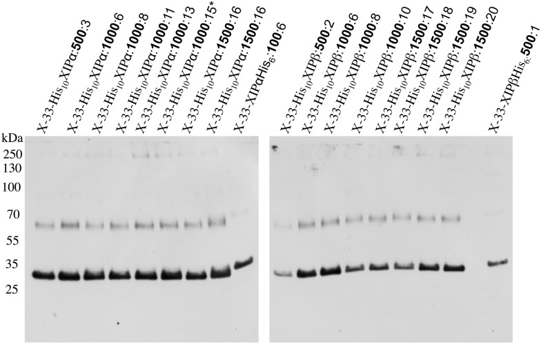
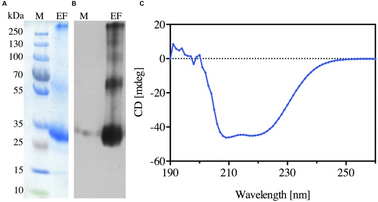
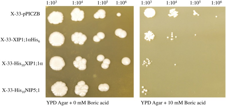
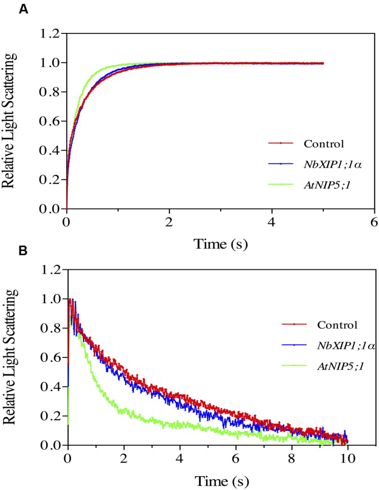
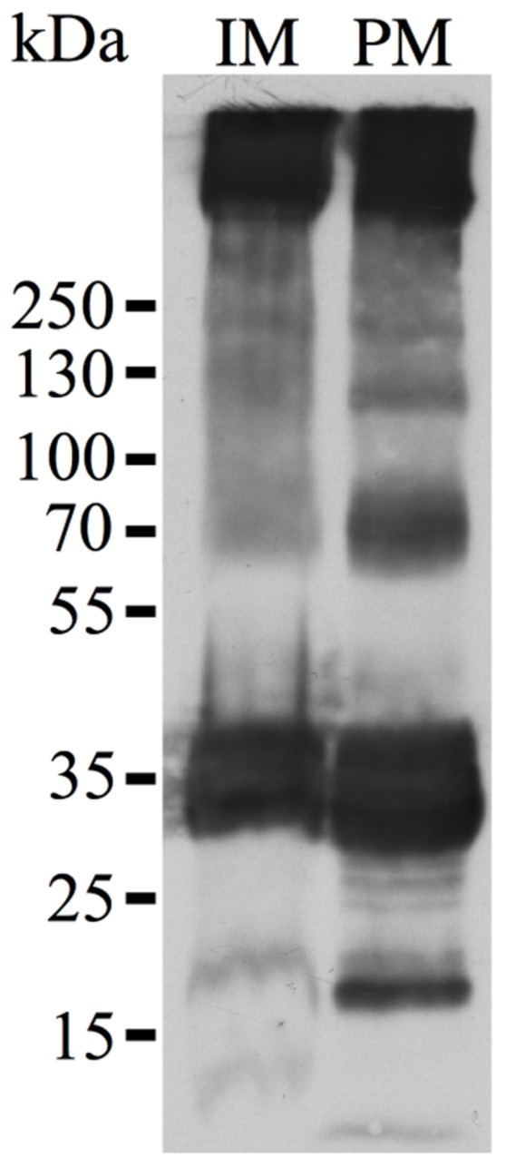
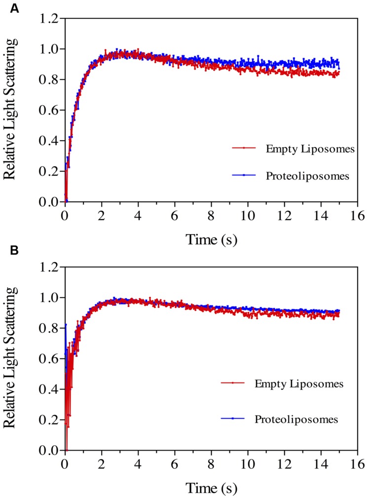
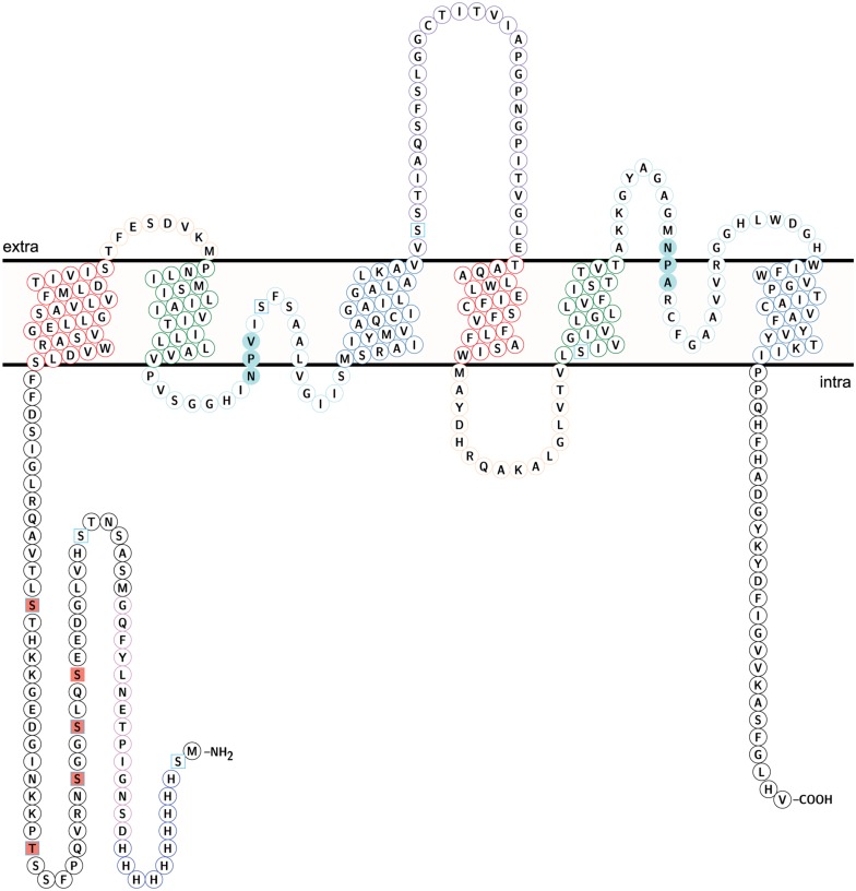
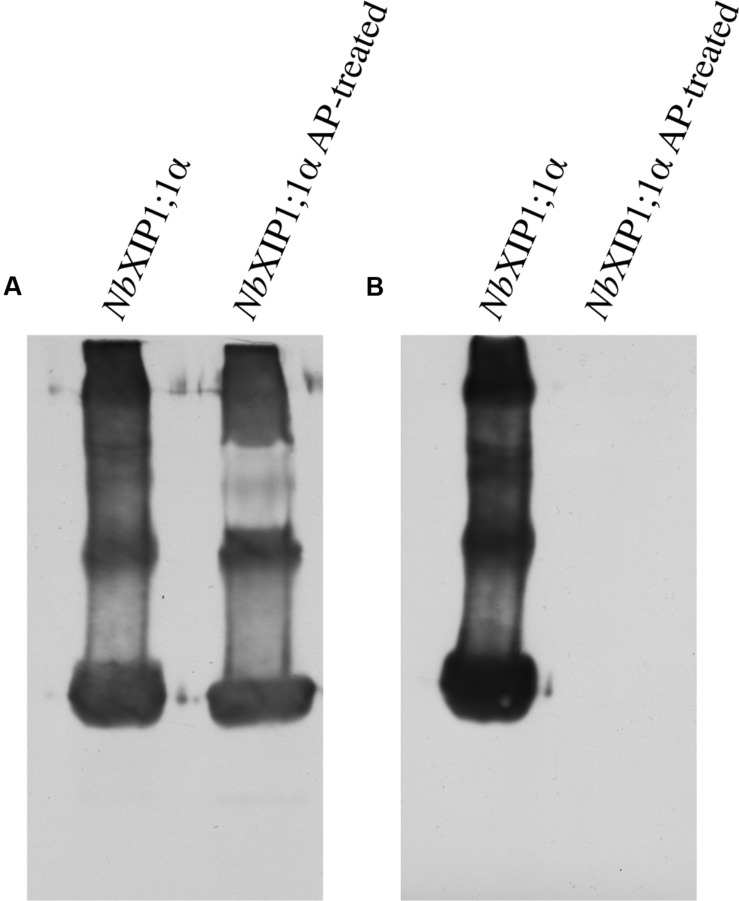
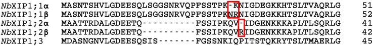
Aquaporins (AQPs) are membrane channel proteins that transport water and uncharged solutes across different membranes in organisms in all kingdoms of life. In plants, the AQPs can be divided into seven different subfamilies and five of these are present in higher plants. The most recently characterized of these subfamilies is the XIP subfamily, which is found in most dicots but not in monocots. In this article, we present data on two different splice variants (α and β) of NbXIP1;1 from Nicotiana benthamiana. We describe the heterologous expression of NbXIP1;1α and β in the yeast Pichia pastoris, the subcellular localization of the protein in this system and the purification of the NbXIP1;1α protein. Furthermore, we investigated the functionality and the substrate specificity of the protein by stopped-flow spectrometry in P. pastoris spheroplasts and with the protein reconstituted in proteoliposomes. The phosphorylation status of the protein and localization of the phosphorylated amino acids were verified by mass spectrometry. Our results show that NbXIP1;1α is located in the plasma membrane when expressed in P. pastoris, that it is not permeable to water but to boric acid and that the protein is phosphorylated at several amino acids in the N-terminal cytoplasmic domain of the protein. A growth assay showed that the yeast cells expressing the N-terminally His-tagged NbXIP1;1α were more sensitive to boric acid as compared to the cells expressing the C-terminally His-tagged isoform. This might suggest that the N-terminal His-tag functionally mimics the phosphorylation of the N-terminal domain and that the N-terminal domain is involved in gating of the channel.

Keywords: AQP; NIP; Nicotiana benthamiana; Pichia pastoris; XIP; boric acid; phosphorylation.


Figures

FIGURE 1
FIGURE 1
Phylogenetic tree of XIPs. The tree shows the phylogenetic relationship between XIPs in Nicotiana benthamiana, and selected XIPs of N. tabaccum, S. tuberosum, S. lycopersicum, and I. nil, when XIP sequences from P. patens and S. moellendorffii are used to root the tree. The numbers at the nodes represents the bootstrap support in %. The XIPs of N. benthamiana, N. tabaccum, S. tumberosum, S. lycopersicum, and I. nil represent a subset the XIP IV group of clade B, while P. patens and S. moellendorffii sequences belong to the basal XIP I group (Venkatesh et al., 2015).
FIGURE 2
FIGURE 2
Recombinant N-terminally His-tagged NbXIP1;1 expression in P. pastoris clones selected at different zeocin levels. Western blot showing the recombinant expression levels in crude cell extracts of representative X-33 N-terminally His-tagged NbXIP1;1 proteins in clones selected at 500 μg/mL, 1000 μg/mL or 1500 μg/mL (μg/mL zeocin resistance levels in bold). C-terminally His-tagged NbXIP1;1 clones were used as reference. The X-33His10XIP1;1α:1000:15 clone was used for large scale purification.
FIGURE 3
FIGURE 3
Purification of the recombinant N-terminally His-tagged NbXIP1;1α protein. (A) Coomassie stained SDS-PAGE gel showing the elution fraction (EF) from the Ni-NTA Agarose beads. (B) Western blot of the elution fraction (EF) showing the purified N-terminally His-tagged NbXIP1;1α protein. (C) Far-UV CD spectrum of NbXIP1;1α. The spectrum was measured at 22°C in 20 mM BTP-HCl pH 8.6, 100 mM NaCl, 12 mM N-nonyl-β-D-glucopyranoside and 2 mM β-mercaptoethanol.
FIGURE 4
FIGURE 4
Boric acid sensitivity of P. pastoris cells expressing NbXIP1;1α. To assess the functionality of the heterologously expressed NbXIP1;1α protein, a growth assay on plates containing 0 mM or 10 mM boric acid was performed. Induced cells, diluted to OD600 = 1, were spotted onto the plates in a serial dilution (indicated). X-33 clones transformed with either AtNIP5;1 or empty pPICZB were included as controls. Growth was recorded after 5 days at 28°C.
FIGURE 5
FIGURE 5
Substrate specific studies in P. pastoris spheroplasts by stopped-flow spectrometry. Stopped-flow traces showing kinetics of osmotic water permeation (A) and glycerol transport (B) in control spheroplasts (red), spheroplasts expressing NbXIP1;1α (blue) and spheroplasts expressing AtNIP5;1 (green). (A) The traces (mean of at least 10 traces) were fitted to single exponential equations. The rate constant and the standard error of estimate (SEE) values for the fitted curves were 2.77 ± 0.04 s–1 (control); 3.20 ± 0.03 s–1 (NbXIP1;1α) and 4.90 ± 0.04 s–1 (AtNIP5;1). (B) The result shows a fast increase in light scattering followed by a gradual decrease in light scattering corresponding to a fast vesicle shrinkage due to water efflux and a gradual vesicle swelling due to glycerol and water influx, respectively. The rate constant and SEE values for the fitted curves after fitting the traces after 0.39 s (mean of at least seven traces) to single exponential equations were 0.213 ± 0.004 s–1 (control); 0.301 ± 0.005 s–1 (NbXIP1;1α) and 0.742 ± 0.015 s–1 (AtNIP5;1).
FIGURE 6
FIGURE 6
Plasma membrane localization of NbXIP1;1α in P. pastoris. Western blot analysis of membrane fractions of P. pastoris cells expressing the N-terminally His-tagged NbXIP1;1α protein. In accordance with previous results using the anti-(His)4 antibody (Nordén et al., 2011), the blot shows a pattern that is characteristic of the expressed protein. IM, internal membranes fraction. PM, plasma membrane fraction. The fractions were diluted to the same volume and an equal volume was loaded in the two lanes.
FIGURE 7
FIGURE 7
Boric acid permeability in proteoliposomes. Stopped-flow traces showing kinetics of boric acid transport in phosphorylated (A) and dephosphorylated (B) NbXIP1;1α protein reconstituted in E. coli lipid vesicles supplemented with 20% cholesterol. Control liposomes (red) and proteoliposomes (blue). (A) The traces (mean of eight traces) were fitted to double exponential equations. The rate constant and SEE values for the fitted curves were control (R1 = 1.47 ± 0.03 s–1; R2 = 0.11 ± 0.01 s–1) and NbXIP1;1α (R1 = 1.62 ± 0.05 s–1; R2 = 0.20 ± 0.03 s–1). (B) The rate constant and SEE values for the fitted curves after fitting the traces (mean of 15 and 29 traces for control liposomes and proteoliposomes, respectively) to double exponential equations were control (R1 = 1.44 ± 0.07 s–1; R2 = 0.11 ± 0.04 s–1) and NbXIP1;1α (R1 = 1.02 ± 0.10 s–1; R2 = 0.26 ± 0.07 s–1).
FIGURE 8
FIGURE 8
Topology of NbXIP1;1α protein showing identified phosphorylation sites. Identified phospho sites – square salmon fill. Predicted phospho sites – square frame in sky blue. The coloring of helices and loops reflect a direct repeat in the gene/protein. Transmembrane helix (TM) 1 and TM 4 – frame in red. TM2 and TM 5 – frame in green. TM 3 and TM 6 – frame in steel blue. Loop A and Loop D – frame in peach. Loop B and loop E – frame in pale turquoise. Loop C – frame in medium purple. NPV/NPA box – pale turquoise fill. H3-H12 – Histidine tag in blue. D13-G26 – TEV protease cleavage site and linker in violet. The topology model was done in Protter (Omasits et al., 2014).
FIGURE 9
FIGURE 9
Phosphorylation in purified NbXIP1;1α protein. (A) Western blot showing purified NbXIP1;1α and purified NbXIP1;1α treated with alkaline phosphatase (AP) in 5 mM Tris-HCl, pH 7.9, 10 mM NaCl, 1 M MgCl2 and 0.1 mM DTT at 30°C for 20 h. Blot was probed with mouse anti-(His)4 and Horse Radish Peroxidase (HRP) – conjugated polyclonal goat anti-mouse IgG as primary and secondary antibodies, respectively. (B) Western blot showing purified NbXIP1;1α and purified NbXIP1;1α treated with AP as in (A) and probed with Zn2+-Phostag BTL-111-bound HRP-Streptavidin.
FIGURE 10
FIGURE 10
Close up of the splice sites in the amino acid sequences of NbXIP1 splice variants. Splice sites in NbXIP1;1 splice variants and NbXIP1;2 splice variants are in red boxes.

References

    1. Agemark M., Kowal J., Kukulski W., Nordén K., Gustavsson N., Johanson U., et al. (2012). Reconstitution of water channel function and 2D-crystallization of human aquaporin 8. Biochim. Biophys. Acta 1818 839–850. 10.1016/j.bbamem.2011.12.006 - DOI - PubMed
    1. Agre P., Kozono D. (2003). Aquaporin water channels: molecular mechanisms for human diseases. FEBS Lett. 555 72–78. 10.1016/s0014-5793(03)01083-4 - DOI - PubMed
    1. Anderberg H. I., Kjellbom P., Johanson U. (2012). Annotation of Selaginella moellendorffii major intrinsic proteins and the evolution of the protein family in terrestrial plants. Front. Plant Sci. 3:33 10.3389/fpls.2012.00033 - DOI - PMC - PubMed
    1. Ariani A., Gepts P. (2015). Genome-wide identification and characterization of aquaporin gene family in common bean (Phaseolus vulgaris L.). Mol. Genet. Genom. 290 1771–1785. 10.1007/s00438-015-1038-2 - DOI - PubMed
    1. Azad A. K., Katsuhara M., Sawa Y., Ishikawa T., Shibata H. (2008). Characterization of four plasma membrane aquaporins in tulip petals: a putative homolog is regulated by phosphorylation. Plant Cell Physiol. 49 1196–1208. 10.1093/pcp/pcn095 - DOI - PubMed

LinkOut - more resources