. 2016 Jul 5:14:54.
doi: 10.1186/s12915-016-0279-9.

Global identification of hnRNP A1 binding sites for SSO-based splicing modulation

Affiliations

Global identification of hnRNP A1 binding sites for SSO-based splicing modulation

Gitte H Bruun et al. BMC Biol. .

Abstract

Background: Many pathogenic genetic variants have been shown to disrupt mRNA splicing. Besides splice mutations in the well-conserved splice sites, mutations in splicing regulatory elements (SREs) may deregulate splicing and cause disease. A promising therapeutic approach is to compensate for this deregulation by blocking other SREs with splice-switching oligonucleotides (SSOs). However, the location and sequence of most SREs are not well known.

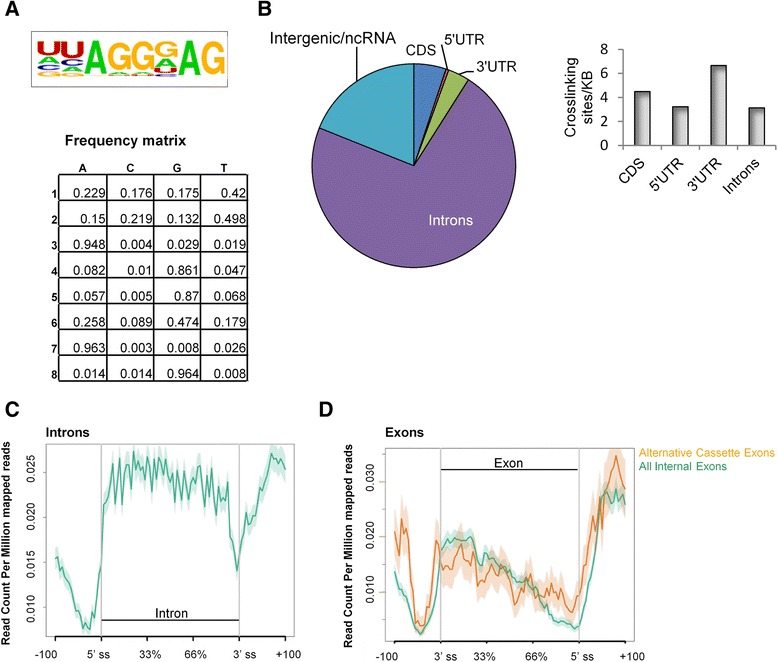
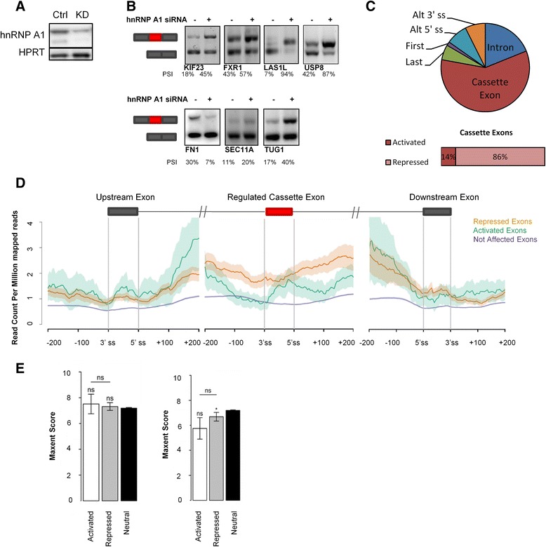
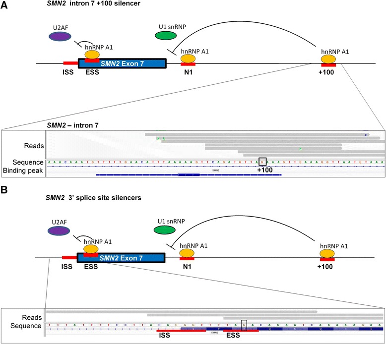
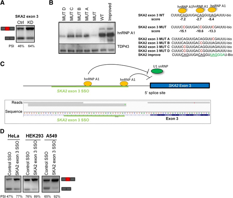
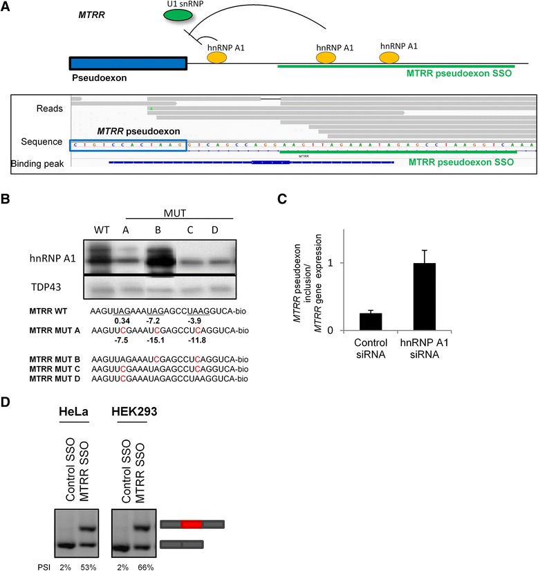
Results: Here, we used individual-nucleotide resolution crosslinking immunoprecipitation (iCLIP) to establish an in vivo binding map for the key splicing regulatory factor hnRNP A1 and to generate an hnRNP A1 consensus binding motif. We find that hnRNP A1 binding in proximal introns may be important for repressing exons. We show that inclusion of the alternative cassette exon 3 in SKA2 can be significantly increased by SSO-based treatment which blocks an iCLIP-identified hnRNP A1 binding site immediately downstream of the 5' splice site. Because pseudoexons are well suited as models for constitutive exons which have been inactivated by pathogenic mutations in SREs, we used a pseudoexon in MTRR as a model and showed that an iCLIP-identified hnRNP A1 binding site downstream of the 5' splice site can be blocked by SSOs to activate the exon.

Conclusions: The hnRNP A1 binding map can be used to identify potential targets for SSO-based therapy. Moreover, together with the hnRNP A1 consensus binding motif, the binding map may be used to predict whether disease-associated mutations and SNPs affect hnRNP A1 binding and eventually mRNA splicing.

Keywords: Alternative splicing; Cross-linking immunoprecipitation (CLIP); Pseudoexons; RNA-seq; Splicing silencer; Splicing splice-switching oligonucleotides (SSOs); Surface plasmon resonance imaging (SPRi); hnRNP A1; iCLIP.


Figures

Fig. 1
Fig. 1
hnRNP A1 iCLIP identified UAGG as the hnRNP A1 binding motif. a Consensus hnRNP A1 binding motifs were generated based on the identified hnRNP A1 binding peaks. The most highly enriched motif and the frequency matrix are shown. b The distribution of hnRNP A1 iCLIP crosslinking sites in different genomic regions (pie chart). hnRNP A1 iCLIP crosslinking sites are most prevalent in introns. However, when accounting for the relative size of each genomic region, there is an enrichment of hnRNP A1 crosslinking sites in the 3′ UTR (bar chart). CDS coding sequence. c The distribution of hnRNP A1 binding peaks across introns, including 100 bases of the adjacent exons. hnRNP A1 binds deep introns more than proximal introns close to exons. Shaded region corresponds to the 95 % confidence interval. d The distribution of hnRNP A1 binding peaks across exons including 100 bp of the adjacent introns. Internal exons (green) defined as all exons except the first and last exon, and cassette exons (orange) defined as alternative internal exons. hnRNP A1 binding peaks are highly enriched downstream of the 5′ splice site, more so for cassette exons than internal exons in general. Shaded region corresponds to the 95 % confidence interval
Fig. 2
Fig. 2
hnRNP A1 binds downstream of the 5′ splice site of repressed exons. a Validation of hnRNP A1 knockdown by western blotting. b RT-PCR validation of hnRNP A1-regulated splicing events identified by RNA sequencing of hnRNP A1-depleted HeLa cells. Representative of two replicates. PSI percent spliced in; semi-quantification of bands on agarose gels by ImageJ. c Types of alternative splicing events regulated by hnRNP A1 identified by hnRNP A1 knockdown and RNA sequencing. Most splicing events regulated by hnRNP A1 are splicing of cassette exons. Most of the regulated cassette exons are repressed by hnRNP A1. d Distribution of hnRNP A1 iCLIP tags in cassette exons activated (green) or repressed (orange) by hnRNP A1 and the upstream and downstream exons. The analysis was based on 18 activated exons and 110 repressed exons found by RNA sequencing of hnRNP A1-depleted HeLa cells (n = 3). The hnRNP A1 crosslinking site density is higher across cassette exons repressed by hnRNP A1 than cassette exons activated by hnRNP A1, suggesting that hnRNP A1 exon repression is a direct mechanism, while hnRNP A1 exon activation is an indirect mechanism. There is no difference between the distribution of hnRNP A1 iCLIP tags in upstream and downstream exons of hnRNP A1-activated and hnRNP A1-repressed exons. The region immediately downstream of the 5′ splice site seems to be important for hnRNP A1-mediated exon repression. The level of hnRNP A1 crosslinking sites in exons not affected by hnRNP A1 knockdown is shown in purple. Shaded region reflects the 95 % confidence interval. e Maxent score for 3′ splice sites and the 5′ splice sites of hnRNP A1-activated, -repressed, or neutral cassette exons. *p value < 0.05. Wilcoxon rank sum test, p = 0.02278. ns non-significant. Error bars are standard error of mean
Fig. 3
Fig. 3
hnRNP A1 iCLIP reads in the SMN2 gene pinpoint well-known hnRNP A1 binding sites. hnRNP A1 iCLIP identifies well-known hnRNP A1 binding sites in SMN2 exon 7. The intronic splicing silencer (ISS) located at the 3′ splice site. The SMN2-specific exon splicing silencer (ESS) in exon 7, the N1 intronic silencer, and the +100 intronic silencer. a hnRNP A1 iCLIP identifies an hnRNP A1 binding site in SMN2 at the hnRNP A1-dependent silencer in intron 7 + 100. The highly identical SMN1 and SMN2 genes differ at this position (framed: SMN1 + 100A, SMN2 + 100G); previous studies showed the +100G is necessary for strong splicing silencer activity. The iCLIP crosslinking site, i.e., the protein binding site, is assumed to be at the beginning of the sequencing reads (gray) when performing iCLIP. The hnRNP A1 binding peak is represented by a blue line. Only two hnRNP A1 reads were identified in SMN1 at this location (not shown). Screenshot from IGV. b hnRNP A1 iCLIP reads in SMN2 at the hnRNP A1-dependent silencer at the exon 7 3′ splice site (ISS) and at the hnRNP A1-dependent silencer generated by the C > T variation (framed) in SMN2 (ESS). No reads were found in SMN1 at this position (not shown). To ensure that the higher number of hnRNP A1 reads in SMN2 than in SMN1 was not due to higher gene expression, we performed SMN1 and SMN2-specific RT-qPCR (data not shown). This proved that the expression of SMN1 and SMN2 was similar in these cells, and thus was likely not the reason for the higher number of hnRNP A1 reads in SMN2
Fig. 4
Fig. 4
SSOs targeting an hnRNP A1 binding site downstream of SKA2 exon 3 improve exon inclusion. a RT-PCR of SKA2 exon 3 splicing in HeLa cells with (KD) or without (Ctrl) hnRNP A1 knockdown. hnRNP A1 knockdown increases SKA2 exon 3 inclusion. PSI percent spliced in; semi-quantification of bands on agarose gels by ImageJ. b Western blot of proteins purified by RNA-affinity chromatography of biotin-conjugated RNA oligonucleotides covering the three putative hnRNP A1 binding sites near the 5′ splice site of SKA2 exon 3. hnRNP A1 binding motifs are underscored, and mutations disrupting the hnRNP A1 binding motifs are shown in red. Introduction of the mutations reduces hnRNP A1 binding, while modifying one of the hnRNP A1 motifs to match our generated consensus binding motif (green) improves hnRNP A1 binding. The score of the putative hnRNP A1 motifs were calculated based on our generated scoring matrix (Additional file 1: Figure S1). Representative of two experiments. c hnRNP A1 iCLIP reads at the 5′ splice site of SKA2 exon 3. hnRNP A1 may bind downstream of the 5′ splice site to repress 5′ splice site recognition. The target site for the SKA2 exon 3 SSO is shown in green. The gene is on the antisense strand. Screenshot from IGV. d Transfection of SSOs targeting the hnRNP A1 binding site downstream of the 5′ splice site in SKA2 exon 3 into HeLa, HEK293, or A549 cells improves exon inclusion. The intensity of the bands was semi-quantified using ImageJ. PSI percent spliced in
Fig. 5
Fig. 5
SSOs targeting hnRNP A1 binding sites downstream of the MTRR pseudoexon improve pseudoexon inclusion. a Model of the hnRNP A1-mediated repression of the MTRR pseudoexon based on the hnRNP A1 iCLIP reads. The significant hnRNP A1 binding peak downstream of the 5′ splice site is indicated. hnRNP A1 may bind downstream of the 5′ splice site to repress splice site recognition by U1 snRNP. The target site for the MTRR SSO (green) covers three hnRNP A1 binding motifs. b Western blot with hnRNP A1 or as control TDP43 antibody of proteins purified by RNA-affinity chromatography of biotin-conjugated RNA oligonucleotides covering the downstream region of the MTRR 5′ splice site. Disruption of the hnRNP A1 binding motifs reduces hnRNP A1 binding. The motifs are scored using our generated scoring matrix (Additional file 1: Figure S1). The proximal hnRNP A1 motif is required for hnRNP A1 binding. hnRNP A1 binding motifs are underscored, and mutations disrupting the motifs are red. Representative of three experiments. c RT-qPCR analysis of the inclusion of the endogenous MTRR pseudoexon in control and hnRNP A1 knockdown HeLa cells. MTRR pseudoexon inclusion increases after hnRNP A1 knockdown. d RT-PCR of SSO-treated HeLa or Hek293 cells. SSO-mediated blocking of the hnRNP A1 binding sites near the MTRR pseudoexon improves endogenous MTRR pseudoexon inclusion. SSO transfections were done in duplicate and gel bands were semi-quantified using ImageJ. PSI percent spliced in

References

    1. Lim KH, Ferraris L, Filloux ME, Raphael BJ, Fairbrother WG. Using positional distribution to identify splicing elements and predict pre-mRNA processing defects in human genes. Proc Natl Acad Sci U S A. 2011;108(27):11093–8. doi: 10.1073/pnas.1101135108. - DOI - PMC - PubMed
    1. Sterne-Weiler T, Howard J, Mort M, Cooper DN, Sanford JR. Loss of exon identity is a common mechanism of human inherited disease. Genome Res. 2011;21(10):1563–71. doi: 10.1101/gr.118638.110. - DOI - PMC - PubMed
    1. Lorson CL, Hahnen E, Androphy EJ, Wirth B. A single nucleotide in the SMN gene regulates splicing and is responsible for spinal muscular atrophy. Proc Natl Acad Sci U S A. 1999;96(11):6307–11. doi: 10.1073/pnas.96.11.6307. - DOI - PMC - PubMed
    1. Cartegni L, Hastings ML, Calarco JA, de Stanchina E, Krainer AR. Determinants of exon 7 splicing in the spinal muscular atrophy genes, SMN1 and SMN2. Am J Hum Genet. 2006;78(1):63–77. doi: 10.1086/498853. - DOI - PMC - PubMed
    1. Kashima T, Rao N, David CJ, Manley JL. hnRNP A1 functions with specificity in repression of SMN2 exon 7 splicing. Hum Mol Genet. 2007;16(24):3149–59. doi: 10.1093/hmg/ddm276. - DOI - PubMed

Publication types

MeSH terms

Substances