. 2016 Jul 6:7:12026.
doi: 10.1038/ncomms12026.

A combined cryo-EM and molecular dynamics approach reveals the mechanism of ErmBL-mediated translation arrest

Affiliations

A combined cryo-EM and molecular dynamics approach reveals the mechanism of ErmBL-mediated translation arrest

Stefan Arenz et al. Nat Commun. .

Abstract

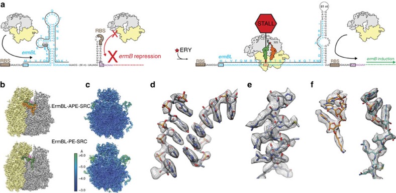
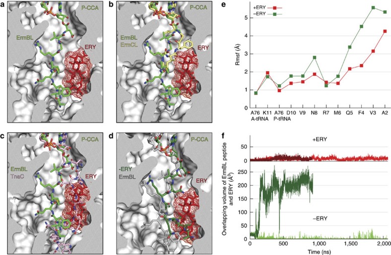
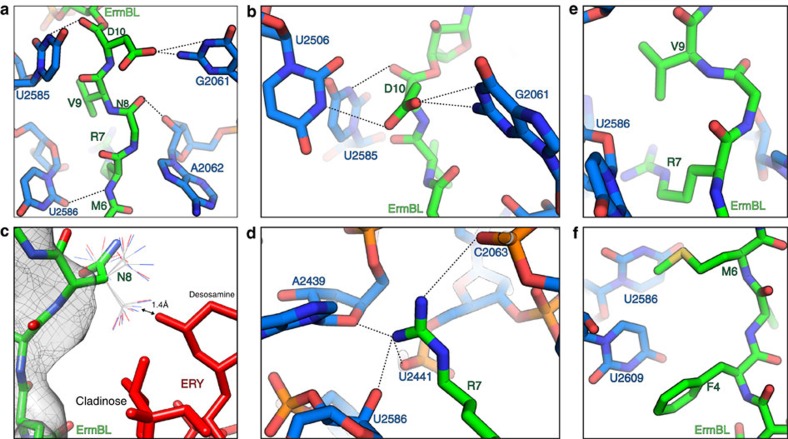
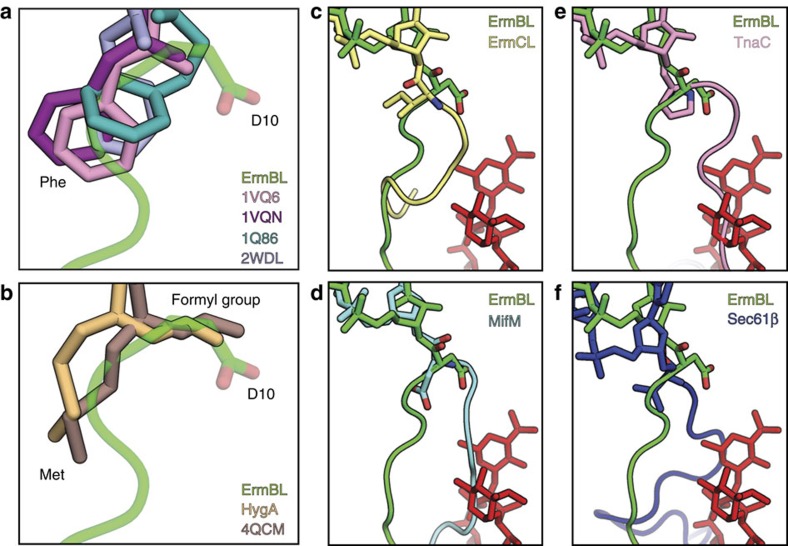
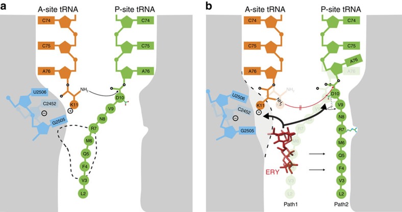
Nascent polypeptides can induce ribosome stalling, regulating downstream genes. Stalling of ErmBL peptide translation in the presence of the macrolide antibiotic erythromycin leads to resistance in Streptococcus sanguis. To reveal this stalling mechanism we obtained 3.6-Å-resolution cryo-EM structures of ErmBL-stalled ribosomes with erythromycin. The nascent peptide adopts an unusual conformation with the C-terminal Asp10 side chain in a previously unseen rotated position. Together with molecular dynamics simulations, the structures indicate that peptide-bond formation is inhibited by displacement of the peptidyl-tRNA A76 ribose from its canonical position, and by non-productive interactions of the A-tRNA Lys11 side chain with the A-site crevice. These two effects combine to perturb peptide-bond formation by increasing the distance between the attacking Lys11 amine and the Asp10 carbonyl carbon. The interplay between drug, peptide and ribosome uncovered here also provides insight into the fundamental mechanism of peptide-bond formation.


Figures

Figure 1
Figure 1. Cryo-EM structure of the ErmBL-SRC.
(a) Schematic for ermBL-dependent regulation of ermB translation in the presence of ERY. (b) Cryo-EM maps of the ErmBL-APE-SRC and ErmBL-PE-SRC, with 30S (yellow), 50S (grey), A-tRNA (orange), P-tRNA (green) and E-tRNA (magenta). (c) Transverse sections of the cryo-EM maps of the ErmBL-APE-SRC and ErmBL-PE-SRC, coloured according to local resolution. (df) Examples of electron density (grey mesh) of the ErmBL-APE-SRC map illustrating (d) base separation within an rRNA helix, (e) side chains of ribosomal protein α-helix and (f) aminoacylated-CCA end of the A-tRNA (orange) and ErmBL peptide attached to the CCA end of the P-tRNA (green).
Figure 2
Figure 2. Path of ErmBL within the ribosomal tunnel.
(a,b) Path of (a) ErmBL (green) and (b) ErmCL (yellow) within the ribosomal tunnel with bound ERY (red mesh). (c) Path of TnaC (pink) compared with the path of ErmBL (green) within the ribosomal tunnel with superimposed ERY (red mesh). (d) Path of ErmBL after 700-ns simulation time in absence of ERY (green) compared with ErmBL as seen in the cryo-EM structure (grey). (e) Fluctuations of the tRNA A76 nucleotides and ErmBL residues in presence (red) and absence (green) of ERY obtained from the simulations. (f) Overlap between the ErmBL peptide and the volume occupied by ERY in the course of the simulations.
Figure 3
Figure 3. Interaction of the ErmBL nascent chain within the ribosomal tunnel.
(af) Interactions of ErmBL (green) with 23S rRNA nucleotides (blue). In c, different rotamers of the N8 side chain are illustrated and ERY is shown in red.
Figure 4
Figure 4. Rotation of the C-terminal amino acid and an alternate path of ErmBL.
(a,b) Orientation of ErmBL D10 compared with (a) CC-Phe-caproic-acid-biotin (CC-pcb) tRNA analogues (pink, PDB1VQ6; purple, PDB1VQN; and teal, PDB1Q86 (ref. 74)), and Phe-tRNA (blue, PDB2WDL33) and with (b) T. thermophilus 70S ribosomes bearing fMet-tRNA (brown, PDB4QCM and yellow, PDB 4Z3R75). (cf) Alternate path of ErmBL (green) compared with (c) that of ErmCL (yellow, PDB 3J7Z13), (d) MifM (light blue, PDB3J9W18), (e) TnaC (pink, PDB4UY8 (ref. 14)) and (f) Sec61β (dark blue, PDB 3J92 (ref. 20)) in relation to the position of ERY (red).
Figure 5
Figure 5. A-site tRNA accommodation at the PTC of the ErmBL-APE-SRC.
(ad) Atomic model for the CCA end of the A-site tRNA (orange) and (a) electron density (grey mesh) in the ErmBL-APE-SRC compared with (b) unaccommodated (pink, PDB1VQ6 (ref. 30)) and (c) accommodated states (brown, PDB4QCM; purple, PDB1VQN30) and (d) with both. (e) Positions of the α-amino group of the amino acid attached to the A-site tRNA in the ErmBL-APE-SRC compared with its position in the pre-peptide-bond formation state. (fh) Position of 23S rRNA nucleotide U2506 (blue) in (f) ErmBL-PE-SRC and (g) ErmBL-APE-SRC including electron density (grey mesh) and (h) compared with the unaccommodated and accommodated states of the PTC. (i) Comparison of 23S rRNA nucleotide U2585 in ErmBL-APE-SRC (blue), compared with the unaccommodated and accommodated states of the PTC. The nascent chain and A-tRNA in ErmBL are shown in green and orange, respectively.
Figure 6
Figure 6. Perturbation of the P-tRNA by ErmBL prevents peptide-bond formation.
(ad) Atomic model for the CCA end of the P-site tRNA (green) and (a,c,d) electron density (grey mesh) in the ErmBL-APE-SRC compared with (b) P-tRNAs in the pre-peptide-bond formation state (brown; PDB4QCM; blue, PDB2WDK33), with (c,d) zoom onto the ribose of A76. (e,f) Relative orientation of the A-site α-amino group and the P-site carbonyl carbon in (e) ErmBL-APE-SRC and (f) the pre-catalysis state. (g) Deviation of the ribose of A76 from the pre-attack state as a function of simulation time. (h) Conformation of the PTC in the ErmBL-SRC overlayed with the network of hydrogen bonds, creating a proton wire that enables peptide-bond formation. Cryo-EM map (grey mesh) with models for 23S rRNA (blue), L27 (brown) and P-tRNA (green).
Figure 7
Figure 7. Influence of the A-site amino acid and ERY on PTC activity.
(a) Movement of the U2504–U2506 backbone in the simulations; histograms of the progression along the most dominant motion for each simulation. WT simulations (upper panel): with ERY (red); without ERY (green). K11A mutation simulations (lower panel): with ERY (orange); without ERY (cyan). (b) Comparison of two ErmBL simulation states in presence (red) or absence (green) of ERY. (c) Histograms of the distance d1 between the mean position of D10 and crevice nucleotides U2504, U2506 and C2452. (d) Histograms of distance d2 between the attacking amide group and the D10 carbonyl carbon. (e) Comparison of two simulation states of wild-type ErmBL (K11; red) with the ErmBL K11A mutant (orange) in presence of ERY. The simulation state of wild-type ErmBL in absence of ERY (green in b) is superimposed (white). (f) Histograms of the interaction enthalpy between the A-site K11 (upper panel) or A11 (lower panel) and the crevice nucleotides U2504, U2506 and C2452. (g) Toe-printing assay to monitor translation of ErmBL variants in presence or absence (−) of ERY. Each template contains a Lys substitution in the ErmBL motif. The A-site K of the wild-type motif RNVDK was changed to all other 19 sense amino acids (X). Arrows indicate positions of ribosome accumulation with the indicated substitution/amino acid in the A-site. The blue arrow points to ribosome accumulation at the Asp of the ErmBL RNVDK motif. The orange arrow indicates the position of ribosome accumulation due to Ile depletion. The blue bars represent quantifications of ribosome stalling of the A-site mutants relative to the wild-type ErmBL motif containing Lys. The uncropped gel images are depicted in Supplementary Fig. 10.
Figure 8
Figure 8. Model for ErmBL-mediated translation arrest.
(a) Canonical peptide-bond formation in absence of ERY. Here the path of the peptide (Path1) overlaps with the binding site of the drug (broken line). (b) Stalling in the presence of ERY. The drug restrains the conformation of the ErmBL peptide (Path2). Relative to a, the C-terminal Asp10 side chain is in a rotated position and the P-site tRNA A76 ribose is displaced. The ERY perturbs the A-site crevice nucleotides (blue), which through electrostatic interactions keeps Lys11 away from the P-site. We propose that the increased distance between the attacking amide group of the Lys11 and the carbonyl carbon of Asp10 hinders peptide-bond formation.

References

    1. Simonovic M. & Steitz T. A. A structural view on the mechanism of the ribosome-catalyzed peptide bond formation. Biochim. Biophys. Acta 1789, 612–623 (2009). - PMC - PubMed
    1. Wilson D. N. & Beckmann R. The ribosomal tunnel as a functional environment for nascent polypeptide folding and translational stalling. Curr. Opin. Struct. Biol. 21, 1–10 (2011). - PubMed
    1. Ito K. & Chiba S. Arrest peptides: cis-acting modulators of translation. Annu. Rev. Biochem. 82, 171–202 (2013). - PubMed
    1. Ramu H., Mankin A. & Vazquez-Laslop N. Programmed drug-dependent ribosome stalling. Mol. Microbiol. 71, 811–824 (2009). - PubMed
    1. Vázquez-Laslop N., Ramu H. & Mankin A. S. in Ribosomes. Structure, Function, Evolution eds Rodnina M. V., Wintermeyer W., Green R. 377–392Springer (2011).

Publication types

MeSH terms

LinkOut - more resources