Review
. 2016 Sep;17(9):537-52.
doi: 10.1038/nrm.2016.74. Epub 2016 Jul 6.

Mechanistic insights into selective autophagy pathways: lessons from yeast

Affiliations
Review

Mechanistic insights into selective autophagy pathways: lessons from yeast

Jean-Claude Farré et al. Nat Rev Mol Cell Biol. 2016 Sep.

Abstract

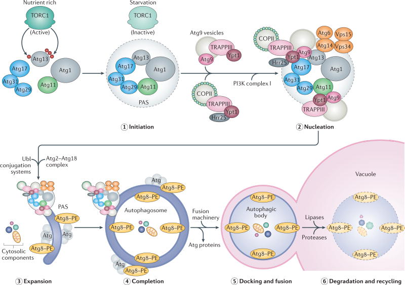
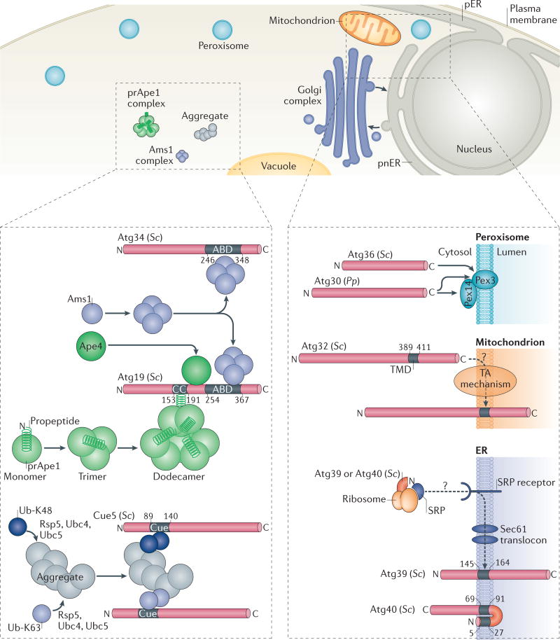
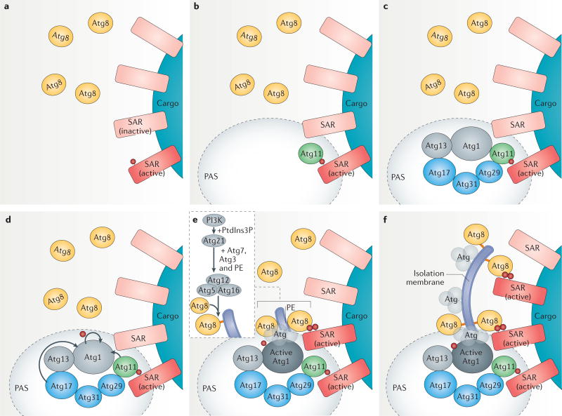
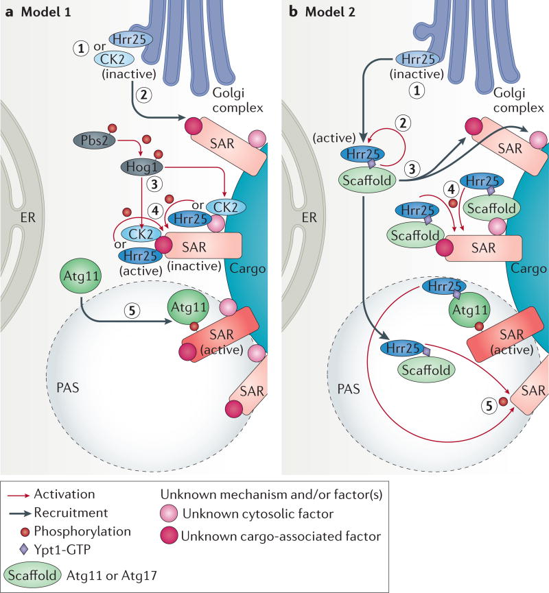
Autophagy has burgeoned rapidly as a field of study because of its evolutionary conservation, the diversity of intracellular cargoes degraded and recycled by this machinery, the mechanisms involved, as well as its physiological relevance to human health and disease. This self-eating process was initially viewed as a non-selective mechanism used by eukaryotic cells to degrade and recycle macromolecules in response to stress; we now know that various cellular constituents, as well as pathogens, can also undergo selective autophagy. In contrast to non-selective autophagy, selective autophagy pathways rely on a plethora of selective autophagy receptors (SARs) that recognize and direct intracellular protein aggregates, organelles and pathogens for specific degradation. Although SARs themselves are not highly conserved, their modes of action and the signalling cascades that activate and regulate them are. Recent yeast studies have provided novel mechanistic insights into selective autophagy pathways, revealing principles of how various cargoes can be marked and targeted for selective degradation.


Conflict of interest statement

Competing interests statement

The authors declare no competing interests.

Figures

Figure 1
Figure 1. Steps in autophagy
Autophagy is inhibited under nutrient-rich conditions via the hyperphosphorylation of autophagy-related 13 (Atg13) by target of rapamycin complex 1 (TORC1) kinase; this process prevents a tight interaction between Atg1 kinase and Atg17 (REF. 41). Starvation or rapamycin treatment activates autophagy by inhibiting TORC1, leading to the hypophosphorylation of Atg13, which can then interact with Atg1 and Atg17. The first two steps, initiation (step 1) and nucleation (step 2), involve the recruitment of cytosolic components of the core autophagic machinery to the phagophore assembly site (PAS) in yeast (omegasomes in mammals). In yeast, the non-selective autophagy-specific PAS is organized partly by the scaffold components Atg11 and Atg17, with Atg17 itself being part of a tripartite Atg17–Atg29–Atg31 subcomplex,. Scaffold components then recruit additional proteins, including transport protein particle III (TRAPPIII) and Ypt1 (a Rab1 family GTPase), which bring coat protein complex II (COPII) and Atg9 vesicles, to initiate the expansion (step 3) of a double-membrane phagophore. This expansion also involves the activity of phosphoinositide 3-kinase (PI3K) complex I (consisting of Atg6, Atg14, vacuolar protein sorting 34 (Vps34) and Vps15), which generates the phosphatidylinositol 3-phosphate required to recruit other factors involved in phagophore elongation, such as the Atg2–Atg18 complex as well as the ubiquitin-like (Ubl) conjugation systems, Atg8–phosphatidylethanolamine (PE) (Atg8–PE) and Atg5–Atg12–Atg16 (depicted as grey Atg molecules in contact with Atg8–PE); see also FIG. 3 for details on PAS assembly and isolation membrane formation. As a result of this membrane expansion, cargo destined for autophagy is surrounded and engulfed into a double-membrane vesicle called the autophagosome (step 4),–, Autophagosomes are then transported to lysosomes (or vacuoles in yeast and plants). Docking and fusion (step 5) of the outer autophagosomal membrane with that of the lysosome (vacuole) releases the autophagic body into the lysosomal (vacuolar) lumen, where hydrolases degrade and recycle (step 6) the macromolecular components for cellular use.
Figure 2
Figure 2. Selective autophagy pathways and cargo recognition by selective autophagy receptors
The upper panel of the figure shows the specific cargoes, such as oligomeric α-mannosidase 1 (Ams1) or precursor of vacuolar aminopeptidase 1 (prApe1), protein aggregates or organelles (peroxisomes, mitochondria, perinuclear ER (pnER) or peripheral ER (pER), or fragments of the nucleus), that are subject to selective autophagy. The lower left panel depicts soluble selective autophagy receptors (SARs). The prApe1 dodecamer is bound by the coiled-coil (CC) domain of autophagy-related 19 (Atg19). Ams1 oligomerizes and associates with Atg19 through the Ams1-binding domain (ABD). prApe1, Ams1 and Atg19 assemble into a large complex called the cytoplasm-to-vacuole targeting (Cvt) complex. Atg34, an Atg19 paralogue, is also a receptor for Ams1 (REF. 24), but not for Ape1 or aspartyl aminopeptidase 4 (Ape4). Ape4 also binds Atg19. Coupling of ubiquitin conjugation to endoplasmic reticulum (ER) degradation 5 (Cue5) binds aggregates through direct interaction of its Cue domain with lysine 63 (K63)- and lysine 48(K48)-linked ubiquitin (Ub) chains that are covalently attached to cargoes by the E3 ubiquitin ligase Rsp5 and the E2 ubiquitin-conjugating enzyme Ubc4 or Ubc5. The lower right panel depicts membrane-associated SARs. Pexophagy receptors of Saccharomyces cerevisiae (Sc) Atg36 and Pichia pastoris (Pp) Atg30 recognize peroxisomal membrane proteins (Pex14 and/or Pex3). The mitophagy receptor Atg32 is embedded in the mitochondrial outer membrane via a single α-helical transmembrane domain (TMD) and probably (indicated by a question mark in the figure) the action of the tail-anchored (TA) mechanism, which refers to the protein machinery that inserts proteins possessing a carboxy-terminal TMD into the membrane such that, topologically, the amino terminus of the protein is cytosolic and the C terminus of the protein is lumenal. The ER-phagy receptors Atg39 and Atg40 have one TMD and two TMDs, respectively, and might insert into the ER membrane co-translationally via the signal recognition particle (SRP), the SRP receptor and the Sec61 translocon.
Figure 3
Figure 3. Phagophore assembly site and isolation membrane formation in selective autophagy activation
a | Phagophore assembly site (PAS) formation starts by activation of selective autophagy receptors (SARs) bound to cargoes. The activation mechanism involves phosphorylation by casein kinases (phosphorylation is indicated by a red ball). b | An activated SAR binds the scaffold protein autophagy-related 11 (Atg11) to initiate PAS formation. c | Atg11 binds the SAR–cargo complex, recruits the Atg17 scaffold complex (composed of Atg17, Atg31 and Atg29) via Atg29, as well as the Atg1 kinase complex (composed of Atg1 and Atg13). d | Atg11 bound to the cargo–SAR complex and the Atg17 complex then activates the Atg1 kinase, which autophosphorylates itself as well as other Atg proteins. e | Activated Atg1 kinase recruits other core autophagy proteins, resulting in the recruitment of Atg8, which is then conjugated to phosphatidylethanolamine (PE) to begin phagophore expansion from the PAS. A second ubiquitin-like conjugate, Atg12–Atg5, forms a complex with Atg16, and is necessary for the recruitment of Atg8 to the PAS and its conjugation to PE (it acts as the E3 ubiquitin ligase). The Atg12–Atg5–Atg16 complex itself is recruited to the PAS by the phosphatidylinositol-3-phosphate (PtdIns3P)-binding protein Atg21, and its localization relies on PtdIns3P synthesis at the PAS by the PI3K complex I (see also FIG. 1). Notably, Atg21 is required mostly for selective autophagy pathways and not for non-selective autophagy. Atg8–PE also interacts with neighbouring SARs activated by phosphorylation. f | Isolation membrane expansion then continues around the cargo, engaging other activated SARs.
Figure 4
Figure 4. Hypothetical models for activation of selective autophagy receptors by casein kinases
Yeast selective autophagy receptors (SARs) are generally phosphorylated and casein kinases (Hrr25 or casein kinase 2 (CK2)) have been shown to play a role in the phosphorylation of SARs involved in the mitophagy, pexophagy and Cvt pathways. The SARs for pexophagy as well as the cytoplasm-to-vacuole targeting (Cvt) cargo, precursor of vacuolar aminopeptidase 1 (prApe1), are phosphorylated by the CK1δ homologue, Hrr25, whereas the mitophagy SAR is phosphorylated by CK2. At least two hypothetical models could explain the phosphorylation and activation mechanism of SARs. However, the order and subcellular location of these steps are not currently known. In Model 1 (part a), inactive Hrr25 and CK2 (step 1) are recruited to inactive SARs (step 2) by an unknown factor (or factors) (shown as pink-shaded circles) and by unknown mechanisms and activated close to the SARs (step 3). Activation of CK2 might depend on the MAPKs of the high osmolarity glycerol (HOG) pathway, Hog1 and Pbs2 (REFS 61,72). Activated CK2 and Hrr25 then phosphorylate and activate SARs (step 4), resulting in the recruitment of autophagy-related 11 (Atg11) (step 5). In model 2 (part b), the inactive Hrr25 (step 1) is activated in the cytosol (step 2) and then recruited to the SAR or its vicinity via unknown factors (step 3), as well as to the phagophore assembly site (PAS), in a manner dependent on an activated Ypt1 (Ypt1-GTP) and the scaffold protein Atg17 (REF. 49) or possibly also Atg11 (as Atg11 is known to recruit Ypt1 to the PAS). Consequently, before PAS formation, the first SAR (shown at the top) will be phosphorylated by the active Hrr25 localized proximal to the SAR by Ypt1-GTP, the scaffold protein and unknown factors associated either with the SAR itself or the cargo (step 4). The direct interaction of the phosphorylated SAR (active) and the scaffold protein Atg11 will initiate PAS formation. Finally, PAS-localized Hrr25 will further propagate the phosphorylating signal and activates other SARs (step 5).

References

    1. Ryter SW, Cloonan SM, Choi AM. Autophagy: a critical regulator of cellular metabolism and homeostasis. Mol. Cells. 2013;36:7–16. - PMC - PubMed
    1. Choi AM, Ryter SW, Levine B. Autophagy in human health and disease. N. Engl. J. Med. 2013;368:651–662. - PubMed
    1. Gozuacik D, Kimchi A. Autophagy as a cell death and tumor suppressor mechanism. Oncogene. 2004;23:2891–2906. - PubMed
    1. Frake RA, Ricketts T, Menzies FM, Rubinsztein DC. Autophagy and neurodegeneration. J. Clin. Invest. 2015;125:65–74. - PMC - PubMed
    1. Rubinsztein DC, Marino G, Kroemer G. Autophagy and aging. Cell. 2011;146:682–695. - PubMed

Substances