Case Reports
. 2016 Sep;57(9):e191-4.
doi: 10.1111/epi.13457. Epub 2016 Jul 6.

Transient and chronic seizure-induced inflammation in human focal epilepsy

Affiliations
Case Reports

Transient and chronic seizure-induced inflammation in human focal epilepsy

Tracy Butler et al. Epilepsia. 2016 Sep.

Abstract

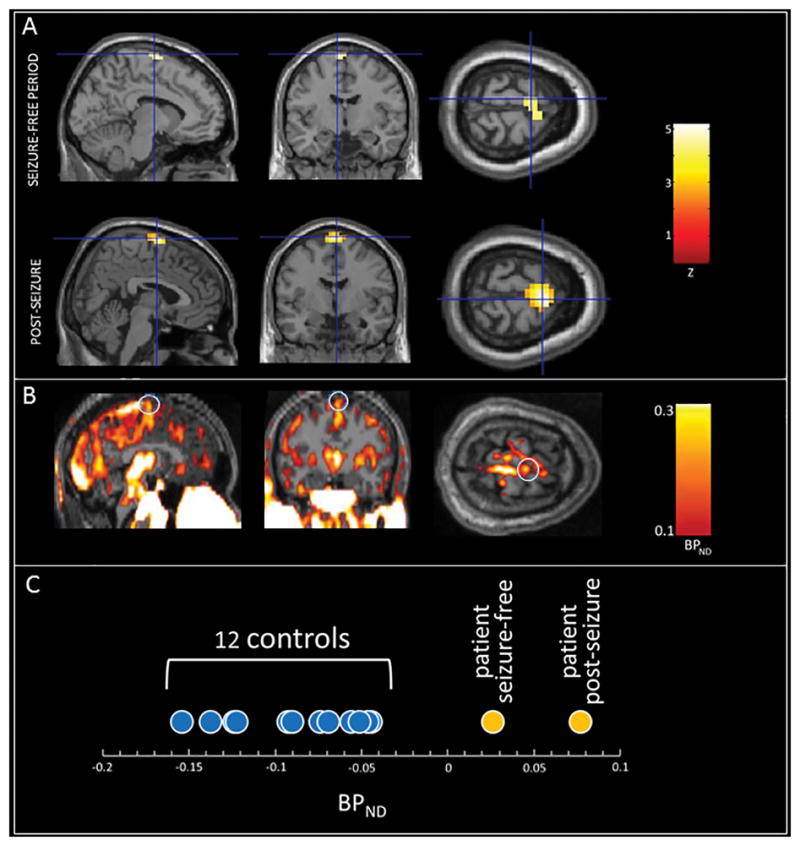
In animal models, inflammation is both a cause and consequence of seizures. Less is known about the role of inflammation in human epilepsy. We performed positron emission tomography (PET) using a radiotracer sensitive to brain inflammation in a patient with frontal epilepsy ~36 h after a seizure as well as during a seizure-free period. When statistically compared to a group of 12 matched controls, both of the patient's scans identified a frontal (supplementary motor area) region of increased inflammation corresponding to his clinically defined seizure focus, but the postseizure scan showed significantly greater inflammation intensity and spatial extent. These results provide new information about transient and chronic neuroinflammation in human epilepsy and may be relevant to understanding the process of epileptogenesis and guiding therapy.

Keywords: Focal cortical dysplasia; Frontal lobe epilepsy; Microglia; Positron emission tomography; Supplementary motor area; Translocator protein.


Conflict of interest statement

No authors have any financial interests relevant to this report to disclose.

Figures

Figure 1
Figure 1
Abnormally increased neuroinflammation (TSPO expression by activated microglial) as assessed using [11C]PK11195 PET in a patient with clinically-defined frontal lobe epilepsy. A: Z-maps (the patient’s PET images statistically compared to 12 age-matched controls) obtained during a seizure-free period (top) and ~36 hours after a typical seizure (bottom.) Note the nearly identical location of abnormally increased TSPO expression in the midline SMA region in both scans, with greater intensity and spatial extent in the post-seizure scan. Crosshairs are at the voxel of peak intensity in each scan. Images are thresholded at Z=2.5 and overlaid onto a template T1 MRI. B: The patient's post-seizure BPND image, reflecting TSPO expression, overlaid on his native T1 MRI, without statistical comparison to controls. Note radiotracer uptake in the region identified in the post-seizure Z-map (white circles) as well as in multiple other cortical and subcortical regions including thalamus, brainstem, posterior superior sagittal sinus and extracranial tissues, all of which are also present in controls, making focally abnormal uptake more difficult to appreciate on visual inspection. C: Average BPND for each subject within the region of abnormality (bilateral SMA) identified in the comparison between the patient’s post-seizure image and the group of controls. Note significantly higher BPND in the patient’s two scan as compared to the twelve controls.

References

    1. Besson P, Andermann F, Dubeau F, et al. Small focal cortical dysplasia lesions are located at the bottom of a deep sulcus. Brain. 2008;131:3246–3255. - PubMed
    1. Butler T, Ichise M, Teich AF, et al. Imaging inflammation in a patient with epilepsy due to focal cortical dysplasia. J Neuroimaging. 2013;23:129–131. - PMC - PubMed
    1. Gershen LD, Zanotti-Fregonara P, Dustin IH, et al. Neuroinflammation in temporal lobe epilepsy measured using positron emission tomographic imaging of translocator protein. JAMA neurology. 2015;72:882–888. - PMC - PubMed
    1. Gowers W. Epilepsy and other chronic convulsive diseases: their causes, symptoms, and treatment. Churchill; London: 1881.
    1. Hirvonen J, Kreisl WC, Fujita M, et al. Increased in vivo expression of an inflammatory marker in temporal lobe epilepsy. J Nucl Med. 2012;53:234–240. - PMC - PubMed

Publication types