. 2016 Jul 5;5(7):e003531.
doi: 10.1161/JAHA.116.003531.

Zofenopril Protects Against Myocardial Ischemia-Reperfusion Injury by Increasing Nitric Oxide and Hydrogen Sulfide Bioavailability

Affiliations

Zofenopril Protects Against Myocardial Ischemia-Reperfusion Injury by Increasing Nitric Oxide and Hydrogen Sulfide Bioavailability

Erminia Donnarumma et al. J Am Heart Assoc. .

Abstract

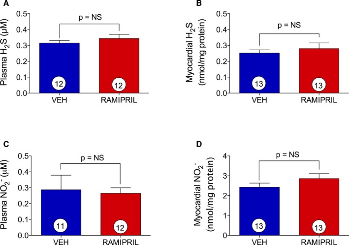
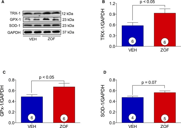
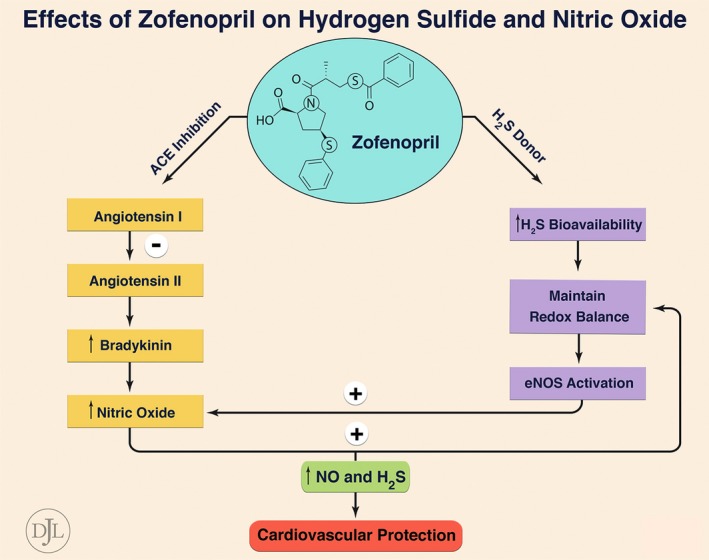
Background: Zofenopril, a sulfhydrylated angiotensin-converting enzyme inhibitor (ACEI), reduces mortality and morbidity in infarcted patients to a greater extent than do other ACEIs. Zofenopril is a unique ACEI that has been shown to increase hydrogen sulfide (H2S) bioavailability and nitric oxide (NO) levels via bradykinin-dependent signaling. Both H2S and NO exert cytoprotective and antioxidant effects. We examined zofenopril effects on H2S and NO bioavailability and cardiac damage in murine and swine models of myocardial ischemia/reperfusion (I/R) injury.

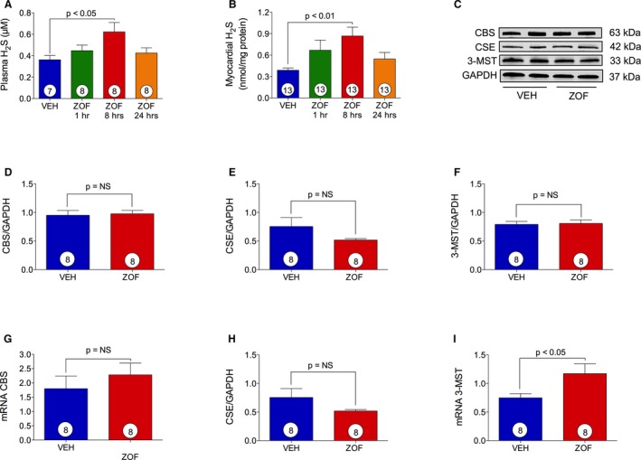
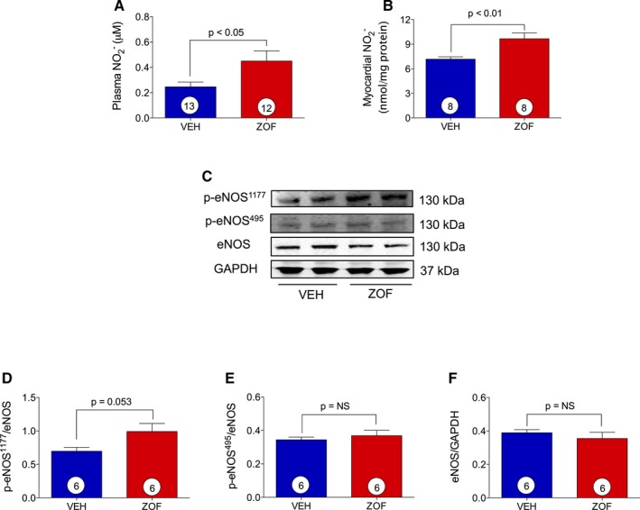
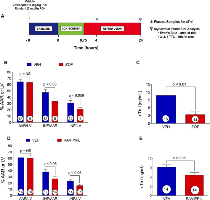
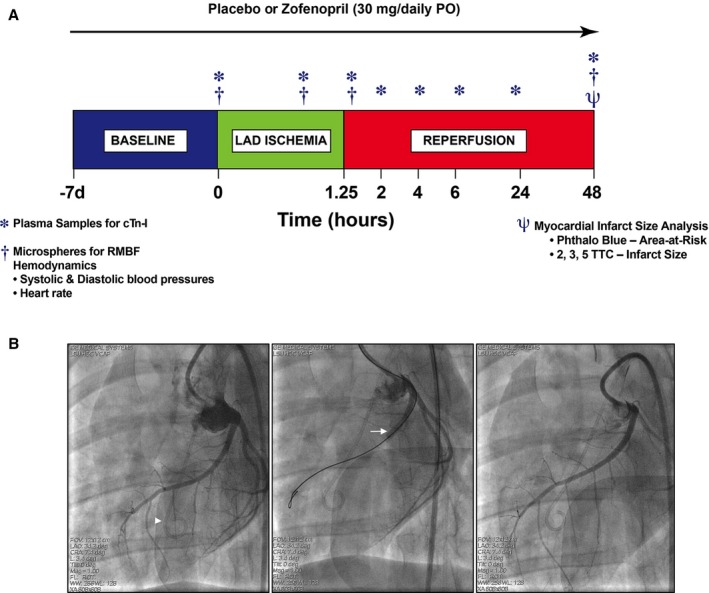
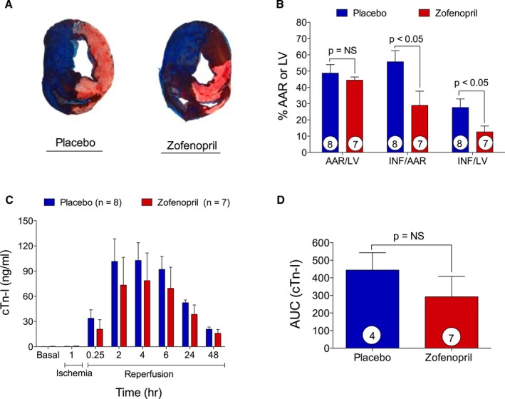
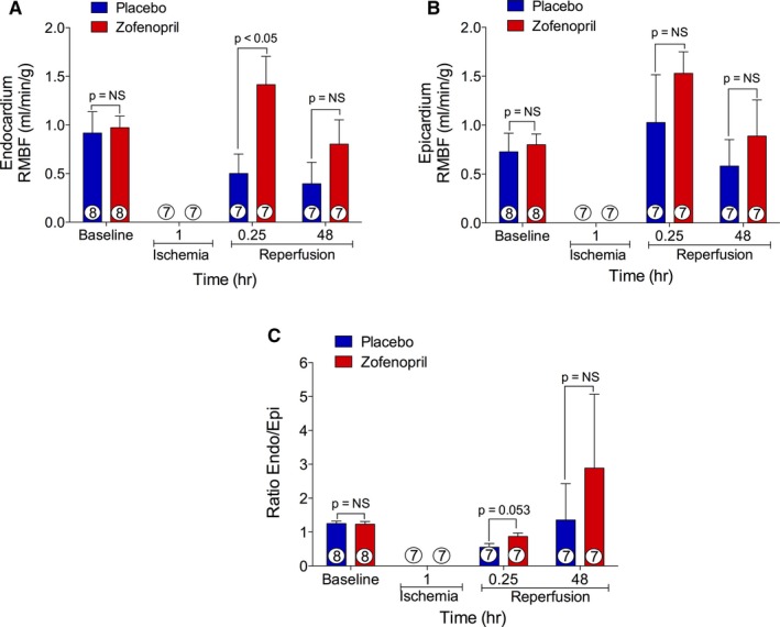
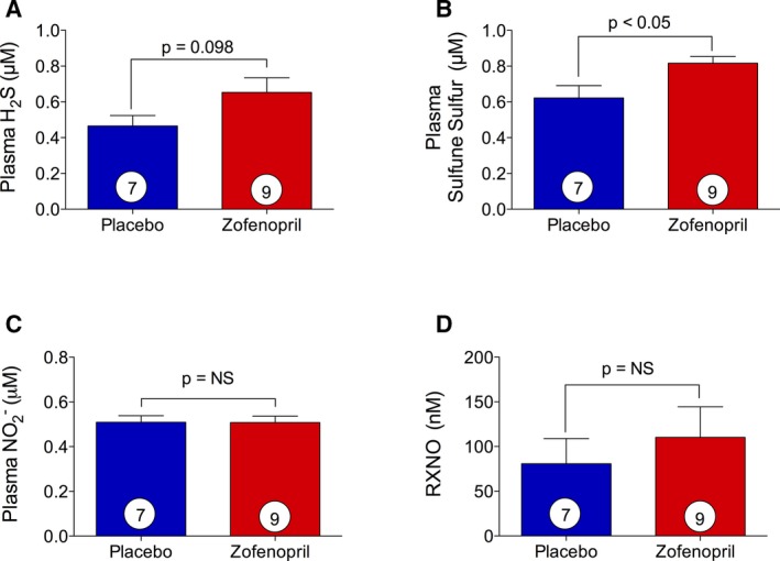
Methods and results: Zofenopril (10 mg/kg PO) was administered for 1, 8, and 24 hours to establish optimal dosing in mice. Myocardial and plasma H2S and NO levels were measured along with the levels of H2S and NO enzymes (cystathionine β-synthase, cystathionine γ-lyase, 3-mercaptopyruvate sulfur transferase, and endothelial nitric oxide synthase). Mice received 8 hours of zofenopril or vehicle pretreatment followed by 45 minutes of ischemia and 24 hours of reperfusion. Pigs received placebo or zofenopril (30 mg/daily orally) 7 days before 75 minutes of ischemia and 48 hours of reperfusion. Zofenopril significantly augmented both plasma and myocardial H2S and NO levels in mice and plasma H2S (sulfane sulfur) in pigs. Cystathionine β-synthase, cystathionine γ-lyase, 3-mercaptopyruvate sulfur transferase, and total endothelial nitric oxide synthase levels were unaltered, while phospho-endothelial nitric oxide synthase(1177) was significantly increased in mice. Pretreatment with zofenopril significantly reduced myocardial infarct size and cardiac troponin I levels after I/R injury in both mice and swine. Zofenopril also significantly preserved ischemic zone endocardial blood flow at reperfusion in pigs after I/R.

Conclusions: Zofenopril-mediated cardioprotection during I/R is associated with an increase in H2S and NO signaling.

Keywords: antihypertensive agent; hydrogen sulfide; myocardial ischemia; nitric oxide; oxidant stress; troponin.


Figures

Figure 1
Figure 1
Zofenopril increases H2S bioavailability. VEH indicates vehicle; ZOF, zofenopril. Effect of vehicle and zofenopril treatment (10 mg/kg PO) at 1, 8, or 24 hours on H2S levels in mice plasma (A) and heart tissue (B). Zofenopril administration induced a significant increase in circulating and cardiac H2S levels at 8 hours of treatment as compared with vehicle. C, Immunoblots for cystathionine γ‐lyase (CSE), cystathionine β‐synthase (CBS), and 3‐mercaptopyruvate sulfur transferase (3‐MST) with relative optical densitometry (D through F). 8 hours of zofenopril treatment did not cause any change in CBS (D), CSE (E), and 3‐MST (F) protein expression. G through I, mRNA level of H2S‐producing enzymes after zofenopril therapy for 8 hours. Zofenopril treatment did not affect CBS (G) or CSE (H) gene expression, but it induced a significant increase in 3‐MST (I) mRNA levels compared with vehicle. Results are expressed as mean±SEM. Number in the circle inside the bar denotes the number of animals used per each group.
Figure 2
Figure 2
Zofenopril effect on NO bioavailability and myocardial p‐eNOS 1177, p‐eNOS 495, and eNOS expression. VEH indicates vehicle; ZOF, zofenopril. Zofenopril administered at 8 hours (10 mg/kg PO) resulted in significant increase of nitrite levels in both (A) plasma and (B) heart tissue compared with vehicle. C, Immunoblots for p‐eNOS 1177, p‐eNOS 495, and eNOS with relative optical densitometry (D through F). Results are expressed as mean±SEM. Number inside the circles in the bar denotes the number of animals used per group.
Figure 3
Figure 3
Ramipril effect on H2S and NO bioavailability. H2S and NO determination in mice plasma (A and C) and myocardial tissue (B and D) after 8 hours of vehicle (VEH) or ramipril (3 mg/kg PO) treatment. Ramipril did not affect basal levels of H2S and NO. Results are expressed as mean±SEM. Number in the circle inside the bar denotes the number of animals used per group. NO indicates nitric oxide.
Figure 4
Figure 4
Induction of myocardial antioxidant protein. VEH indicates vehicle; ZOF, zofenopril. A, Immunoblots for Trx‐1, GPx‐1, and SOD‐1 with relative optical densitometry (B through D). Zofenopril administration promoted a significant upregulation of antioxidant proteins Trx‐1 (B) and GPx‐1 (C) and an induction of SOD‐1 (D). Results are expressed as mean±SEM. Number in the circle inside the bar denotes the number of animals used per group.
Figure 5
Figure 5
Zofenopril and ramipril effect on a murine model of myocardial ischemia reperfusion (I/R) injury. VEH indicates vehicle; ZOF, zofenopril. A, Murine model of myocardial I/R protocol. B, Area‐at‐risk (AAR) per left ventricle (LV) and infarct (INF) per AAR and LV. Zofenopril administration (10 mg/kg PO) 8 hours before induction of myocardial I/R injury significantly reduced INF/AAR compared with vehicle. There was no difference in AAR/LV between vehicle‐ and zofenopril‐treated animals. C, Circulating cTn‐I levels measured at 4 hours of reperfusion were significantly diminished by zofenopril pretreatment. D, Ramipril administration (3 mg/kg PO) 8 hours before induction of I/R injury significantly reduced the % INF/AAR and % INF/LV. E, cTn‐I release at 4 hours of reperfusion was significantly reduced by ramipril administration compared with vehicle. Results are expressed as mean±SEM. Number in the circle inside the bar denotes the number of animals used per group.
Figure 6
Figure 6
Experimental protocol for in vivo myocardial ischemia/reperfusion (I/R) injury in swine. A, Female Yucatan pigs were subjected to 75 minutes (1.25 hours) of myocardial ischemia by occluding left anterior descending coronary artery (LAD) via a balloon catheter placement and 48 hours of reperfusion. Zofenopril (30 mg/daily PO) or placebo therapy was initiated at 7 days prior I/R injury and continued for 2 days after ischemia. At baseline, 60 minutes of LAD occlusion, 15 minutes of reperfusion, and 48 hours of reperfusion, microspheres labeled with samarium, europium, lutetium, or lanthanum were injected to measure regional myocardial blood flow (RMBF). At baseline, 60 minutes of ischemia, and 15 minutes, 2, 4, 6, 24, and 48 hours of reperfusion, plasma samples were collected for measurement of cardiac troponin‐I release. At day 2 of reperfusion, heart tissue was collected for infarct size determination. Baseline plasma samples were used for assessment of circulating levels of H2S, sulfane sulfur, NO 2 , and S‐nitrosothiols (RXNO). B, Angiographic left anterior oblique caudal images of the LAD at baseline (left), during 75 minutes occlusion (middle), and at 15 minutes reperfusion (right). Intracoronary occlusion was achieved by inflation of an angioplasty balloon catheter (arrow) deployed in the proximal LAD distal to the first anterior septal branch. Proximal LAD deployment of the balloon produced an area‐at‐risk (AAR) region ≈45% of the left ventricular (LV) mass. The AAR was distributed primarily in the anterior LV free wall and includes a portion of the anterior septum. For measurement of regional blood flow, microspheres were injected into the LV cavity through a pigtail catheter (arrowhead).
Figure 7
Figure 7
Reduction of infarct size by zofenopril pretreatment in a swine model of myocardial ischemia/reperfusion (I/R). A, Representative mid‐ventricular heart slices from placebo‐ and zofenopril‐treated animals. Area‐at‐risk (AAR) per left ventricle (LV) and infarct (INF) per AAR and LV were determined with a dual stain technique. There was no difference in AAR/LV between placebo‐ and zofenopril‐treated groups. B, Zofenopril pretreatment significantly reduced INF/AAR and INF/LV compared with placebo. C and D, Plasma levels of cTn‐I measured at time 0, 60 minutes of ischemia, and 15 minutes and 2, 4, 6, 24, and 48 hours of reperfusion and cumulative release of cTn‐I. Zofenopril reduced cTn‐I release during reperfusion with no significant extent. Results are expressed as mean±SEM. Number in the circle inside the bar denotes the number of animals used per group.
Figure 8
Figure 8
Regional myocardial blood flow. A, Endocardial myocardium blood flow, (B) epicardial myocardium blood flow, and (C) the ratio endocardial/epicardial blood flow in the ischemic zone at baseline, 60 minutes of left anterior descending coronary artery occlusion, and 15 minutes and 48 hours of reperfusion. Zofenopril resulted in a significantly greater endocardial blood flow at 15 minutes of reperfusion compared with placebo. Number inside the bar denotes the number of animals used per group.
Figure 9
Figure 9
Effect of zofenopril on circulating H2S, NO 2 , sulfane sulfur and S‐nitrosothiols (RXNO). At day 7 of placebo or zofenopril treatment of swine, plasma samples were harvested prior surgical procedure of myocardial ischemia reperfusion (I/R) to assess H2S, sulfane sulfur, NO, and RXNO levels. A, Zofenopril therapy did not significantly increase plasma H2S availability. B, Data for levels of sulfane sulfur demonstrate a significant increase in zofenopril‐treated animals compared with placebo. C, Zofenopril treatment did not alter circulating nitrite levels. D, Zofenopril treatment for 7 days did not alter circulating RXNO levels. Results are expressed as mean±SEM. Number in the circle inside the bar denotes the number of animals used per group.
Figure 10
Figure 10
Effect of zofenopril on H2S and NO bioavailability. By inhibiting myocardial angiotensin converting enzyme (ACE) activity, zofenopril reduces the generation of angiotensin II and increases levels of bradykinin (BK). BK, through stimulation of endothelial B2 receptors, promotes the release of NO, prostacyclin, and endothelium‐derived hyperpolarizing factor (EDHF), which in turn leads to cardioprotection. On the other hand, zofenopril, by releasing H2S, enhances tissue antioxidant defense and promotes eNOS activation, leading to increased levels of NO. Therefore, ACE inhibition, H2S, and NO account for zofenopril‐mediated cardioprotective effects.

References

    1. Indications for ACE inhibitors in the early treatment of acute myocardial infarction: systematic overview of individual data from 100,000 patients in randomized trials. ACE Inhibitor Myocardial Infarction Collaborative Group. Circulation. 1998;97:2202–2212. - PubMed
    1. Flather MD, Yusuf S, Kober L, Pfeffer M, Hall A, Murray G, Torp‐Pedersen C, Ball S, Pogue J, Moye L, Braunwald E. Long‐term ACE‐inhibitor therapy in patients with heart failure or left‐ventricular dysfunction: a systematic overview of data from individual patients. ACEInhibitor Myocardial Infarction Collaborative Group. Lancet. 2000;355:1575–1581. - PubMed
    1. Ma TK, Kam KK, Yan BP, Lam YY. Renin‐angiotensin‐aldosterone system blockade for cardiovascular diseases: current status. Br J Pharmacol. 2010;160:1273–1292. - PMC - PubMed
    1. Rodrigues EJ, Eisenberg MJ, Pilote L. Effects of early and late administration of angiotensin‐converting enzyme inhibitors on mortality after myocardial infarction. Am J Med. 2003;115:473–479. - PubMed
    1. Hansen ML, Gislason GH, Kober L, Schramm TK, Folke F, Buch P, Abildstrom SZ, Madsen M, Rasmussen S, Torp‐Pedersen C. Different angiotensin‐converting enzyme inhibitors have similar clinical efficacy after myocardial infarction. Br J Clin Pharmacol. 2008;65:217–223. - PMC - PubMed

Publication types

MeSH terms