. 2016 Aug 19;84(9):2689-96.
doi: 10.1128/IAI.00414-16. Print 2016 Sep.

Infectivity of Plasmodium falciparum in Malaria-Naive Individuals Is Related to Knob Expression and Cytoadherence of the Parasite

Affiliations

Infectivity of Plasmodium falciparum in Malaria-Naive Individuals Is Related to Knob Expression and Cytoadherence of the Parasite

Danielle I Stanisic et al. Infect Immun. .

Abstract

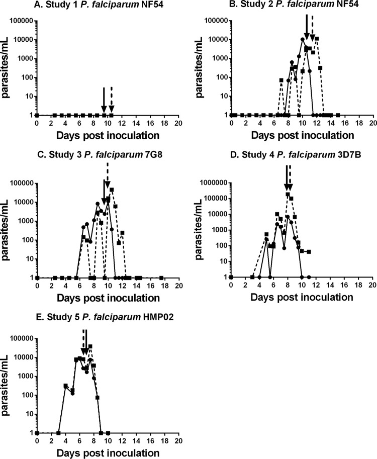
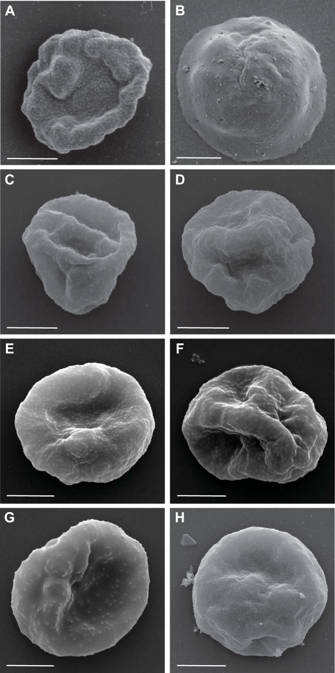
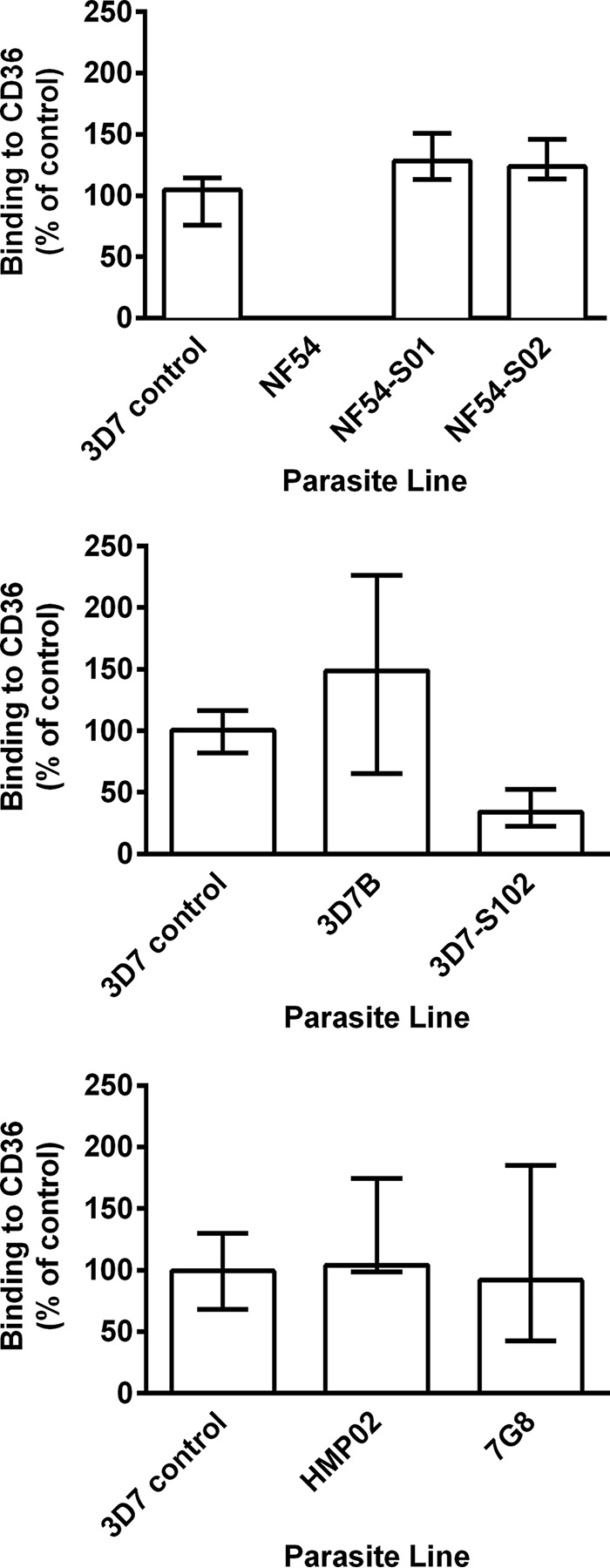
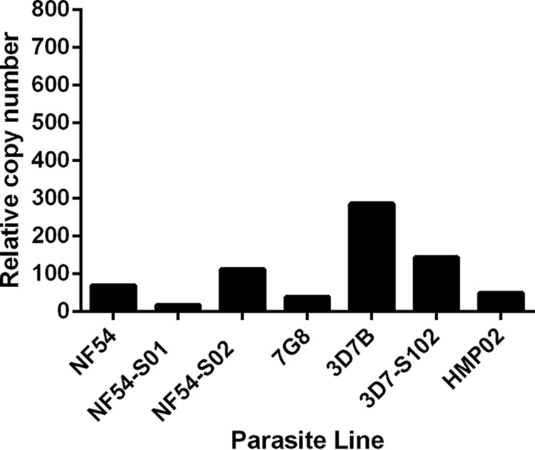
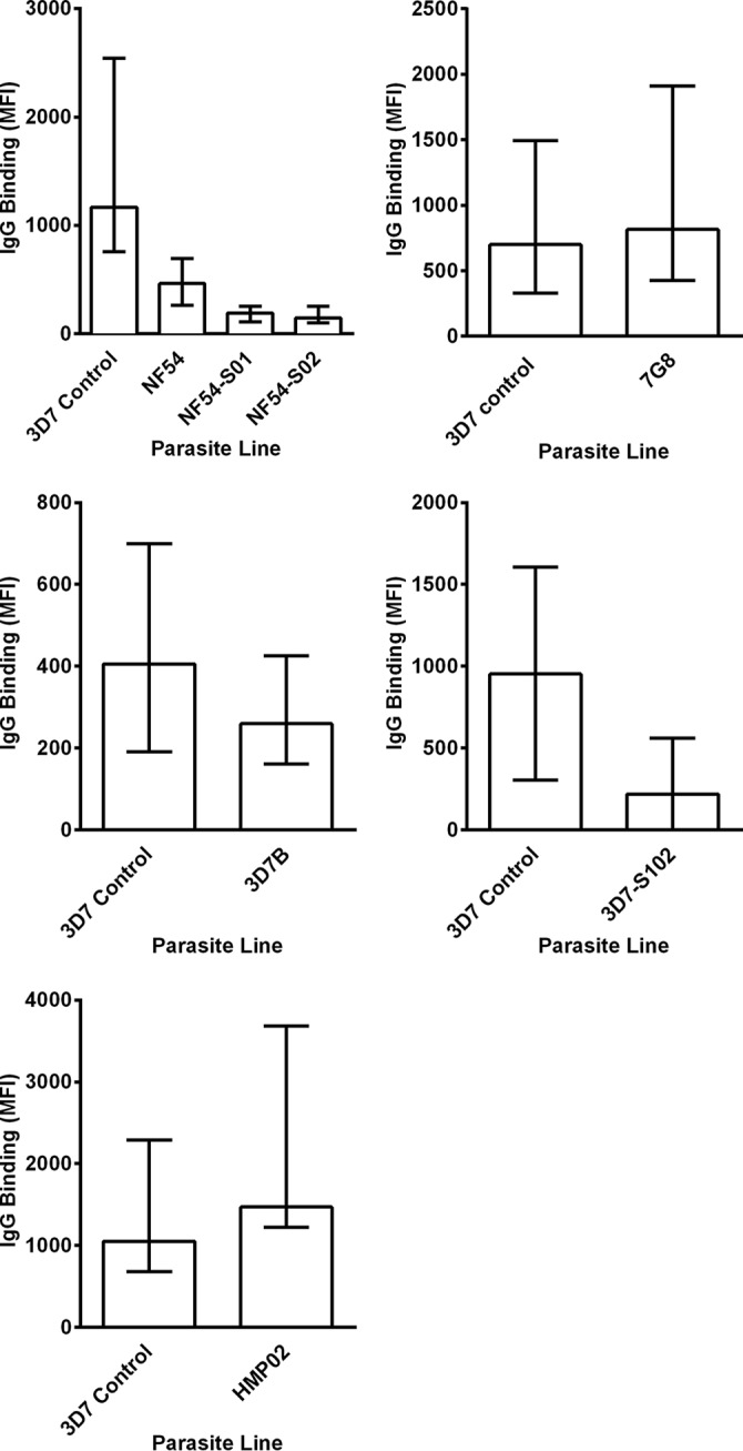
Plasmodium falciparum is the most virulent human malaria parasite because of its ability to cytoadhere in the microvasculature. Nonhuman primate studies demonstrated relationships among knob expression, cytoadherence, and infectivity. This has not been examined in humans. Cultured clinical-grade P. falciparum parasites (NF54, 7G8, and 3D7B) and ex vivo-derived cell banks were characterized. Knob and knob-associated histidine-rich protein expression, CD36 adhesion, and antibody recognition of parasitized erythrocytes (PEs) were evaluated. Parasites from the cell banks were administered to malaria-naive human volunteers to explore infectivity. For the NF54 and 3D7B cell banks, blood was collected from the study participants for in vitro characterization. All parasites were infective in vivo However, infectivity of NF54 was dramatically reduced. In vitro characterization revealed that unlike other cell bank parasites, NF54 PEs lacked knobs and did not cytoadhere. Recognition of NF54 PEs by immune sera was observed, suggesting P. falciparum erythrocyte membrane protein 1 expression. Subsequent recovery of knob expression and CD36-mediated adhesion were observed in PEs derived from participants infected with NF54. Knobless cell bank parasites have a dramatic reduction in infectivity and the ability to adhere to CD36. Subsequent infection of malaria-naive volunteers restored knob expression and CD36-mediated cytoadherence, thereby showing that the human environment can modulate virulence.


Figures

FIG 1
FIG 1
Course of parasitemia in study participants inoculated with P. falciparum cell banks. Shown are the parasite levels in study participants following inoculation with the different P. falciparum cell bank parasites. (A) Study 1, P. falciparum NF54 (cultured cell bank). (B) Study 2, P. falciparum NF54 (cultured cell bank). (C) Study 3, P. falciparum 7G8 (cultured cell bank). (D) Study 4, P. falciparum 3D7B (cultured cell bank). (E) Study 5, P. falciparum HMP02 (ex vivo cell bank). Arrows indicate the times of administration of drug treatment. Note the different y-axis scale for P. falciparum 3D7B. In each study, n = 2.
FIG 2
FIG 2
Surface characteristics of PEs from different P. falciparum parasite lines. Shown are scanning electron micrographs of the P. falciparum 3D7 control (A), P. falciparum NF54 (cultured cell bank) (B), P. falciparum NF54-S01 (derived ex vivo from S01 at the time of drug treatment) (C), P. falciparum NF54-S02 (derived ex vivo from S02 at the time of drug treatment) (D), P. falciparum 3D7B (cultured cell bank) (E), P. falciparum 3D7-S102 (derived ex vivo from S102 at the time of drug treatment) (F), P. falciparum 7G8 (cultured cell bank) (G), and P. falciparum HMP02 (ex vivo cell bank) (H). Representative images are shown, and the scale bars all represent 2 μm.
FIG 3
FIG 3
Binding of P. falciparum PEs from different parasite lines to CD36. Shown is the adhesion to recombinant CD36 of P. falciparum NF54 (cultured cell bank) and P. falciparum NF54-S01 and NF54-S02 (derived ex vivo from S01 and S02 at the time of drug treatment) (top), P. falciparum 3D7B (cultured cell bank) and P. falciparum 3D7-S102 (derived ex vivo from S102 at the time of drug treatment) (middle), and P. falciparum HMP02 (ex vivo cell bank) and P. falciparum 7G8 (cultured cell bank) (bottom). Values are expressed as percentages of the P. falciparum 3D7 control parasite binding to CD36. Assays were performed twice independently, and bars represent median values and interquartile ranges of experimental replicates of samples tested in triplicate.
FIG 4
FIG 4
Expression of kahrp in different P. falciparum parasite lines. Shown are kahrp expression levels in P. falciparum cell bank and ex vivo-derived parasites relative to those of the single-copy fructose-bisphosphate aldolase gene.
FIG 5
FIG 5
Antibody recognition of the surface of P. falciparum PEs from different parasite lines. Shown is the binding of antibodies in serum samples from Papua New Guinean adults to surface antigens expressed by PEs from P. falciparum NF54 (cultured cell bank), P. falciparum NF54-S01 and NF54-S02 (derived ex vivo from S01 and S02 at the time of drug treatment) (top left), P. falciparum 7G8 (cultured cell bank) (top right), P. falciparum 3D7B (cultured cell bank) (middle left), P. falciparum 3D7-S102 (derived ex vivo from S102 at the time of drug treatment) (middle right), and P. falciparum HMP02 (ex vivo cell bank) (bottom). The IgG binding level is expressed as the geometric mean fluorescence intensity (MFI) in all of the graphs, and the bars represent the median values and interquartile ranges of samples tested in duplicate (n = 10 for all P. falciparum cell banks). Minimal reactivity was observed among serum samples from nonexposed Melbourne controls.

Similar articles

Cited by

References

    1. Hanson J, Lam SW, Mahanta KC, Pattnaik R, Alam S, Mohanty S, Hasan MU, Hossain A, Charunwatthana P, Chotivanich K, Maude RJ, Kingston H, Day NP, Mishra S, White NJ, Dondorp AM. 2012. Relative contributions of macrovascular and microvascular dysfunction to disease severity in falciparum malaria. J Infect Dis 206:571–579. doi:10.1093/infdis/jis400. - DOI - PubMed
    1. Pologe LG, Pavlovec A, Shio H, Ravetch JV. 1987. Primary structure and subcellular localization of the knob-associated histidine-rich protein of Plasmodium falciparum. Proc Natl Acad Sci U S A 84:7139–7143. doi:10.1073/pnas.84.20.7139. - DOI - PMC - PubMed
    1. Quadt KA, Barfod L, Andersen D, Bruun J, Gyan B, Hassenkam T, Ofori MF, Hviid L. 2012. The density of knobs on Plasmodium falciparum-infected erythrocytes depends on developmental age and varies among isolates. PLoS One 7:e45658. doi:10.1371/journal.pone.0045658. - DOI - PMC - PubMed
    1. Langreth SG, Reese RT, Motyl MR, Trager W. 1979. Plasmodium falciparum: loss of knobs on the infected erythrocyte surface after long-term cultivation. Exp Parasitol 48:213–219. doi:10.1016/0014-4894(79)90101-2. - DOI - PubMed
    1. Li A, Mansoor AH, Tan KS, Lim CT. 2006. Observations on the internal and surface morphology of malaria infected blood cells using optical and atomic force microscopy. J Microbiol Methods 66:434–439. doi:10.1016/j.mimet.2006.01.009. - DOI - PubMed

Publication types

LinkOut - more resources