. 2016 Aug 19;84(9):2662-70.
doi: 10.1128/IAI.00254-16. Print 2016 Sep.

A Nonoligomerizing Mutant Form of Helicobacter pylori VacA Allows Structural Analysis of the p33 Domain

Affiliations

A Nonoligomerizing Mutant Form of Helicobacter pylori VacA Allows Structural Analysis of the p33 Domain

Christian González-Rivera et al. Infect Immun. .

Abstract

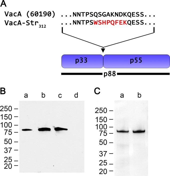
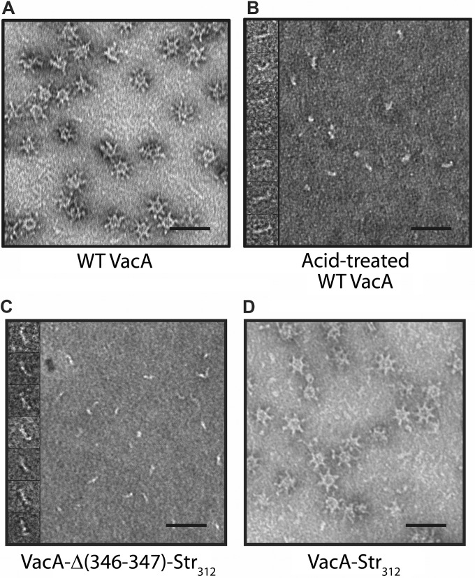
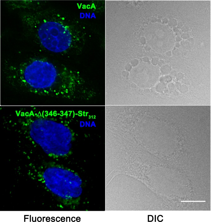
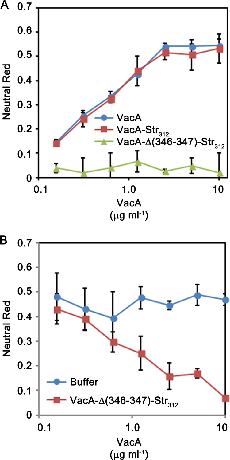
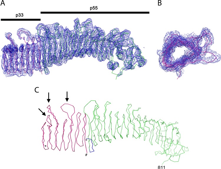
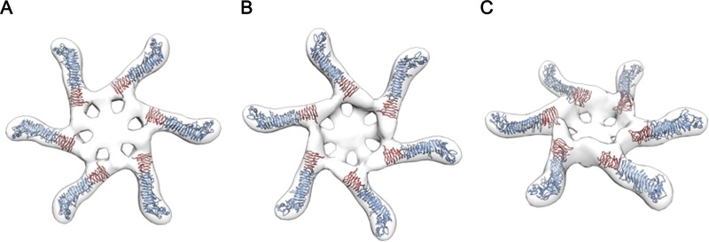
Helicobacter pylori secretes a pore-forming VacA toxin that has structural features and activities substantially different from those of other known bacterial toxins. VacA can assemble into multiple types of water-soluble flower-shaped oligomeric structures, and most VacA activities are dependent on its capacity to oligomerize. The 88-kDa secreted VacA protein can undergo limited proteolysis to yield two domains, designated p33 and p55. The p33 domain is required for membrane channel formation and intracellular toxic activities, and the p55 domain has an important role in mediating VacA binding to cells. Previous studies showed that the p55 domain has a predominantly β-helical structure, but no structural data are available for the p33 domain. We report here the purification and analysis of a nonoligomerizing mutant form of VacA secreted by H. pylori The nonoligomerizing 88-kDa mutant protein retains the capacity to enter host cells but lacks detectable toxic activity. Analysis of crystals formed by the monomeric protein reveals that the β-helical structure of the p55 domain extends into the C-terminal portion of p33. Fitting the p88 structural model into an electron microscopy map of hexamers formed by wild-type VacA (predicted to be structurally similar to VacA membrane channels) reveals that p55 and the β-helical segment of p33 localize to peripheral arms but do not occupy the central region of the hexamers. We propose that the amino-terminal portion of p33 is unstructured when VacA is in a monomeric form and that it undergoes a conformational change during oligomer assembly.


Figures

FIG 1
FIG 1
Introduction of a strep tag into VacA. The p88 VacA protein secreted by H. pylori strain 60190 (GenBank U05676) contains two domains, designated p33 and p55. H. pylori 60190 was engineered as described in Materials and Methods to allow production of VacA proteins with a strep tag introduced at the junction between p33 and p55 domains. (A) The figure compares the amino acid sequence of the p33-p55 junction in WT VacA with the corresponding sequence in a modified VacA protein containing the strep tag (WSHPQFEK) introduced at amino acid 312. (B) H. pylori strains were grown in broth culture, and unconcentrated broth culture supernatants were analyzed by immunoblotting using polyclonal anti-VacA serum. Lanes: a, strain 60190, producing WT VacA; b, strain producing VacA with a strep tag introduced at position 312 (VacA-Str312); c, strain producing VacA-Δ(346-347)-Str312; d, vacA-null mutant. (C) SDS-PAGE and Coomassie blue staining of strep-tagged VacA proteins, purified as described in Materials and Methods. Lanes: a, VacA-Str312; b, VacA-Δ(346-347)-Str312.
FIG 2
FIG 2
EM analysis of purified wild-type VacA and VacA-Δ(346-347)-Str312. VacA proteins were purified from H. pylori broth culture supernatant as described in Materials and Methods. (A to D) EM analysis of purified WT VacA at neutral pH (A), WT VacA exposed to low-pH conditions as described in Materials and Methods (B), VacA-Δ(346-347)-Str312 at neutral pH (C), and VacA-Str312 at neutral pH (D). The insets show high-magnification images of representative particles. Scale bar, 50 nm for all panels.
FIG 3
FIG 3
Internalization of VacA-Δ(346-347)-Str312 by gastric epithelial cells. AGS cells were treated for 4 h in the presence of 5 mM ammonium chloride with 5 μg/ml of either acid-activated WT VacA (top panels) or VacA-Δ(346-347)-Str312 (bottom panels), each labeled with Alexa Fluor 488. The cells were then washed with PBS, fixed with 4% paraformaldehyde, and stained with Hoechst to label DNA. Fluorescence and DIC images are shown. Scale bar, 10 μm.
FIG 4
FIG 4
Analysis of vacuolating toxic activity. H. pylori 60190 (producing WT VacA), a strain producing VacA-Str312, and a strain producing VacA-Δ(346-347)-Str312 were grown in broth cultures, and VacA proteins were purified as described in Materials and Methods. (A) HeLa cells were incubated with the indicated final concentrations of purified VacA proteins, and cell vacuolation was quantified by neutral red uptake assay (as determined by the optical density at 600 nm [OD600]). WT VacA and VacA-Str312 exhibited cell-vacuolating activity (corresponding to high neutral red uptake values), whereas VacA-Δ(346-347)-Str312 lacked vacuolating activity. (B) WT VacA (10 μg/ml) was incubated with the indicated concentrations of VacA-Δ(346-347)-Str312 or buffer only, and the mixtures were then added to HeLa cells in the presence of 5 mM ammonium chloride. Cell vacuolation was quantified by neutral red uptake assay (i.e., based on the OD600). VacA-Δ(346-347)-Str312 inhibited the activity of WT VacA.
FIG 5
FIG 5
Density map of VacA-Δ(346-347)-Str312. VacA-Δ(346-347)-Str312 was crystallized, and the crystals were analyzed as described in Materials and Methods. (A) The structure of the p55 domain is colored green, and the structure of a portion of the p33 domain is colored red. A 2Fo−Fc map contoured at 1.5 sigma reveals the presence of an extended β-helix structure in the p33 domain. Although the junction of p33 with the p55 fragment is not resolved, the density map suggests the presence of a β-helix segment in p33 consisting of 7 rungs and about 165 amino acids (red coloration). (B) Rotated view of the β-helix segment within the p33 domain. (C) Line representation of the VacA-Δ(346-347)-Str312 structure. The portion of p55 corresponding to the previously determined structure (residues 355 to 811) (52) is shown in green, and additional p55 residues mapped in the present study are shown are shown in blue. A β-helical portion of the p33 domain is shown in red. *, N-terminal end of p33 β-helix; #, C-terminal end of p33 β-helix; +, N-terminal end of p55 domain. Arrows designate distinctive loops in the p33 domain.
FIG 6
FIG 6
Model depicting assembly of VacA monomers into hexameric structures. The density map of VacA-Δ(346-347)-Str312 was fitted into an EM map of a VacA hexamer (31). The figure illustrates bottom, top, and side views of the hexamer. Blue indicates p55 domains, and red indicates an ∼165-amino-acid β-helical segment within the p33 domain.

Similar articles

Cited by

References

    1. Atherton JC, Blaser MJ. 2009. Coadaptation of Helicobacter pylori and humans: ancient history, modern implications. J Clin Invest 119:2475–2487. doi:10.1172/JCI38605. - DOI - PMC - PubMed
    1. Cover TL, Blaser MJ. 2009. Helicobacter pylori in health and disease. Gastroenterology 136:1863–1873. doi:10.1053/j.gastro.2009.01.073. - DOI - PMC - PubMed
    1. Gilbreath JJ, Cody WL, Merrell DS, Hendrixson DR. 2011. Change is good: variations in common biological mechanisms in the epsilonproteobacterial genera Campylobacter and Helicobacter. Microbiol Mol Biol Rev 75:84–132. doi:10.1128/MMBR.00035-10. - DOI - PMC - PubMed
    1. Rieder G, Fischer W, Haas R. 2005. Interaction of Helicobacter pylori with host cells: function of secreted and translocated molecules. Curr Opin Microbiol 8:67–73. doi:10.1016/j.mib.2004.12.004. - DOI - PubMed
    1. Zanotti G, Cendron L. 2014. Structural and functional aspects of the Helicobacter pylori secretome. World J Gastroenterol 20:1402–1423. doi:10.3748/wjg.v20.i6.1402. - DOI - PMC - PubMed

LinkOut - more resources