. 2016 Jul 19;113(29):E4133-42.
doi: 10.1073/pnas.1600299113. Epub 2016 Jul 5.

Dendrimer-RNA nanoparticles generate protective immunity against lethal Ebola, H1N1 influenza, and Toxoplasma gondii challenges with a single dose

Affiliations

Dendrimer-RNA nanoparticles generate protective immunity against lethal Ebola, H1N1 influenza, and Toxoplasma gondii challenges with a single dose

Jasdave S Chahal et al. Proc Natl Acad Sci U S A. .

Erratum in

Abstract

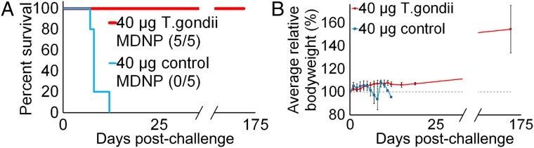
Vaccines have had broad medical impact, but existing vaccine technologies and production methods are limited in their ability to respond rapidly to evolving and emerging pathogens, or sudden outbreaks. Here, we develop a rapid-response, fully synthetic, single-dose, adjuvant-free dendrimer nanoparticle vaccine platform wherein antigens are encoded by encapsulated mRNA replicons. To our knowledge, this system is the first capable of generating protective immunity against a broad spectrum of lethal pathogen challenges, including H1N1 influenza, Toxoplasma gondii, and Ebola virus. The vaccine can be formed with multiple antigen-expressing replicons, and is capable of eliciting both CD8(+) T-cell and antibody responses. The ability to generate viable, contaminant-free vaccines within days, to single or multiple antigens, may have broad utility for a range of diseases.

Keywords: nanoparticle; parasites; replicon; vaccine platform; viruses.


Conflict of interest statement

Conflict of interest statement: A patent has been filed for the nanoparticle vaccine by J.S.C., O.F.K., R.L., H.L.P., and D.G.A.

Figures

Fig. 1.
Fig. 1.
MDNP vaccine platform. (A) Ionizable dendrimer-based nanomaterial, a lipid-anchored PEG, and RNA are combined to form the final vaccine nanoparticle. Additional characterization is provided in Fig. S9. (B) Size distribution of the MDNPs by dynamic light scattering. Error bars ± SD and n = 3. (C) Transmission electron micrograph of MDNPs. (Scale bar: 50 nm.) (D) Firefly luciferase activity in BHK cells mediated by conventional mRNA and replicon RNAs based on VEEV and SFV genomes, assayed 14 h posttransfection. Error bars ± SD and n = 3. (E) Stability of formulated nanoparticles using different nanomaterials over 2 h in 50% AB human serum at 37 °C. MDNPs were formulated to contain an equimolar mixture of RNAs labeled with Alexa Fluor 594 and Alexa Fluor 647. When in close contact (i.e., within the intact MNDP), the RNAs would act as a FRET pair. When the MDNPs disassembled, the released RNAs would no longer close enough to generate a FRET signal. To measure FRET, the samples were excited at 540 nm and the fluorescence intensity was read at 690 and 620 nm. FRET was determined as the ratio of the fluorescence intensities at 690/620 nm. FRET signals were normalized to the signals of free RNA controls. The more stable MDNP nanomaterial was synthesized using 2-tridecyloxirane, whereas the less stable control MDNP used 1,2-Epoxydodecane. Negative controls were free RNA. PBS was used to determine background levels. The FRET signal was normalized to the value of the completely ruptured nanoparticles. Nanoencapsulation in the MDNP provides superior protection because nanoparticles remain intact and do not release their RNA payloads while in whole human serum. Because of this stability, the RNA payloads are protected from endonuclease degradation. Error bars ± SD and n = 4–8. (F) Vaccine stability over long periods of storage. Nanoparticles containing luciferase replicon RNA were created and stored at 4 °C for extended periods of time. HeLa cells were treated with a fixed amount of the nanoparticles, and luciferase expression was assayed after 14 h. After a minimum of 30 d of storage, no statistically significant changes in luciferase transfection efficiency were detected by ANOVA analysis (with Tukey multiple comparison correction), indicating the particles remained stable and the RNA payload remained intact. Error bars ± SD and n = 4.
Fig. S9.
Fig. S9.
Nanomaterial characterization. Electrospray ionization data for the modified dendrimer nanomaterial.
Fig. 2.
Fig. 2.
MDNP RNA vaccines stimulate CD8+ T-cell responses against a model intracellular antigen. (A) Quantification of four independent biological replicates of OT-1 proliferation assay performed 4 d posttransfer/3 d postimmunization. Error bars ± SD. (B) Quantification of six independent biological replicates of OT-1 proliferation assay performed 10 d posttransfer/14 d postimmunization. Error bars ± SD. (C) Intracellular cytokine staining for IFN-γ and IL-2 on splenocytes of VEEV-cOVA MDNP-immunized mice 9 d postinjection, gated on CD8+ cells. Independent results are shown for two unimmunized (Left) and two immunized (Right) mice. Unimm., unimmunized.
Fig. S1.
Fig. S1.
Transgene delivery mediated by MDNP administration of cultured myotubes or by intramuscular injection in vivo. (A) Differentiated C2C12 myoblast cells were differentiated in 96-well dishes for 5 d and treated with 1 μg of mCherry-encoding VEEV replicon encapsulated in MDNP. After 24 h, the MDNP-treated or PBS-treated control wells were imaged by fluorescence microscopy. (Magnification: 100×.) (B) Four micrograms of the indicated luciferase-encoding RNA was injected bilaterally into the thigh muscles, and luciferase activity was measured at 36 and 60 h postinjection by IVIS. Max, maximum; Min, minimum.
Fig. S2.
Fig. S2.
MDNP RNA vaccines stimulate CD8+ T-cell responses against a model intracellular antigen. (A) Expression of cOVA in BHK cells mediated by conventional mRNA and replicon RNAs based on VEEV and SFV genomes, assayed 14 h posttransfection by immunoblot. (B, Top four panels) gating strategy to identify adoptively transferred OT-1 cells. (B, Bottom six panels) Representative results of OT-1 proliferation assay performed 4 d posttransfer/3 d postimmunization. (C) Results of OT-1 proliferation assay performed 4 d posttransfer/3 d postimmunization with 40 μg of VEEV-cOVA MDNPs. Results of two identical independent experiments shown.
Fig. S3.
Fig. S3.
HIV Gag polyprotein expression mediated by VEEV replicon in BHK21 cells. Immunoblotting was performed 14 h posttransfection with commercial transfection reagents with HIV1 Gag-specific goat polyclonal IgG vT-20 (Santa Cruz Biotechnology).
Fig. S4.
Fig. S4.
VEEV-based MDNP immunization protects against lethal EBOV challenge. (A) Expression of EBOV GP in BHK cells mediated by conventional or 3′ UTR-stabilized mRNA and replicon RNAs based on VEEV and SFV genomes, assayed 26 h posttransfection (using commercial transfection agents) by immunoblot. (B) MDNP-encapsulated VEEV RNAs direct synthesis of EBOV GP in culture. BHK21 cell lysates were assayed by immunoblot after 14 h.
Fig. S5.
Fig. S5.
Proportions of SIINFEKL-tetramer–positive staining CD8+ cells measured in blood of immunized mice. (A) Mice were immunized with 40 μg of MDNP-encapsulated cOVA VEEV. Blood was collected on the indicated days postimmunization, and discrete tetramer-positive populations were detected by FACS analysis. Results are expressed as the percentage of total gated CD8+ cells. Error bars ± SD for n = 3. (B) Separate experiment for comparison, essentially identical to A, but mice were immunized in an independent experiment with MDNP-encapsulated cOVA mRNA. Note the different time scale, selected to capture the anticipated earlier peaking of the mRNA-stimulated immune response. (C) Detection of cOVA mRNA in mouse tissues 11 d postimmunization. Mice were necropsied, and the indicated organs and tissues were harvested. Samples were immediately frozen in liquid nitrogen, and cOVA mRNA levels were quantified via a QuantiGene 2.0 kit (Affymetrix) as described in SI Materials and Methods. Mean transcript abundance is expressed relative to signal intensity obtained from samples from an unimmunized control animal. Error bars ± SD for n = 3. Differences between mRNA levels in treated and untreated animals were considered statistically significant if *P < 0.05.
Fig. S6.
Fig. S6.
VEEV replicon stimulates the type I IFN response in mouse cells. B16 cells carrying a SEAP reporter gene under the control of a combined IFN-α/β–inducible ISG54 promoter and ISRE regulatory element and lacking IFN-γ receptor activity [“type I” reporter cells (B16-Blue IFN-α/β cells; Invivogen], or carrying the same reporter construct and lacking type I IFN receptor activity [“type II” reporter cells (B16-Blue IFN-γ cells; Invivogen)] were transfected with 5-methylcytidine/pseudouridine base-modified mRNA as a control (5meC/Ψ mRNA; Trilink) or VEEV replicon RNA using TransIT-mRNA reagents, as described in Materials and Methods. SEAP activity in the culture medium was quantified by colorimetric assay 20 h posttransfection. Error bars ± SD for biological triplicates.
Fig. S7.
Fig. S7.
Single-dose VEEV-based MDNP immunization protects against lethal H1N1 influenza challenge. (A) Immunoblot showing HA expression in VEEV-HA–transfected cells. (B) FACS analysis of surface staining with an HA-specific, fluorophore-conjugated, single-chain antibody confirming surface expression of HA protein. Untransf., untransfected.
Fig. 3.
Fig. 3.
Single-dose VEEV-based MDNP immunization protects against lethal H1N1 influenza challenge. (A) Protection against influenza challenge. Mice were infected with a lethal dose of influenza A/NWS/33 on day 0, and body weight was plotted as a percentage of preinfection weight. Mice were euthanized when body weight dropped below 80%. One hundred percent of control-immunized mice died, and 100% of HA-immunized mice fully recovered and survived for 3 wk with no signs of infection, at which point the experiment was terminated. Error bars ± SD and n = 3. (B) Serum anti-HA IgG in response to infection. HA-immunized surviving mice exhibited higher HA-specific antibody titers 7 d postinfection than control-immunized mice at the time of death. Results for each individual animal are shown. Error bars ± SD of technical duplicates.
Fig. 4.
Fig. 4.
VEEV-based MDNP immunization protects against lethal EBOV challenge. (A) EBOV-GP–specific T-cell responses assayed 9 d postimmunization with 40 μg of VEEV-GP MDNP vaccine. Splenocytes were cultured ex vivo for 6 h in the absence or presence of the EBOV-GP–derived WE15 peptide, and IFN-γ and IL-2 expression in CD8+ and CD4+ cells was assayed by intracellular cytokine staining and FACS. One representative result of three similar independent experiments is shown. (B) EBOV-GP specific IgG antibody titers were determined by ELISA from mice vaccinated with the indicated amounts of Ebola-GP MDNP, 40 μg of cOVA MDNP, or 40 μg of naked VEE-GP on days 0 and 21. A single-dose, prime-only vaccination with 40 μg of EBOV-GP MDNP was also tested. Titers were determined by reciprocal end-point dilution on serum from day 23 postvaccination. Red symbols denote animals that succumbed to EBOV disease. The dashed line represents the assay’s level of detection. (C) Survival of animals following challenge with 1,000 pfu of mouse-adapted EBOV 28 d postvaccination. Control animals are denoted by a solid gray line. Study was concluded 21 d postinfection (n = 10 animals per group).
Fig. 5.
Fig. 5.
VEEV-based MDNP immunization protects against lethal T. gondii infection. (A) Survival curve of animals vaccinated with hexaplex MDNPs containing GRA6, ROP2A, ROP18, SAG1, SAG2A, and AMA1 replicons. Control animals received MDNPs containing an irrelevant antigen (HA). All control animals (n = 5) succumbed to infection, whereas all hexaplex-immunized animals survived (n = 5). (B) Relative body weights of mice (normalized to preinfection weights) were tracked over time.
Fig. S8.
Fig. S8.
Antigen expression. (A) Simultaneous antigen production from multiplexed MDNPs. MDNPs were coformulated with equimolar amounts of VEEV GP and VP40 replicons. BHK cells were treated with 1.25 μg worth of total replicon (0.625 μg per antigen). Western blots performed on cell lysates confirmed the coexpression of both antigens from multiplexed MDNPs. (B) T. gondii antigen expression. BHK cells were transfected with VEEV replicons encoding GRA6, ROP2A, ROP18, SAG1, SAG2A, or AMA1. Antigens were FLAG-tagged to facilitate detection by immunoblot.

References

    1. Arnon R, Ben-Yedidia T. Old and new vaccine approaches. Int Immunopharmacol. 2003;3(8):1195–1204. - PubMed
    1. Azad N, Rojanasakul Y. Vaccine delivery—Current trends and future. Curr Drug Deliv. 2006;3(2):137–146. - PubMed
    1. Koblin BA, et al. NIAID HIV Vaccine Trials Network Safety and immunogenicity of an HIV adenoviral vector boost after DNA plasmid vaccine prime by route of administration: A randomized clinical trial. PLoS One. 2011;6(9):e24517. - PMC - PubMed
    1. Pittman PR. Aluminum-containing vaccine associated adverse events: Role of route of administration and gender. Vaccine. 2002;20(Suppl 3):S48–S50. - PubMed
    1. Scheiermann J, Klinman DM. Clinical evaluation of CpG oligonucleotides as adjuvants for vaccines targeting infectious diseases and cancer. Vaccine. 2014;32(48):6377–6389. - PMC - PubMed

Publication types

MeSH terms