. 2016 Mar;7(3-4):110-124.
doi: 10.18632/genesandcancer.104.

MUC16 contributes to the metastasis of pancreatic ductal adenocarcinoma through focal adhesion mediated signaling mechanism

Affiliations

MUC16 contributes to the metastasis of pancreatic ductal adenocarcinoma through focal adhesion mediated signaling mechanism

Sakthivel Muniyan et al. Genes Cancer. 2016 Mar.

Abstract

MUC16, a heavily glycosylated type-I transmembrane mucin is overexpressed in several cancers including pancreatic ductal adenocarcinoma (PDAC). Previously, we have shown that MUC16 is significantly overexpressed in human PDAC tissues. However, the functional consequences and its role in PDAC is poorly understood. Here, we show that MUC16 knockdown decreases PDAC cell proliferation, colony formation and migration in vitro. Also, MUC16 knockdown decreases the tumor formation and metastasis in orthotopic xenograft mouse model. Mechanistically, immunoprecipitation and immunofluorescence analyses confirms MUC16 interaction with galectin-3 and mesothelin in PDAC cells. Adhesion assay displayed decreased cell attachment of MUC16 knockdown cells with recombinant galectin-1 and galectin-3 protein. Further, CRISPR/Cas9-mediated MUC16 knockout cells show decreased tumor-associated carbohydrate antigens (T and Tn) in PDAC cells. Importantly, carbohydrate antigens were decreased in the region that corresponds to MUC16 and suggests for the decreased MUC16-galectin interactions. Co-immunoprecipitation also revealed a novel interaction between MUC16 and FAK in PDAC cells. Interestingly, we observed decreased expression of mesenchymal and increased expression of epithelial markers in MUC16-silenced cells. Additionally, MUC16 loss showed a decreased FAK-mediated Akt and ERK/MAPK activation. Altogether, these findings suggest that MUC16-focal adhesion signaling may play a critical role in facilitating PDAC growth and metastasis.

Keywords: CRISPR/Cas9; FAK; MUC16; metastasis; pancreatic cancer.


Conflict of interest statement

CONFLICT OF INTEREST

The authors have declared that no competing interests exist.

Figures

Figure 1
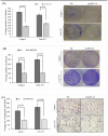
Figure 1. MUC16 knockdown alters in capan-1 and colo-357 PDAC cell proliferation
(A). Immunoblot analyzes were performed to detect the MUC16 expression in MUC16 knock down capan-1 and colo-357 cell lines. A 40μg of total protein was resolved electrophoretically on a 10% SDS gel, transferred on to PVDF membranes and probed with the anti-MUC16 antibody. β-actin was used as a loading control. (B). Immunofluorescent analyzes of MUC16 expression in MUC16 knock down capan-1 and Colo-357 cell lines. MUC16 knockdown colo-357 and capan-1 cells with respective Scr transfected cells were cultured on the coverslip for 48 hours. 48 hours later, cells were fixed and probed with the anti-MUC16 antibody. Primary antibody probing was followed by FITC-conjugated anti-mouse secondary antibody and counterstained with DAPI. All the microscopic pictures are in the similar magnification (x630); scale bar: 50 μm. The MUC16 knock down capan-1 (C), and Colo-357 (D) cells were plated in triplicates in six-well plates at a density of 10×104 cells/well and cultured in 2% serum containing media. The cells were trypsinized and counted every 24 hours for seven days, and the growth curve was plotted for the number of cells counted versus time of incubation. * indicates p < 0.05 and ** indicates p < 0.01.
Figure 2
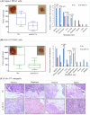
Figure 2. MUC16 expression loss decreases colony formation and migration of PDAC cells
(A). Colony-forming ability of MUC16 knock downed cells in comparison with respective Scr transfected cells were performed in vitro. Both colo-357 and capan-1 cells were plated at a density of 2 × 10³ cells per well in a six-well plate. After 16 hours incubation, the unattached cells were removed by changing the media. The cells were fed fresh media once in three days. Twelve days later, the cells were washed with PBS, stained with crystal violet and photographed. The bar diagram on the left indicates the number of colonies. The panel on the right shows the representative picture of respective groups. (B). MUC16 knockdown colo-357 and capan-1 with respective Scr control cells were plated at a density of 5 × 10² cells/cm2 in soft agar plates. After 24 hours, the dish containing double cells were excluded. Cells were fed with fresh culture media twice a week. By the end of 5 weeks, the colonies formed were stained with 0.1 % crystal violet and counted. The bar diagram on the left indicates the number of colonies. The right panel shows the representative picture of colonies. (C). The migratory ability of MUC16 in colo-357 and capan-1 cells. MUC16 knockdown colo-357 and capan-1 with respective Scr control cells of 1×10⁶ cells were plated on top of the 6-well insert. After 24 hours, the cells that migrated were fixed, stained and counted. The data on the left indicates the average number of PDAC cells per field of view (original magnification ×10). The right panel represents the cells from different groups.
Figure 3
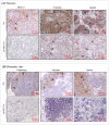
Figure 3. Loss of MUC16 expression decreases the in vivo tumorigenic potential of PDAC cells
(A). MUC16 depleted (sh-MUC16) and scramble vector (Scr) transfected capan-1 cells in 0.05 ml orthotopically injected into the nude mice. Ten mice were assigned to each group. Mice were continuously monitored for growth and weight. After 21 days, the tumors were resected and weighed. All the major organs were dissected, and metastatic tumor growth was analyzed by both visually and microscopically in H & E staining. Box plot on the left panel indicates the orthotopic tumor growth. The tumor weights of each mouse are represented by a dot. Right panel indicates the number of metastasis incidence from respective group. (B). MUC16 depleted (sh-MUC16) and scramble vector (Scr) transfected colo-357 cells were orthotopically injected into the nude mice. Eight and seven mice were assigned to Scr and sh-MUC16 group, respectively. Mice were continuously monitored for growth and weight. After 60 days, the tumors were resected and weighed. All the major organs were dissected, and metastatic tumor growth was analyzed by both visually and microscopically in H & E staining. Box plot on the left panel indicates the orthotopic tumor growth. The tumor weights of each individual mice are represented as a dot. Right panel indicates the number of metastasis incidence from respective group. (C). Hematoxylin and eosin stain of xenograft in primary and metastatic sites of colo-357 xenografts in orthotopic model. Colo-357 xenografts were harvested and processed for H and E staining on primary as well as metastatic sites. All the micrographs are in the same magnification (original × 10).
Figure 4
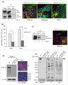
Figure 4. Immunohistochemical analyses of MUC16 and metastatic markers in primary and metastatic sites of colo-357 xenografts
(A). Immunohistochemical staining for MUC16, vimentin and MMP9 in colo-357 cells implanted pancreas. Black arrow heads indicates the immunostaining of MUC16 (cell surface), vimentin (cytoplasm) and MMP9 (cytoplasm and extracellular). (B). Immunohistochemical staining for MUC16 in metastatic site of colo-357 cells implanted mice. Black arrow in the representative image indicates the cell surface immunostaining of MUC16. Nuclei are stained with hematoxylin (blue). All the micrographs are in the same magnification (original × 20).
Figure 5
Figure 5. MUC16 interact with mesothelin and galectin-3 in PDAC cells
(A). Endogenous MUC16 and mesothelin protein interaction in colo-357 cells were analyzed by co-immunoprecipitation and immunoblotting using specific antibodies. IgG was used as the negative control with the same amount of protein (left panel). Right panel shows the MUC16-mesothelin interaction in colo-357 cells implanted mice pancreas and other metastatic sites. Colocalization were enlarged and presented as inset. All the microscopic pictures are in the similar magnification (scale bar: 100 μm). (B). MUC16 knockdown colo-357 cells were plated in triplicate to galectin-1 and galectin-3 protein-coated 96-well plates. After 1 h incubation at 37°C, unattached cells were washed carefully with PBS twice. The adhered cells were incubated with Calcein-AM dye, and the fluorescence was measured. (C). MUC16 interaction with galectin-3 were determined by immunoprecipitation and Immunofluorescence analyzes. Colo-357 cells were immunoprecipitated using MUC16 antibody and were probed using galectin-3 specific antibodies. IgG was used as the negative control with the same amount of protein (left panel). The right panel shows the MUC16-galectin-3 interaction in primary site of the pancreas. All the microscopic pictures are in the similar magnification (scale bar: 100 μm). (D). Immunoblot analyzes were performed to determine the MUC16 expression level in CRISPR/Cas-mediated MUC16 knockout in capan-1 cells. A 40 μg of total protein was resolved in SDS-PAGE, transferred on to PVDF membranes and probed with the anti-MUC16 antibody. β-actin was used as a loading control (left panel). Right panel show immunofluorescent analyzes of MUC16 expression in parental and MUC16-deleted capan-1 cells. Confocal images are in the similar magnification (scale bar: 100 μm). (E). Immunoblot analysis were carried out to determine the T and Tn antigen level in wildtype and MUC16 CRISPR/Cas-based knockout capan-1 cells. The immunoblot shows a clear decrease in both T- and Tn-antigen level in MUC16 depleted samples. Solid black arrow indicates the MUC16 migration position in PAGE. MSLN: mesothelin; Gal-1: galectin-1; Gal-3: galectin-3; T: Thomsen-Friedenreich and Tn: Thomsen-nouvelle antigens.
Figure 6
Figure 6. Novel interaction of MUC16 with FAK and activates its downstream signaling in PDAC cells
(A). Colo-357 cell lysates were pull down using either MUC16 or FAK or respective IgG antibodies. Immunoblot of bound fractions confirms the protein of interest expression. Reciprocally the MCU16 pulldown samples were probed with anti-FAK antibodies and vice versa. IgG was used as a negative control with the same amount of protein. (B). MUC16 knockdown colo-357 and capan-1 with respective vector control cells were cultured for 48 hours. After 48 hours, MUC16 knockdown with respective vector control cells were collected, lysed and analyzed for p-FAK (Tyr925), FAK, E-cadherin, CK18, N-cadherin, and Zeb-1 protein levels. β-actin protein level was used as loading control. (C). MUC16 shRNA and vector transfected colo-357 and capan-1 cells were maintained under the same condition as described above. The lysates were analyzed for FAK-mediated AKT and ERK/MAPK phosphorylation using immunoblotting analyzes. β-actin protein level was used as loading control.

References

    1. Chaturvedi P, Singh AP, Moniaux N, Senapati S, Chakraborty S, Meza JL, Batra SK. MUC4 mucin potentiates pancreatic tumor cell proliferation, survival, and invasive properties and interferes with its interaction to extracellular matrix proteins. Mol Cancer Res. 2007;5:309–320. - PubMed
    1. Torres MP, Chakraborty S, Souchek J, Batra SK. Mucin-based targeted pancreatic cancer therapy. Curr Pharm Des. 2012;18:2472–2481. - PMC - PubMed
    1. Horn A, Chakraborty S, Dey P, Haridas D, Souchek J, Batra SK, Lele SM. Immunocytochemistry for MUC4 and MUC16 is a useful adjunct in the diagnosis of pancreatic adenocarcinoma on fine-needle aspiration cytology. Arch Pathol Lab Med. 2013;137:546–551. - PMC - PubMed
    1. Kohlgraf KG, Gawron AJ, Higashi M, Meza JL, Burdick MD, Kitajima S, Kelly DL, Caffrey TC, Hollingsworth MA. Contribution of the MUC1 tandem repeat and cytoplasmic tail to invasive and metastatic properties of a pancreatic cancer cell line. Cancer Res. 2003;63:5011–5020. - PubMed
    1. Chugh S, Gnanapragassam VS, Jain M, Rachagani S, Ponnusamy MP, Batra SK. Pathobiological implications of mucin glycans in cancer: Sweet poison and novel targets. Biochim Biophys Acta. 2015;1856:211–225. - PMC - PubMed