Lipoma arborescens arising in the extra-articular bursa of the knee joint
- PMID: 27382924
- PMCID: PMC4935798
- DOI: 10.1051/sicotj/2016019
Lipoma arborescens arising in the extra-articular bursa of the knee joint
Abstract
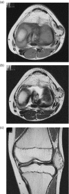
Lipoma arborescens arising in the extra-articular bursa of the knee joint is extremely rare. We describe an 11-year-old boy who complained of a gradual swelling mass of the lateral knee joint. Magnetic resonance imaging (MRI) showed a high signal intensity tumor on T1- and T2-weighted images with a thickened septa and nodular lesion that showed low signal intensity. The radiologist suggested the possible differential diagnosis of well-differentiated liposarcoma. At operation, the tumor was found under the iliotibial tract and was not in contact with the knee joint. Histopathologically, this lesion was diagnosed as lipoma arborescens arising in the extra-articular bursa of the knee joint. On MRI, the appearance of lipoma arborescens arising in the extra-articular bursa of the knee joint differed from that of conventional intra-articular lipoma arborescens. In this report, we describe a case of extra-articular lipoma arborescens of the knee joint bursa and discuss the diagnosis and etiology.
© The Authors, published by EDP Sciences, 2016.
Figures


References
-
- Mulder JD, Schutte HE, Kroon HM, Taconis WK (1993) Radiologic Atlas of Bone Tumors. Amsterdam, Elsevier Science Publishers.
-
- Laorr A, Peterfy CG, Tirman PF, Rabassa AE (1995) Lipoma arborescens of the shoulder: magnetic resonance imaging findings. Can Assoc Radiol J 46(4), 311–313. - PubMed
-
- Nisolle JF, Blouard E, Baudrez V, Boutsen Y, De Cloedt P, Esselinckx W (1999) Subacromial-subdeltoid lipoma arborescens associated with a rotator cuff tear. Skeletal Radiol 28(5), 283–285. - PubMed
-
- Levadoux M, Gadea J, Flandrin P, Carlos E, Aswad R, Panuel M (2000) Lipoma arborescens of the elbow: a case report. J Hand Surg Am 25(3), 580–584. - PubMed
-
- Noel ER, Tebib JG, Dumontet C, Colson F, Carret JP, Vauzelle JL, Bouvier M (1987) Synovial lipoma arborescens of the hip. Clin Rheumatol 6(1), 92–96. - PubMed
LinkOut - more resources
Full Text Sources
Other Literature Sources
