. 2016 Jul 6;36(27):7298-313.
doi: 10.1523/JNEUROSCI.4064-15.2016.

Rab35 Functions in Axon Elongation Are Regulated by P53-Related Protein Kinase in a Mechanism That Involves Rab35 Protein Degradation and the Microtubule-Associated Protein 1B

Affiliations

Rab35 Functions in Axon Elongation Are Regulated by P53-Related Protein Kinase in a Mechanism That Involves Rab35 Protein Degradation and the Microtubule-Associated Protein 1B

David Villarroel-Campos et al. J Neurosci. .

Abstract

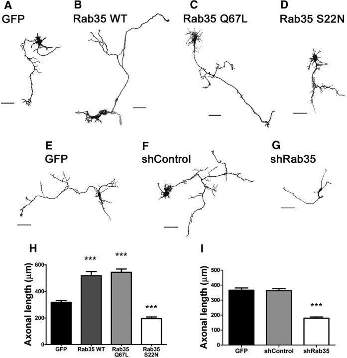
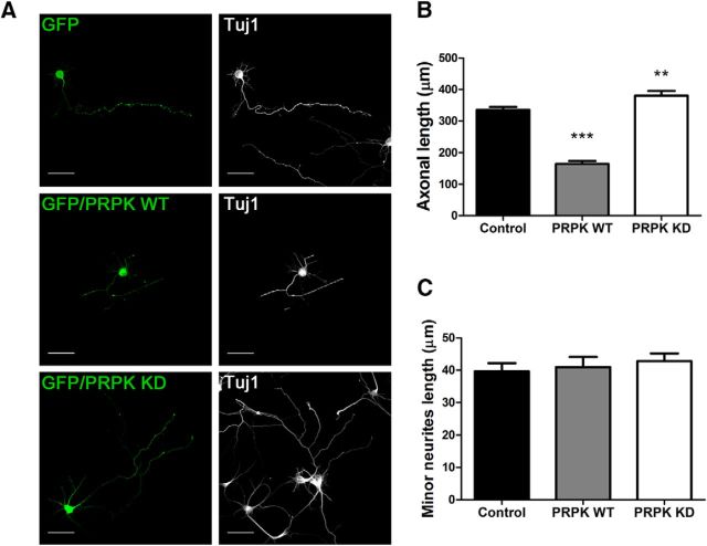
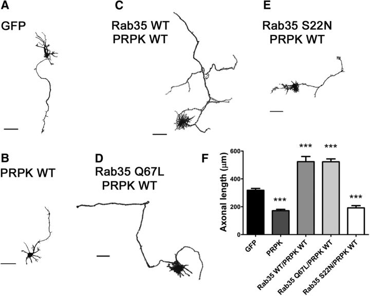
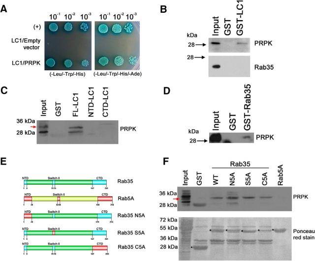
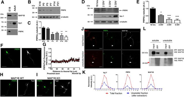
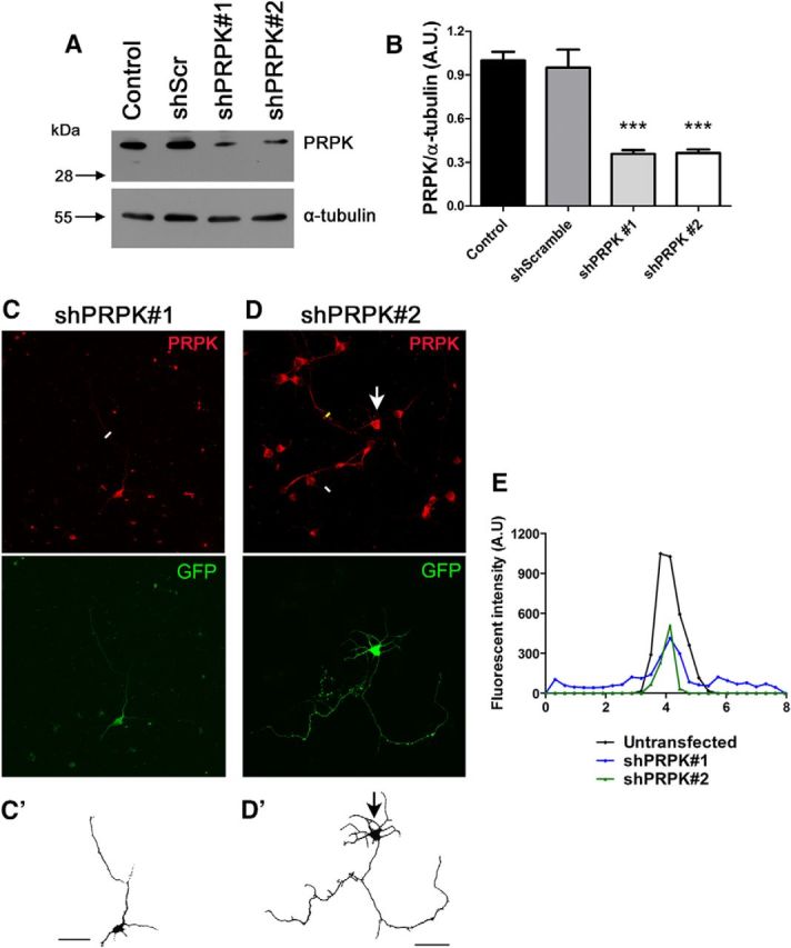
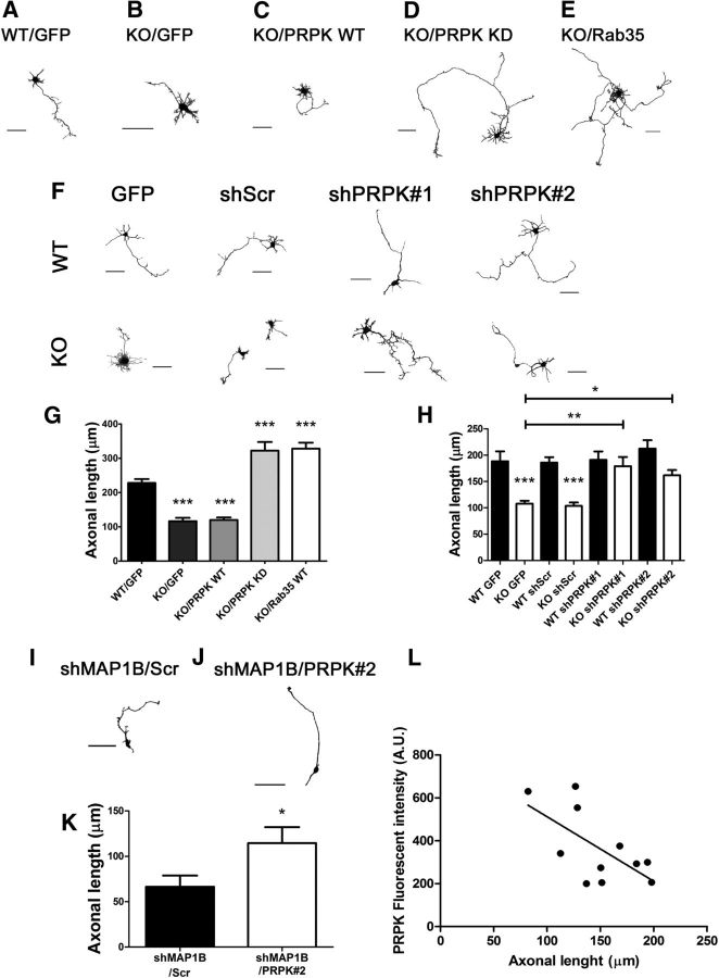
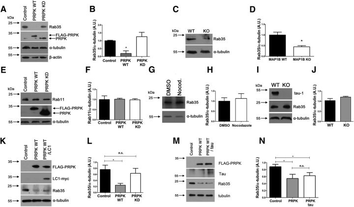
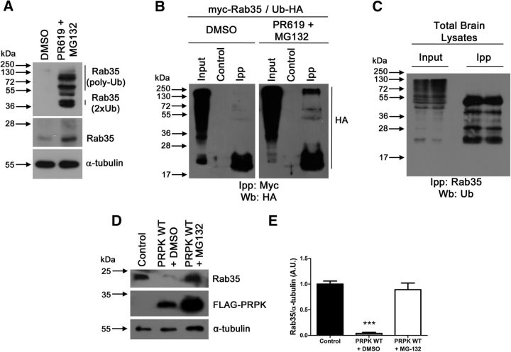
Rab35 is a key protein for cargo loading in the recycling endosome. In neuronal immortalized cells, Rab35 promotes neurite differentiation. Here we describe that Rab35 favors axon elongation in rat primary neurons in an activity-dependent manner. In addition, we show that the p53-related protein kinase (PRPK) negatively regulates axonal elongation by reducing Rab35 protein levels through the ubiquitin-proteasome degradation pathway. PRPK-induced Rab35 degradation is regulated by its interaction with microtubule-associated protein 1B (MAP1B), a microtubule stabilizing binding protein essential for axon elongation. Consistently, axon defects found in MAP1B knock-out neurons were reversed by Rab35 overexpression or PRPK inactivation suggesting an epistatic relationship among these proteins. These results define a novel mechanism to support axonal elongation, by which MAP1B prevents PRPK-induced Rab35 degradation. Such a mechanism allows Rab35-mediated axonal elongation and connects the regulation of actin dynamics with membrane trafficking. In addition, our study reveals for the first time that the ubiquitin-proteasome degradation pathway regulates a Rab GTPase.

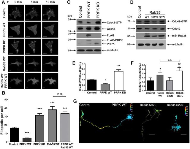
Significance statement: Rab35 is required for axonal outgrowth. We define that its protein levels are negatively regulated by p53-related protein kinase (PRPK). We show that microtubule-associated protein 1B (MAP1B) interacts with PRPK, preventing PRPK-dependent Rab35 proteasome degradation. We demonstrate that Rab35 regulates Cdc42 activity in neurons. This is the first evidence showing that a Rab protein is regulated by degradation dependent on the ubiquitin-proteasome system.

Keywords: MAP1B; Rab35; axon development; p53-related protein kinase; ubiquitin proteosome.


Figures

Figure 1.
Figure 1.
Rab35 promotes axonal growth. AD, Cultured hippocampal neurons expressing GFP (control; A), Rab35 WT (B), Rab35 Q67L (C) or Rab35 S22N (D). Cells were fixed at 3 DIV. Images were segmented and the color inverted. Scale bar, 50 μm. E–G, Cultured hippocampal neurons expressing GFP (control; E), shControl (F) or shRab35(g). Cells were fixed at 3 DIV. Images were segmented and the colors inverted. Scale bar, 50 μm. H, Quantification of axon length in neurons transfected as in AD. n = 50 neurons from eight different cultures; one-way ANOVA with Dunnett's post-test (***p < 0.001). I, Quantification of axon length in neurons transfected as in EG. n = 50 neurons from three different cultures; one-way ANOVA with Dunnett's post-test (***p < 0.001).
Figure 2.
Figure 2.
PRPK negatively regulates axon elongation. A, Cultured hippocampal neurons transfected with GFP (control), GFP/PRPK WT, or GFP/PRPK KD. Cells were fixed and immunostained after 3 DIV. Tuj1 staining is also shown. Scale bar, 50 μm. B, Quantification of axon length in cells transfected as in A. n = 50 neurons from three different cultures; one-way ANOVA with Dunnett's post-test (**p < 0.01, ***p < 0.001). C, Quantification of minor neurite length in cells transfected as in A. n = 50 neurons from three different cultures; one-way ANOVA with Dunnett's post-test (no statistically significant differences were observed).
Figure 3.
Figure 3.
Rab35 rescues PRPK-induced inhibition of axon growth. AE, Cultured hippocampal neurons at 3 DIV expressing GFP (A) or PRPK WT (B) as controls, or coexpressing PRPK WT with either Rab35 WT (C), Rab35 Q67L (D), or Rab35 S22N (E). Images processed as in Figure 5. Scale bar, 50 μm. F, Quantification of axon length in neurons transfected as in AE. n = 30 neurons from eight different cultures; one-way ANOVA with Dunnett's post-test (***p < 0.001).
Figure 4.
Figure 4.
Epistatic relationship of PRPK and Rab35 in actin-related processes. A, Still images taken from time-lapse recordings over a 10 min period, from undifferentiated N1E-115 cells transfected with EGFP-Lifeact (Control) or cotransfected with EGFP-Lifeact and either PRPK WT, PRPK KD, or Rab35 WT. Additionally, cells expressing EGFP-Lifeact, PRPK WT, and Rab35 were included. Movies were acquired at 2 frames/min. The first, middle, and last frames of each movie were deconvoluted, and are presented from left to right. Scale bar, 15 μm. B, Quantification of the mean number of filopodia per cell during the entire recording period. n = 20 cells, except for Rab35 WT, where n = 18; one-way ANOVA with Dunnett's post-test (***p < 0.001). C, Cdc42 activity pull-down assay in COS7 cells expressing PRPK WT or PRPK KD. Untransfected cells were used as a control. D, Cdc42 activity pull-down assay in COS7 cells expressing Rab35 WT, S22N, or the Q67L mutant. Untransfected cells were used as a control. E, Quantification of the Cdc42-GTP/Cdc42 ratio from C. n = 3; one-way ANOVA with Dunnett's post-test (*p < 0.05, **p < 0.01). F, Quantification of the Cdc42-GTP/Cdc42 ratio from D. n = 3; one-way ANOVA with Bonferroni's post-test (*p < 0.05, **p < 0.01; indicating differences with control condition. #p < 0.05, ##p < 0.01; indicating differences with S22N condition). G, Two DIV cultured hippocampal neurons expressing a FRET biosensor for Cdc42 alone (control condition) or coexpressing PRPK WT, Rab35 Q67L, or Rab35 S22N. FRET maps of representative neurons are depicted with a thermal scale. Scale bar, 40 μm.
Figure 5.
Figure 5.
PRPK interacts with LC1 and Rab35. A, Y2H binding assay with AH109 yeast cotransformed with pGBKT7-LC1 and pGADT7-PRPK. Yeast carrying pGADT7-T and pGBKT7–p53 were used as a positive control (+); yeast cotransformed with pGBKT7-LC1 and empty pGADT7 were used as a negative control. Yeast from all three cotransformations were grown in low-stringency TDO (-Leu/-Trp/-His) and high-stringency QDO (-Leu/-Trp/-His/-Ade) media supplemented with X-α-Gal. Decreasing dilutions were seeded and allowed to grow for 4 d. BD, GST pull-down assay from (E18) rat brain protein extracts. GST-LC1 and GST alone were incubated with brain supernatants, and the bound fraction was analyzed by immunoblotting for PRPK (B, top) and Rab35 (B, bottom). GST pull-down assays using full-length LC1, and N- and C-terminal domains were incubated with brain supernatants and the bound fraction analyzed by immunoblotting for PRPK (C). GST-Rab35 and GST alone were incubated with brain supernatants, and the bound fraction was analyzed by immunoblotting for PRPK (D). E, Schematic representation of Rab35 (swapping motif in light blue) and Rab5 (swapping motif in red) protein domains. Swapping mutants for Rab35 (N5A, S5A, and C5A). The numbers refer to the amino acid positions in mouse Rab35 and their substitution by the corresponding motif of Rab5. F, GST pull-down assays using proteins described in E were incubated with brain supernatants, and the bound fraction was analyzed by immunoblotting against PRPK (top). Purified proteins are indicated with an asterisk in the Ponceau red-stained membrane (bottom).
Figure 6.
Figure 6.
Characterization of PRPK expression. A, PRPK expression levels were assessed by immunoblotting of E18 embryonic and adult whole rat brain protein extracts. MAP1B was used as an embryonic marker and Syt1 as an adult marker. B, PRPK postnatal expression pattern was evaluated by immunoblotting of whole rat brain protein extracts obtained between P0 and P21. An additional E18 embryonic time-point was included. C, Quantification of relative PRPK postnatal expression from B, (n = 3). Data expressed as the normalized ratio of PRPK to α-tubulin; one-way ANOVA with Dunnett's post-test (**p < 0.01). D, PRPK expression in cultured hippocampal neurons was determined by immunoblotting, between 18 h in vitro and 5 DIV. tau-1 epitope expression was also assessed, as it is a widely used axonal marker, and its relative abundance increases following culture maturation. E, Quantification of relative PRPK expression from cultured neurons in D (n = 3). Data expressed as the normalized ratio of PRPK to α-tubulin; one-way ANOVA with Dunnett's post-test (*p < 0.05, ***p < 0.001). F, Two DIV cultured hippocampal neuron stained with anti-PRPK and anti-Tuj1. Scale bar, 50 μm. G, Quantification of anti-PRPK fluorescence intensity along the last 100 μm of the axon as in F, standardized against anti-Tuj1 fluorescence intensity. The axon tip is toward the right. H, I, Cultured hippocampal neurons from MAP1B WT (H) or KO (I) embryonic brains at 2 DIV stained with anti-PRPK and anti-Tuj1. Scale bar, 50 μm. J, Hippocampal neurons stained with anti-(Tuj1, anti-PRPK, and anti-MAP1B. Control neurons (without extraction of the soluble tubulin fraction) are shown in the top, and the axon is indicated with arrowheads. Neurons in the bottom were subjected to extraction of the soluble tubulin fraction, and the axon is indicated with arrows. The yellow strips indicate the axonal region where the fluorescence intensity profile was analyzed. Scale bar, 50 μm. K, Fluorescence intensity profile for each probe in control neurons (total fraction, red line) and after extraction of the soluble fraction (insoluble fraction, blue line). L, Immunoprecipitation from 4 DIV WT neurons using antibody anti-MAP1B and blotting against PRPK. Protein extracts correspond to both soluble and insoluble fractions. The red arrow highlights the PRPK band.
Figure 7.
Figure 7.
PRPK knockdown in neuroblastoma and primary neurons. A, N1E-115 cells were transfected with PRPK WT or were cotransfected with PRPK WT and either shScr, shPRPK#1, or shPRPK#2. Untransfected cells were included as an additional control. After 48 h, PRPK expression levels were assessed by immunoblotting using anti-PRPK and α-tubulin. B, Quantification of PRPK expression from A. n = 3; one-way ANOVA with Dunnett's post-test (***p < 0.001). C, D, Cultured hippocampal WT neurons at 2 DIV, expressing shPRPK#1 (C) or shPRPK#2 (D). Transfected neurons are shown in C′ and D′; a neuron in D′ is also indicated by an arrow. Scale bar, 50 μm. E, PRPK fluorescence intensity along the white lines in C and D (axonal sections of neurons expressing shPRPK#1 or #2) and yellow line (axonal sections of an untransfected neuron).
Figure 8.
Figure 8.
Axon growth deficiencies in MAP1B KO and PRPK expressing neurons are rescue by Rab35 activity or PRPK inactivation. A, Cultured hippocampal neurons from WT brains transfected with GFP alone as control. B, Cultured hippocampal neurons from MAP1B KO brains transfected with GFP alone as control. CE, Neurons from MAP1B KO brains cotransfected with GFP and PRPK WT (C), PRPK KD (D), or Rab35 WT (E). Cells were fixed at 2 DIV, images were segmented, and the color inverted. Scale bar, 40 μm. F, Cultured hippocampal neurons from MAP1B WT or KO embryonic brains transfected with GFP alone (control) or with shScr, shPRPK#1, or shPRPK#2. Cells were fixed at 2 DIV, images were segmented, and the color inverted. Scale bar, 50 μm. G, Quantification of axon length in neurons transfected from A to E. n = 30 neurons from seven independent cultures; one-way ANOVA with Dunnett's post-test (***p < 0.001). H, Quantification of axon length in neurons from F. n = 20 neurons from three independent cultures, except the shPRPK#1 condition where n = 9 for WT and 15 for KO; one-way ANOVA with Tukey's post-test (*p < 0.05, **p < 0,01, ***p < 0.001). I, J, Two DIV cultured WT neurons expressing shMAP1B and either shScr (F) or shPRPK#2 (G). Scale bar, 50 μm. K, Axonal length quantification from neurons in F and G. n = 8 neurons; unpaired Student's t test (*p < 0.05). L, Axonal length quantification in wild-type neurons expressing different levels of PRPK. Primary neurons were transfected with shRNA against PRPK (n = 11) and their axonal length plotted according to the PRPK decrease of fluorescence intensity induced by knockdown approach. (Pearson r = −0.6382; p = 0.0346).
Figure 9.
Figure 9.
PRPK regulates Rab35 expression levels. A, Rab35 expression levels evaluated by immunoblotting in COS7 cells transfected with PRPK WT or PRPK KD. Untransfected cells were used as a control. B, Quantification of Rab35 expression shown in A. n = 3; one-way ANOVA with Dunnett's post-test (*p < 0.05). C, Rab35 expression levels in embryonic brain from MAP1B WT or KO mice. D, Quantification of Rab35 expression shown in B. n = 3; unpaired Student's t test (*p < 0.05). E, Rab11 expression in COS7 cells expressing PRPK WT or PRPK KD. Untransfected cells were used as a control. F, Quantification of Rab11 expression shown in E. n = 3; one-way ANOVA with Dunnett's post-test (no statistically significant differences were observed). G, Two DIV cultured neurons were treated with DMSO or nocodazole 20 μm for 4 h, and then processed for immunoblotting. Rab35 expression remains unaffected between conditions. H, Quantification of Rab35 expression shown in C. n = 3; unpaired Student's t test (no statistically significant differences were observed). I, Brain protein extracts derived from WT and tau KO mice were analyzed with anti tau-1 and anti Rab35. In the KO brain, tau-1 immunoreactivity was absent, and Rab35 levels were unmodified. J, Quantification of Rab35 expression from A. n = 3; unpaired Student's t test (no statistically significant differences were observed). K, COS7 cells expressing PRPK WT alone or cotransfected with PRPK WT and LC1-myc were used to assess Rab35 expression levels. L, Quantification of Rab35 expression. n = 3; unpaired Student's t test (*p < 0.05). M, COS7 cells expressing PRPK WT alone or cotransfected with PRPK WT and Tau were used to assess Tab35 expression levels. N, Quantification of Rab35 expression. n = 3; unpaired Student's t test (*p < 0.05).
Figure 10.
Figure 10.
PRPK promotes Rab35 degradation. A, Rab35 ubiquitination levels evaluated by immunoblotting in COS7 cells treated with DMSO or PR619/MG132 (50 μm for 2 h and 20 μm for 4 h, respectively). B, The ubiquitination levels of Rab35 were analyzed in COS7 cells treated as (A) expressing Rab35-Myc and Ub-HA by immunoprecipitation with anti-myc antibody followed by immunoblotting against Ub-HA. Anti-β-galactosidase antibody was used as control. C, The endogenous ubiquitination levels of Rab35 were analyzed in E18 mice brain by immunoprecipitation with anti-Rab35 antibody followed by immunoblotting against ubiquitin. D, Effect of DMSO or MG-132 (20 μm for 4 h) on Rab35 expression in COS7 cells expressing PRPK WT. Untransfected cells were used as a control. E, Quantification of Rab35 expression shown in D. n = 3; one-way ANOVA with Dunnett's post-test (***p < 0.001).

Similar articles

Cited by

References

    1. Abe Y, Matsumoto S, Wei S, Nezu K, Miyoshi A, Kito K, Ueda N, Shigemoto K, Hitsumoto Y, Nikawa J, Enomoto Y. Cloning and characterization of a p53-related protein kinase expressed in interleukin-2-activated cytotoxic T-cells, epithelial tumor cell lines, and the testes. J Biol Chem. 2001;276:44003–44011. doi: 10.1074/jbc.M105669200. - DOI - PubMed
    1. Abe Y, Takeuchi T, Imai Y, Murase R, Kamei Y, Fujibuchi T, Matsumoto S, Ueda N, Ogasawara M, Shigemoto K, Kito K. A small Ras-like protein Ray/Rab1c modulates the p53-regulating activity of PRPK. Biochem Biophys Res Commun. 2006;344:377–385. doi: 10.1016/j.bbrc.2006.03.071. - DOI - PubMed
    1. Barr F, Lambright DG. Rab GEFs and GAPs. Curr Opin Cell Biol. 2010;22:461–470. doi: 10.1016/j.ceb.2010.04.007. - DOI - PMC - PubMed
    1. Bouquet C, Soares S, von Boxberg Y, Ravaille-Veron M, Propst F, Nothias F. Microtubule-associated protein 1B controls directionality of growth cone migration and axonal branching in regeneration of adult dorsal root ganglia neurons. J Neurosci. 2004;24:7204–7213. doi: 10.1523/JNEUROSCI.2254-04.2004. - DOI - PMC - PubMed
    1. Brown A, Slaughter T, Black MM. Newly assembled microtubules are concentrated in the proximal and distal regions of growing axons. J Cell Biol. 1992;119:867–882. doi: 10.1083/jcb.119.4.867. - DOI - PMC - PubMed

MeSH terms

Substances

LinkOut - more resources